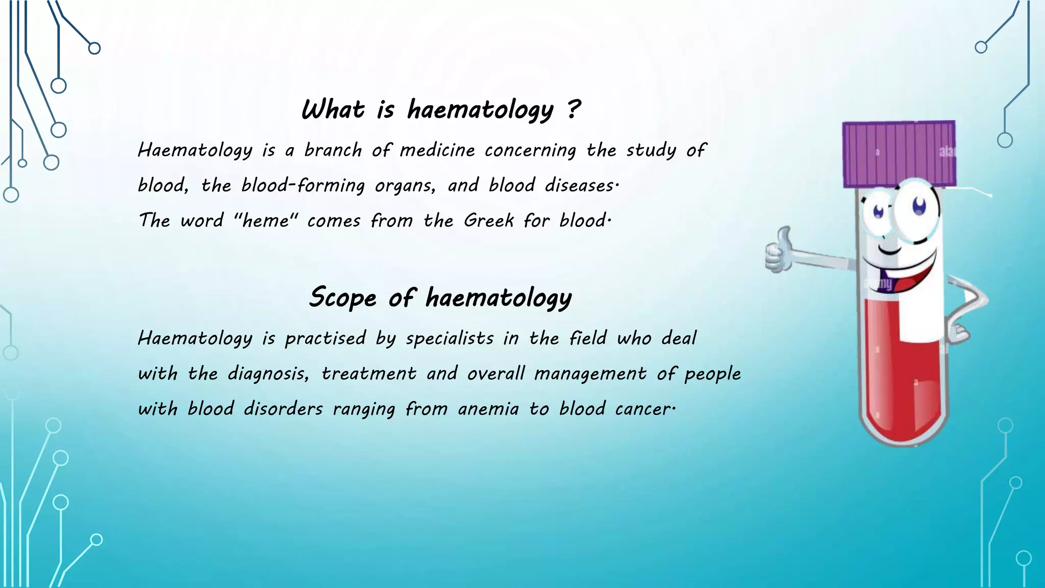
SlideShare a Scribd company logo
What is haematology ?
Haematology is a branch of medicine concerning the study of
blood, the blood-forming organs, and blood diseases.
The word "heme" comes from the Greek for blood.
Scope of haematology
Haematology is practised by specialists in the field who deal
with the diagnosis, treatment and overall management of people
with blood disorders ranging from anemia to blood cancer.
Training and work
• Physicians working in the field of hematology are
called hematologists.
• Initially, hematologists complete a four-year
medical degree and this is followed by three or
four years in an internship or residency program.
• Thereafter, they spend two or three more years
learning how to diagnose and treat blood disorders.
• B.Sc (Clinical Lab Technology)
• M.Sc (Medical Lab technology)
• DMLT
• PGDCLT
Extensive, first-hand practical experience in a
recognised training centre provides hematologists with
expertise in the following areas:
• The cause of abnormalities in blood formation and
other blood disorders
• The diagnosis of various blood related disorders and
cancers using laboratory tests and workups
• The care and treatment of patients with blood
diseases or hematological diseases
Some of the diseases treated by haematologists include:
• Iron deficiency anaemia and other types of anaemia such as sickle cell anaemia or
trauma-related anaemia
• Polycythaemia or excess production of red blood cells
• Myelofibrosis
• Leukaemia
• Platelet and bleeding disorders such as haemophilia, idiopathic thrombocytopenic
purpura and Von Willebrand disease
• The myelodysplastic syndromes
• Hemoglobinopathies such as thalassemia and sickle cell disease
• Multiple myeloma
• Malignant lymphomas
• Blood transfusion
• Bone marrow stem cell transplantation
SICKLE CELL
ANAEMIA
SICKLE CELL ANAEMIA
• Sickle cell anaemia is one of a group of inherited disorders known as
sickle cell disease.
• It affects the shape of red blood cells, which carry oxygen to all parts
of the body.
• Red blood cells are usually round and flexible, so they move easily
through blood vessels. In sickle cell anemia, some red blood cells are
shaped like sickles or crescent moons.
• These sickle cells also become rigid and sticky, which can slow or block
blood flow.
• There's no cure for most people with sickle cell anemia.
• Treatments can relieve pain and help prevent complications associated
with the disease.
Signs and symptoms
Signs and symptoms of sickle cell anemia usually appear around 6 months of age. They vary
from person to person and may change over time. Signs and symptoms can include:
• Anemia
• Episodes of pain
• Swelling in hands and feet
• Frequent infections
• Delayed growth or puberty
• Vision problems
Causes
Sickle cell anemia is caused by a change in the gene that tells the body to make the iron-rich
compound in red blood cells called hemoglobin. Hemoglobin enables red blood cells to carry
oxygen from the lungs throughout the body. The hemoglobin associated with sickle cell anemia
causes red blood cells to become rigid, sticky and misshapen. For a child to be affected, both
mother and father must carry one copy of the sickle cell gene — also known as sickle cell trait
— and pass both copies of the altered form to the child.
If only one parent passes the sickle cell gene to the child, that child will have the
sickle cell trait. With one typical hemoglobin gene and one altered form of the
gene, people with the sickle cell trait make both typical hemoglobin and sickle cell
hemoglobin.
Their blood might contain some sickle cells, but they generally don't have
symptoms. They're carriers of the disease, however, which means they can pass the
gene to their children.
Risk factors
For a baby to be born with sickle cell anemia, both parents must carry a sickle cell
gene. In the United States, sickle cell anemia most commonly affects people of
African, Mediterranean and Middle Eastern descent.
Complications
Sickle cell anemia can lead to a host of complications, including:
• Stroke
• Acute chest syndrome
• Pulmonary hypertension
• Organ damage
• Blindness
• Leg ulcer
• Splenic sequestration
• Gall stones
• Pregnancy complications
• Priapism
• Deep vein thrombosis
Prevention
If you carry the sickle cell trait, seeing a genetic counselor before
trying to conceive can help you understand your risk of having a
child with sickle cell anemia.
A genetic counselor can also explain possible treatments, preventive
measures and reproductive options.
POLYCYTHAEMIA
POLYCYTHEMIA
• Polycythemia refers to an increase in the number of red blood
cells in the body.
• The extra cells cause the blood to be thicker, and this, in
turn, increases the risk of other health issues, such as blood
clots.
• Polycythemia can have different causes, each of which has its
own treatment options. The treatment of polycythemia
involves treating any underlying conditions, if possible, and
finding ways to bring blood cell levels down.
Types of polycythemia
There are two types of polycythemia, which have different causes.
1. Primary polycythemia
• Primary polycythemia is also called polycythemia vera (PV).
• PV is a rare, slow growing blood cancer that is a type of condition known as a
myeloproliferative neoplasm.
• PV causes the bone marrow to create excess precursor blood cells that develop and
function abnormally, leading to the production of too many red blood cells.
• A person with PV may also have increased numbers of other blood cells, such as white
blood cells or platelets.
2. Secondary polycythemia
• Secondary polycythemia can occur if the increase in red blood cells is not due
to the myeloproliferative disease of PV.
• The overproduction of blood cells in secondary polycythemia is limited to the
red blood cells.
Causes of secondary polycythemia
• being at a very high altitude
• obstructive sleep apnea
• certain types of tumor
• heart or lung disease that causes a low oxygen level in the body
Risk factors
• Some people may be more at risk than others of primary polycythemia. The National
Center for Advancing Translational Science note that most cases of PV appear as a
person ages, usually around the age of 60 years.
• They also note that it is more common in men than in women.
• PV is not necessarily hereditary, and most people with the disease do not have a family
history of PV.
• However, there appears to be a connection to a certain genetic mutation.
• The Leukemia & Lymphoma Society note that almost all individuals with PV have a
mutation in the Janus kinase 2 (JAK2) gene.
• However, the precise role that it plays in the condition is still unclear.
• Other gene mutations, such as that of the TET2 gene, may also have an association
with this condition.
• Most of the time, these genes are not hereditary, but in some rare cases, they may
pass from a parent to their child in the sperm or egg.
• Anyone with a family history of PV may still wish to see a doctor for an evaluation.
SYMPTOMS
• dizziness or vertigo
• headaches
• excessive sweating
• itchy skin
• ringing in the ears
• blurred vision
• fatigue
• reddish or purplish skin on the palms, earlobes, and nose
• bleeding or bruising
• a burning sensation in the feet
• abdominal fullness
• frequent nosebleeds
• bleeding gums
COMPLICATIONS
• enlarged spleen
• blood clots
• angina
• stroke
• peptic ulcers
• heart disease
• gout
• other blood disorders, such as myelofibrosis or
leukaemia
Diagnosis
If doctors suspect that a person has polycythemia, they will order several tests to help identify
the underlying issue.
1. Blood tests
Blood tests, such as a complete blood count, will reveal any increase in red blood cells in the
bloodstream, as well as any abnormal levels of platelets and white blood cells. If PV seems
possible, a doctor will order more specific blood tests.
2. Bone marrow biopsy
If necessary, the doctor may also want to remove some bone marrow on which to perform tests
in a lab.A bone marrow biopsy involves taking a small sample of the bone marrow with a needle
for examination under a microscope.
3. Genetic tests
Although genetic causes of PV are more rare, doctors may also want to analyze the person’s
bone marrow for genetic mutations that have links to PV.They may also recommend examining
cells in the blood for JAK2 mutation
Treatment
Treatment for polycythemia will depend on the underlying cause of the condition.
In people with secondary polycythemia resulting from causes such as obstructive sleep
apnea, treating the underlying cause should help resolve the high red blood cell count.
PV, however, is a chronic condition that has no known cure. The treatment for PV aims
to manage the condition by reducing both the red blood cell count and the risk of
complications, such as blood clots. If the platelet count is too high, there are ways to
treat this as well.
Treatment options for PV can include:
Phlebotomy
To help manage the condition, doctors will look to reduce the red blood cell count
through a process called phlebotomy, which manually removes blood through one of the
veins.
Depending on the individual case, doctors will recommend removing a certain amount of
blood at set intervals to get the red blood cell count closer to normal levels.
Drugs to reduce blood cells
Sometimes, phlebotomy is not enough to control blood cell growth. Doctors
may then recommend myelosuppressive drugs to help control blood cell
counts.
These drugs include hydroxyurea (Hydrea), which suppresses the bone marrow
to keep it from producing as many blood cells.
JAK2 inhibitors
In cases where a person does not respond well to other medications, doctors
may recommend drugs that inhibit the JAK2 enzyme, which the JAK2 gene
is responsible for producing. These include ruxolitinib (Jakafi).
Other medications
Doctors will also often recommend other drugs to help control symptoms.
These include aspirin and antihistamines. Taking low dose aspirin helps reduce
the risk of clotting in many people with PV, and it may also alleviate
troubling symptoms, such as headaches. Doctors may recommend
antihistamine drugs to help relieve itchiness.
LEUKEMIA
What is leukemia?
• Leukemia is cancer of the body's blood-forming tissues, including the
bone marrow and the lymphatic system.
• Many types of leukemia exist. Some forms of leukemia are more
common in children. Other forms of leukemia occur mostly in adults.
• Leukemia usually involves the white blood cells.
• Your white blood cells are potent infection fighters — they normally
grow and divide in an orderly way, as your body needs them.
• But in people with leukemia, the bone marrow produces an excessive
amount of abnormal white blood cells, which don't function properly.
• Treatment for leukemia can be complex — depending on the type of
leukemia and other factors.
• But there are strategies and resources that can help make your
treatment successful.
Symptoms
Leukemia symptoms vary, depending on the type of leukemia. Common leukemia
signs and symptoms include:
• Fever or chills
• Persistent fatigue, weakness
• Frequent or severe infections
• Losing weight without trying
• Swollen lymph nodes, enlarged liver or spleen
• Easy bleeding or bruising
• Recurrent nosebleeds
• Tiny red spots in your skin (petechiae)
• Excessive sweating, especially at night
• Bone pain or tenderness
How leukemia forms ?
• In general, leukemia is thought to occur when some blood cells
acquire changes (mutations) in their genetic material or DNA.
• A cell's DNA contains the instructions that tell a cell what to
do. Normally, the DNA tells the cell to grow at a set rate and
to die at a set time.
• In leukemia, the mutations tell the blood cells to continue
growing and dividing.
• When this happens, blood cell production becomes out of
control.
• Over time, these abnormal cells can crowd out healthy blood
cells in the bone marrow, leading to fewer healthy white blood
cells, red blood cells and platelets, causing the signs and
symptoms of leukemia.
TYPES OF LEUKEMIA
The first type of classification is by how fast the leukemia progresses:
1. Acute leukemia. In acute leukemia, the abnormal blood cells are immature blood cells
(blasts). They can't carry out their normal functions, and they multiply rapidly, so the disease
worsens quickly. Acute leukemia requires aggressive, timely treatment.
2. Chronic leukemia. There are many types of chronic leukemias. Some produce too many cells
and some cause too few cells to be produced. Chronic leukemia involves more-mature blood
cells. These blood cells replicate or accumulate more slowly and can function normally for a
period of time. Some forms of chronic leukemia initially produce no early symptoms and can go
unnoticed or undiagnosed for years.
The second type of classification is by type of white blood cell affected:
1. Lymphocytic leukemia. This type of leukemia affects the lymphoid cells (lymphocytes), which
form lymphoid or lymphatic tissue. Lymphatic tissue makes up your immune system.
2. Myelogenous (my-uh-LOHJ-uh-nus) leukemia. This type of leukemia affects the myeloid
cells. Myeloid cells give rise to red blood cells, white blood cells and platelet-producing cells.
The major types of leukemia are:
1. Acute lymphocytic leukemia (ALL). This is the most common type of leukemia in
young children. ALL can also occur in adults.
2. Acute myelogenous leukemia (AML). AML is a common type of leukemia. It
occurs in children and adults. AML is the most common type of acute leukemia in
adults.
3. Chronic lymphocytic leukemia (CLL). With CLL, the most common chronic adult
leukemia, you may feel well for years without needing treatment.
4. Chronic myelogenous leukemia (CML). This type of leukemia mainly affects
adults. A person with CML may have few or no symptoms for months or years
before entering a phase in which the leukemia cells grow more quickly.
5. Other types. Other, rarer types of leukemia exist, including hairy cell leukemia,
myelodysplastic syndromes and myeloproliferative disorders.
Risk factors
Factors that may increase your risk of developing some types of leukemia
include:
1. Previous cancer treatment. People who've had certain types of
chemotherapy and radiation therapy for other cancers have an increased risk
of developing certain types of leukemia.
2. Genetic disorders. Genetic abnormalities seem to play a role in the
development of leukemia. Certain genetic disorders, such as Down syndrome,
are associated with an increased risk of leukemia.
3. Exposure to certain chemicals. Exposure to certain chemicals, such as
benzene — which is found in gasoline and is used by the chemical industry —
is linked to an increased risk of some kinds of leukemia.
4. Smoking. Smoking cigarettes increases the risk of acute myelogenous
leukemia.
5. Family history of leukemia. If members of your family have been diagnosed
with leukemia, your risk of the disease may be increased.
BLEEDING DISORDER
What id bleeding disorder ?
• A bleeding disorder, sometimes called a coagulopathy,
is a condition that makes you more likely to bleed
than the average person.
• The body cannot form a clot properly. This involves
one of the parts of our coagulation (bleeding and
clotting) system.
• The coagulation system should be in balance—not too
much bleeding but also not too much clotting.
• One part of the process involves the platelets, a
type of blood cell.
• The second part is found in the liquid portion of
blood, called coagulation factors. People sometimes
refer to people with bleeding disorders as
“free bleeders.”
SYMPTOMS
• Frequent or prolonged nosebleeds
• Recurrent or prolonged bleeding of the
gums
• Excessive menstrual periods referred to
as menorrhagia as well as
excessive bleeding postpartum (after
delivery)
• Prolonged bleeding after a needle stick
(blood draw or injection like a vaccine)
• Excessive bleeding during or after surgery
that requires additional efforts to
stop bleeding
• Large palpable bruises
Causes
• Inherited: Some bleeding disorders like hemophilia and Von Willebrand disease are inherited,
meaning the person is born with the disease.
• Liver disease: The coagulation factors are made predominantly in the liver. People with
severe liver disease are unable to produce enough coagulation factors and therefore are
more likely to experience bleeding.
• Vitamin K deficiency: Several of the coagulation factors require vitamin K to function
correctly so people who are vitamin K deficient are more likely to have bleeding.
• Anticoagulation therapy: People on anticoagulation therapy (medications that prevent
clotting) are at increased risk of bleeding.
• Platelet disorders: If the platelets cannot function properly, they cannot form a proper
clot, which may result in bleeding.
Types of Bleeeding disorders
1. Hemophilia:
• Probably the most well known bleeding disorder is hemophilia.
• People with hemophilia are missing one of their coagulation
factors.
• The name of their type of hemophilia depends on what factor
is missing.
• The most common type is hemophilia A; these patients are
missing factor 8 from their coagulation system.
• There is also hemophilia B (factor 9 deficiency)1 and hemophilia
C (factor 11 deficiency).
• Hemophilia is inherited (passed down in families).
• It involves the Y chromosome so men are predominantly
affected.
• People with hemophilia can have significant bleeding
from injuries or spontaneous (without injury) bleeding.
2. Von Willebrand disease:
• Von Willebrand disease (VWD) is the most common bleeding disorder in the world.
• It is estimated that 1% of the population has some form of Von Willebrand disease.
• VWD is also inherited but both men and women can be equally affected.
• The amount of bleeding can vary greatly depending on what type of VWD a person inherits.
3. Liver disease:
• When the liver is not functioning properly, it is unable to produce coagulation factors.
• Without these factors, patients can have significant bleeding.
4. Low platelet count (thrombocytopenia):
• There are numerous reasons for why the platelet count is low including leukemia, a side
effect of chemotherapy, and immune thrombocytopenia (where the immune system
destroys the platelets).
5. Platelet function disorders:
• In addition to low platelet numbers causing bleeding, if the platelets do not function
properly, bleeding may occur
TREATMENT
1. Factor replacement: Patients with hemophilia can infuse themselves with
factor replacement products that can treat bleeding episodes. Nowadays, many patients
with hemophilia infuse factor replacements on a regular basis to prevent bleeding episodes.
2. Desmopressin (also identified by the brand name Stimate): Desmopressin is a replacement
product for the hormone vasopressin. Taking desmopressin results in a temporary increase
in von Willebrand antigen and factor 8 which may stop bleeding in patients with
mild hemophilia A or von Willebrand disease.
3. Platelet transfusions: If the platelet count is low or the platelets do not
function correctly, a platelet transfusion may be given to prevent/treat bleeding.
4. Fresh frozen plasma: Coagulation factors are found in the plasma (liquid component
of blood). If a person is missing several different factors, like in liver failure, an infusion
of fresh frozen plasma can be given.
5. Vitamin K: If a patient if Vitamin K deficient, supplementation can be given.
6. Antifibrinolytics: These medications prevent excessive bleeding by stabilizing clots. These
are primarily used to control bleeding in the mouth or heavy menstrual periods.
MULTIPLE MYELOMA
What is multiple myeloma?
• Multiple myeloma also known as Kahler's disease, is a type of blood cancer.
• There's no cure, but treatments can slow its spread and sometimes make symptoms go
away.
• A type of white blood cell called a plasma cell makes antibodies that fight infections in
your body.
• When you have multiple myeloma, these cells multiply the wrong way.
• They let too much protein (called immunoglobulin) into your bones and blood.
• It builds up throughout your body and damages your organs.
• The plasma cells crowd out regular blood cells in your bones.
• They also send out chemicals that trigger other cells to eat away at your bones.
• The weak areas that this creates in your bones are called lytic lesions.
• As multiple myeloma gets worse, the plasma cells spill out of your bone marrow and
spread. This causes more organ damage.
Multiple Myeloma Causes and Risk Factors
Experts aren't sure what causes multiple myeloma. But you're more likely to get it if:
• You're older than 65
• You're male
• You're African American
• You have a family member with it
• You're overweight or obese
• You've been exposed to radiation
• You've had contact with chemicals used in rubber manufacturing, woodworking, or
firefighting; or in herbicides
Multiple Myeloma Symptoms
Early on, you might not notice any symptoms. But over time,
you may have:
• Bone pain
• Weakness and fatigue
• Weight loss and loss of appetite
• Upset stomach
• Constipation
• Confusion
• Frequent infections
• Severe thirst
• Weakness or numbness in your arms and legs
Multiple Myeloma Complications
Multiple myeloma can cause problems including:
1. Bone problems. Your bones can become weaker, leading to fractures.
2. Blood problems. You might get anemia, which means your body doesn't have
enough red blood cells. This can make you tired and pale and cause heart
problems. You might also have too few platelets, which makes it harder for
your blood to clot.
3. Infections. When you have myeloma, your body produces a lot of weak
antibodies that crowd out healthy ones, making it harder for you to fight
infection. A lack of white blood cells can also weaken your immune system.
4. Kidney damage. Myeloma can clog your kidneys so they don't filter the way
they should. This might lead to kidney failure.
Multiple Myeloma Diagnosis
Your doctor may suspect multiple myeloma if you have a blood test for something else and it
shows:
• Too much calcium in your blood (hypercalcemia)
• Too few red blood cells (anemia)
• Kidney problems
• High total protein levels in your blood, but low levels of one called albumin.
To confirm a diagnosis, you might have blood tests including:
1. A complete blood count (CBC). It measures the different kinds of cells in your blood.
2. Blood urea nitrogen (BUN) and creatinine. These check how well your kidneys are working.
Other blood and urine tests check whether your body is making unusual proteins and, if so,
what kinds and how much.
Multiple Myeloma Treatment
• Doctors grade multiple myeloma cases as high,
intermediate, or standard risk, based on genes in the
tumors.
• If you don't have symptoms, your doctor may choose to
watch you closely rather than start treatment right away.
• If you have symptoms, your doctor will work with you to
come up with a treatment plan. It will aim to improve
your quality of life by making you feel better and helping
you get enough nutrition.
• If your case is high-risk, you might consider joining a clinical
trial for an existing or new treatment. Research is looking
for more effective drugs.
BLOOD TRANSFUSION
What is blood transfusion ?
• A blood transfusion is a routine medical procedure in which donated blood is
provided to you through a narrow tube placed within a vein in your arm.
• This potentially life-saving procedure can help replace blood lost due to
surgery or injury.
• A blood transfusion also can help if an illness prevents your body from
making blood or some of your blood's components correctly.
• Blood transfusions usually occur without complications. When complications
do occur, they're typically mild.
Why it's done?
People receive blood transfusions for many reasons — such as surgery, injury, disease
and bleeding disorders.
Blood has several components, including:
• Red cells carry oxygen and help remove waste products
• White cells help your body fight infections
• Plasma is the liquid part of your blood
• Platelets help your blood clot properly
A transfusion provides the part or parts of blood you need, with red blood cells being
the most commonly transfused. You can also receive whole blood, which contains all
the parts, but whole blood transfusions aren't common.
How you prepare ?
Your blood will be tested before a transfusion to determine whether your blood type is
A, B, AB or O and whether your blood is Rh positive or Rh negative. The donated
blood used for your transfusion must be compatible with your blood type.
Tell your health care provider if you've had a reaction to a blood transfusion in the
past.
What you can expect ?
Blood transfusions are usually done in a hospital, an outpatient clinic or a doctor's
office. The procedure typically takes one to four hours, depending on which parts of the
blood you receive and how much blood you need.
Before the procedure
In some cases, you can donate blood for yourself before elective surgery, but most
transfusions involve blood donated by strangers. An identification check will ensure you
receive the correct blood.
During the procedure
An intravenous (IV) line with a needle is inserted into one of your blood vessels. The donated
blood that's been stored in a plastic bag enters your bloodstream through the IV. You'll be seated
or lying down for the procedure, which usually takes one to four hours.
A nurse will monitor you throughout the procedure and take measures of your blood pressure,
temperature and heart rate. Tell the nurse immediately if you develop:
• Fever
• Shortness of breath
• Chills
• Unusual itching
• Chest or back pain
• A sense of uneasiness
After the procedure
The needle and IV line will be removed. You might develop a bruise around the needle site,
but this should go away in a few days.
Contact your health care provider if you develop shortness of breath or chest or back pain
in the days immediately following a blood transfusion.
Results
You might need further blood testing to see how your body is responding to the donor
blood and to check your blood counts.
Some conditions require more than one blood transfusion.
Bloodborne infections
Blood banks screen donors and test donated blood to reduce the risk of transfusion-related
infections, so infections, such as HIV or hepatitis B or C, are extremely rare.
Other serious reactions
Also rare, these include:
1. Acute immune hemolytic reaction. Your immune system attacks the transfused red blood cells
because the donor blood type is not a good match. The attacked cells release a substance
into your blood that harms your kidneys.
2. Delayed hemolytic reaction. Similar to an acute immune hemolytic reaction, this reaction
occurs more slowly. It can take one to four weeks to notice a decrease in red blood cell
levels.
3. Graft-versus-host disease. In this condition, transfused white blood cells attack your bone
marrow. Usually fatal, it's more likely to affect people with severely weakened immune
systems, such as those being treated for leukemia or lymphoma.
BONEMARROW
STEM CELL TRANSPLANTATION
What is bonemarrow transplantation ?
• A bone marrow transplant is a procedure that infuses healthy blood-forming
stem cells into your body to replace your damaged or diseased bone marrow.
• A bone marrow transplant is also called a stem cell transplant.
• You might need a bone marrow transplant if your bone marrow stops working
and does not produce enough healthy blood cells.
• Bone marrow transplants may use cells from your own body (autologous
transplant) or from a donor (allogeneic transplant).
Why it's done ?
A bone marrow transplant may be used to:
• Safely allow treatment of your condition with high doses of chemotherapy or
radiation by replacing or rescuing the bone marrow damaged by treatment
• Replace diseased or damaged marrow with new stem cells
• Provide new stem cells, which can help kill cancer cells directly.
Bone marrow transplants can benefit people with a variety of both cancerous (malignant)
and noncancerous (benign) diseases, including:
• Acute leukaemia
• Adrenoleukodystrophy
• Aplastic anaemia
• Bone marrow failure syndromes
• Chronic leukaemia
• Hemoglobinopathies
• Hodgkin's lymphoma
• Immune deficiencies
• Inborn errors of metabolism
• Multiple myeloma
• Myelodysplastic syndromes
• Neuroblastoma
• Non-Hodgkin's lymphoma
• Plasma cell disorders
• POEMS syndrome
• Primary amyloidosis
RISKS
Possible complications from a bone marrow transplant
include:
• Graft-versus-host disease (allogeneic transplant
only)
• Stem cell (graft) failure
• Organ damage
• Infections
• Cataracts
• Infertility
• New cancers
• Death
How you prepare ?
Pretransplant tests and procedures
You'll undergo a series of tests and procedures to assess your general health and
the status of your condition, and to ensure that you're physically prepared for
the transplant.
The evaluation may take several days or more.
In addition, a surgeon or radiologist will implant a long thin tube (intravenous
catheter) into a large vein in your chest or neck.
The catheter, often called a central line, usually remains in place for the duration
of your treatment.
Your transplant team will use the central line to infuse the transplanted stem
cells, medications and blood products into your body.
Collecting stem cells for autologous transplant
• If a transplant using your own stem cells (autologous
transplant) is planned, you'll undergo a procedure called
apheresis to collect blood stem cells.
• Before apheresis, you'll receive daily injections of growth
factor to increase stem cell production and move stem
cells into your circulating blood so that they can be
collected.
• During apheresis, blood is drawn from a vein and
circulated through a machine. The machine separates
your blood into different parts, including stem cells.
These stem cells are collected and frozen for future use
in the transplant. The remaining blood is returned to
your body.
Collecting stem cells for allogeneic transplant
• If you're having a transplant using stem cells from a donor
(allogeneic transplant), you will need a donor.
• Once a donor is found, stem cells are gathered from that
person for the transplant.
• Stem cells can come from your donor's blood or bone marrow.
Your transplant team decides which is better for you based on
your situation.
• Another type of allogeneic transplant uses stem cells from the
blood of umbilical cords (cord blood transplant).
• Mothers can choose to donate umbilical cords after their babies'
births.
• The blood from these cords is frozen and stored in a cord blood
bank until needed for a bone marrow transplant.
CONDITIONING
After you complete your pretransplant tests and procedures, you begin a process known as
conditioning. During conditioning, you'll undergo chemotherapy and possibly radiation to:
• Destroy cancer cells if you are being treated for a malignancy
• Suppress your immune system
• Prepare your bone marrow for the new stem cells
Side effects of the conditioning process can include:
• Nausea and vomiting
• Diarrhea
• Hair loss
• Mouth sores or ulcers
• Infection
• Bleeding
• Infertility or sterility
• Anemia
• Fatigue
• Cataracts
• Organ complications, such as heart, liver or lung
failure
DIET AND LIFESTYLE FACTORS
Following food safety guidelines to prevent foodborne infections
• Eating a wide variety of healthy foods, including vegetables; fruits; whole grains;
lean meats, poultry and fish; legumes; and healthy fats, such as olive oil
• Limiting salt intake
• Restricting alcohol
• Avoiding grapefruit and grapefruit juice due to their effect on a group of
immunosuppressive medications (calcineurin inhibitors)
• After your bone marrow transplant, regular physical activity helps you control
your weight, strengthen your bones, increase your endurance, strengthen your
muscles and keep your heart healthy. As you recover, you can slowly increase
your physical activity.
• Taking steps to prevent cancer is even more important after your transplant.
Don't smoke.
HAEMATOLOGY INTRODUCTION.pptx

More Related Content

What's hot

PLATELET COUNT by Dr. Pandian M .pptx
PLATELET COUNT by Dr. Pandian M .pptxPLATELET COUNT by Dr. Pandian M .pptx
PLATELET COUNT by Dr. Pandian M .pptx
Pandian M
 
cbc Histogram
cbc Histogramcbc Histogram
cbc Histogram
TONY SCARIA
 
Erythrocyte indices
Erythrocyte  indicesErythrocyte  indices
Erythrocyte indices
Dr. Pritika Nehra
 
Leukemia
LeukemiaLeukemia
Hemolytic anemia (1)
Hemolytic anemia (1)Hemolytic anemia (1)
Hemolytic anemia (1)
vvyvi
 
Introduction to haematology notes for MLT students
Introduction to haematology notes for MLT studentsIntroduction to haematology notes for MLT students
Introduction to haematology notes for MLT students
Vamsi kumar
 
Red Blood Cell disorder
Red Blood Cell  disorderRed Blood Cell  disorder
Red Blood Cell disorder
Hirut Gebremichael
 
morphology of red blood cells
morphology of red blood cellsmorphology of red blood cells
morphology of red blood cells
MLT LECTURES BY TANVEER TARA
 
Introduction to hematology
Introduction to hematologyIntroduction to hematology
Introduction to hematology
Juan Carlos Munévar
 
HEMATOLOGY: Laboratory Tests
HEMATOLOGY: Laboratory TestsHEMATOLOGY: Laboratory Tests
HEMATOLOGY: Laboratory Tests
hm alumia
 
Basics of Haematology
Basics of HaematologyBasics of Haematology
Basics of Haematology
Reyaz Bhat
 
Hematology
HematologyHematology
Hematology
DJ CrissCross
 
introduction of cytopathology
introduction of cytopathologyintroduction of cytopathology
introduction of cytopathology
Appy Akshay Agarwal
 
Blood film examination
Blood film examinationBlood film examination
Blood film examination
ayeayetun08
 
Polycythemia
PolycythemiaPolycythemia
Polycythemia
SUNIL KUMAR PEDDANA
 
Leucopenia 07 (3)
Leucopenia 07 (3)Leucopenia 07 (3)
Leucopenia 07 (3)
AshwineeGC
 
Hematology
HematologyHematology
Hematology
ariva zhagan
 
Blood disorders ppt
Blood disorders pptBlood disorders ppt
Blood disorders ppt
K BHATTACHARJEE
 
Sickle cell anaemia
Sickle cell anaemiaSickle cell anaemia
Sickle cell anaemia
dr pushkar chaudhary
 

What's hot (20)

bone marrow
bone marrowbone marrow
bone marrow
 
PLATELET COUNT by Dr. Pandian M .pptx
PLATELET COUNT by Dr. Pandian M .pptxPLATELET COUNT by Dr. Pandian M .pptx
PLATELET COUNT by Dr. Pandian M .pptx
 
cbc Histogram
cbc Histogramcbc Histogram
cbc Histogram
 
Erythrocyte indices
Erythrocyte  indicesErythrocyte  indices
Erythrocyte indices
 
Leukemia
LeukemiaLeukemia
Leukemia
 
Hemolytic anemia (1)
Hemolytic anemia (1)Hemolytic anemia (1)
Hemolytic anemia (1)
 
Introduction to haematology notes for MLT students
Introduction to haematology notes for MLT studentsIntroduction to haematology notes for MLT students
Introduction to haematology notes for MLT students
 
Red Blood Cell disorder
Red Blood Cell  disorderRed Blood Cell  disorder
Red Blood Cell disorder
 
morphology of red blood cells
morphology of red blood cellsmorphology of red blood cells
morphology of red blood cells
 
Introduction to hematology
Introduction to hematologyIntroduction to hematology
Introduction to hematology
 
HEMATOLOGY: Laboratory Tests
HEMATOLOGY: Laboratory TestsHEMATOLOGY: Laboratory Tests
HEMATOLOGY: Laboratory Tests
 
Basics of Haematology
Basics of HaematologyBasics of Haematology
Basics of Haematology
 
Hematology
HematologyHematology
Hematology
 
introduction of cytopathology
introduction of cytopathologyintroduction of cytopathology
introduction of cytopathology
 
Blood film examination
Blood film examinationBlood film examination
Blood film examination
 
Polycythemia
PolycythemiaPolycythemia
Polycythemia
 
Leucopenia 07 (3)
Leucopenia 07 (3)Leucopenia 07 (3)
Leucopenia 07 (3)
 
Hematology
HematologyHematology
Hematology
 
Blood disorders ppt
Blood disorders pptBlood disorders ppt
Blood disorders ppt
 
Sickle cell anaemia
Sickle cell anaemiaSickle cell anaemia
Sickle cell anaemia
 

Similar to HAEMATOLOGY INTRODUCTION.pptx

Polycythemia
PolycythemiaPolycythemia
Polycythemia
BilalJan15
 
Polycythemia by Dr. Sookun Rajeev Kumar
Polycythemia by Dr. Sookun Rajeev KumarPolycythemia by Dr. Sookun Rajeev Kumar
Polycythemia by Dr. Sookun Rajeev Kumar
Dr. Sookun Rajeev Kumar
 
Polycythemia iquin
Polycythemia iquinPolycythemia iquin
Polycythemia iquiniquin
 
SICKLE- CELL ANEMIA.pptx
SICKLE- CELL ANEMIA.pptxSICKLE- CELL ANEMIA.pptx
SICKLE- CELL ANEMIA.pptx
RohanTeja
 
Haemopoetic system Introduction.pptx
Haemopoetic system Introduction.pptxHaemopoetic system Introduction.pptx
Haemopoetic system Introduction.pptx
SunaynaChoudhary
 
Overview of Anemia
Overview of AnemiaOverview of Anemia
Overview of Anemia
Medicine Points
 
Pseudovasculitides. Mikhail Valivach. 2015
Pseudovasculitides. Mikhail Valivach. 2015Pseudovasculitides. Mikhail Valivach. 2015
Pseudovasculitides. Mikhail Valivach. 2015
Mikhail Valivach
 
hematology and endocrinology
hematology and endocrinologyhematology and endocrinology
hematology and endocrinology
Rasha Ibrahim
 
Aplasticanemia.pptx
Aplasticanemia.pptxAplasticanemia.pptx
Aplasticanemia.pptx
bhavanibb
 
Medical bt seminar.pptx abc
Medical bt  seminar.pptx abcMedical bt  seminar.pptx abc
Medical bt seminar.pptx abc
AakifahAmreen
 
#seminar on how to approach a patient with anemia
#seminar on how to approach a patient with anemia#seminar on how to approach a patient with anemia
#seminar on how to approach a patient with anemia
LuzSan3
 
SICKEL CELL ANEMIA
SICKEL CELL ANEMIA SICKEL CELL ANEMIA
SICKEL CELL ANEMIA
Mansishejwalkar
 
Polycythemia
PolycythemiaPolycythemia
Polycythemia
MR. JAGDISH SAMBAD
 
Polycythemia vera rumana
Polycythemia vera rumanaPolycythemia vera rumana
Polycythemia vera rumana
Rumana Hameed
 
Sickle Cell Anemia. Student Presentation
Sickle Cell Anemia. Student PresentationSickle Cell Anemia. Student Presentation
Sickle Cell Anemia. Student PresentationSEPA_genomics
 
Approach to anaemia
Approach to anaemiaApproach to anaemia
Approach to anaemia
PhocasBIMENYIMANA
 
Investigation in dentistry by nabaa.pptx
Investigation in dentistry by nabaa.pptxInvestigation in dentistry by nabaa.pptx
Investigation in dentistry by nabaa.pptx
nabaan993
 
Global Medical Cures™ | Blood disorders
Global Medical Cures™ | Blood disordersGlobal Medical Cures™ | Blood disorders
Global Medical Cures™ | Blood disorders
Global Medical Cures™
 

Similar to HAEMATOLOGY INTRODUCTION.pptx (20)

Polycythemia
PolycythemiaPolycythemia
Polycythemia
 
Polycythemia by Dr. Sookun Rajeev Kumar
Polycythemia by Dr. Sookun Rajeev KumarPolycythemia by Dr. Sookun Rajeev Kumar
Polycythemia by Dr. Sookun Rajeev Kumar
 
Polycythemia iquin
Polycythemia iquinPolycythemia iquin
Polycythemia iquin
 
SICKLE- CELL ANEMIA.pptx
SICKLE- CELL ANEMIA.pptxSICKLE- CELL ANEMIA.pptx
SICKLE- CELL ANEMIA.pptx
 
Haemopoetic system Introduction.pptx
Haemopoetic system Introduction.pptxHaemopoetic system Introduction.pptx
Haemopoetic system Introduction.pptx
 
Overview of Anemia
Overview of AnemiaOverview of Anemia
Overview of Anemia
 
Pseudovasculitides. Mikhail Valivach. 2015
Pseudovasculitides. Mikhail Valivach. 2015Pseudovasculitides. Mikhail Valivach. 2015
Pseudovasculitides. Mikhail Valivach. 2015
 
hematology and endocrinology
hematology and endocrinologyhematology and endocrinology
hematology and endocrinology
 
polycythemia
polycythemiapolycythemia
polycythemia
 
Aplasticanemia.pptx
Aplasticanemia.pptxAplasticanemia.pptx
Aplasticanemia.pptx
 
Medical bt seminar.pptx abc
Medical bt  seminar.pptx abcMedical bt  seminar.pptx abc
Medical bt seminar.pptx abc
 
#seminar on how to approach a patient with anemia
#seminar on how to approach a patient with anemia#seminar on how to approach a patient with anemia
#seminar on how to approach a patient with anemia
 
SICKEL CELL ANEMIA
SICKEL CELL ANEMIA SICKEL CELL ANEMIA
SICKEL CELL ANEMIA
 
Polycythemia
PolycythemiaPolycythemia
Polycythemia
 
Polycythemia vera rumana
Polycythemia vera rumanaPolycythemia vera rumana
Polycythemia vera rumana
 
Sickle Cell Anemia. Student Presentation
Sickle Cell Anemia. Student PresentationSickle Cell Anemia. Student Presentation
Sickle Cell Anemia. Student Presentation
 
Approach to anaemia
Approach to anaemiaApproach to anaemia
Approach to anaemia
 
Pnh 2
Pnh 2Pnh 2
Pnh 2
 
Investigation in dentistry by nabaa.pptx
Investigation in dentistry by nabaa.pptxInvestigation in dentistry by nabaa.pptx
Investigation in dentistry by nabaa.pptx
 
Global Medical Cures™ | Blood disorders
Global Medical Cures™ | Blood disordersGlobal Medical Cures™ | Blood disorders
Global Medical Cures™ | Blood disorders
 

More from D. JASMINE PRIYA

hemostatic function tests.pptx
hemostatic function tests.pptxhemostatic function tests.pptx
hemostatic function tests.pptx
D. JASMINE PRIYA
 
Compatability testing.pptx
Compatability testing.pptxCompatability testing.pptx
Compatability testing.pptx
D. JASMINE PRIYA
 
Hemolytic disease in new born
Hemolytic disease in new bornHemolytic disease in new born
Hemolytic disease in new born
D. JASMINE PRIYA
 
Histopathology staining method
Histopathology staining methodHistopathology staining method
Histopathology staining method
D. JASMINE PRIYA
 
programmed cell death .pptx
programmed cell death .pptxprogrammed cell death .pptx
programmed cell death .pptx
D. JASMINE PRIYA
 
DNA, RNA, PROTEIN.pptx
DNA, RNA, PROTEIN.pptxDNA, RNA, PROTEIN.pptx
DNA, RNA, PROTEIN.pptx
D. JASMINE PRIYA
 
Sterilization.pptx
Sterilization.pptxSterilization.pptx
Sterilization.pptx
D. JASMINE PRIYA
 
microbiology branches.pptx
microbiology branches.pptxmicrobiology branches.pptx
microbiology branches.pptx
D. JASMINE PRIYA
 
MICROBIOLOGY - INTRODUCTION.pptx
MICROBIOLOGY - INTRODUCTION.pptxMICROBIOLOGY - INTRODUCTION.pptx
MICROBIOLOGY - INTRODUCTION.pptx
D. JASMINE PRIYA
 
CYTOLOGY PPT.pptx
CYTOLOGY PPT.pptxCYTOLOGY PPT.pptx
CYTOLOGY PPT.pptx
D. JASMINE PRIYA
 
BLOOD BANKING.pptx
BLOOD BANKING.pptxBLOOD BANKING.pptx
BLOOD BANKING.pptx
D. JASMINE PRIYA
 
SYSTEMS OF HUMAN BODY AND ITS FUNCTIONS
SYSTEMS OF HUMAN BODY AND ITS FUNCTIONS SYSTEMS OF HUMAN BODY AND ITS FUNCTIONS
SYSTEMS OF HUMAN BODY AND ITS FUNCTIONS
D. JASMINE PRIYA
 
Human anatomy and physiology lesson
Human anatomy and physiology lessonHuman anatomy and physiology lesson
Human anatomy and physiology lesson
D. JASMINE PRIYA
 
Clinical pathology ppt
Clinical pathology pptClinical pathology ppt
Clinical pathology ppt
D. JASMINE PRIYA
 
Histopathology introduction
Histopathology introductionHistopathology introduction
Histopathology introduction
D. JASMINE PRIYA
 

More from D. JASMINE PRIYA (15)

hemostatic function tests.pptx
hemostatic function tests.pptxhemostatic function tests.pptx
hemostatic function tests.pptx
 
Compatability testing.pptx
Compatability testing.pptxCompatability testing.pptx
Compatability testing.pptx
 
Hemolytic disease in new born
Hemolytic disease in new bornHemolytic disease in new born
Hemolytic disease in new born
 
Histopathology staining method
Histopathology staining methodHistopathology staining method
Histopathology staining method
 
programmed cell death .pptx
programmed cell death .pptxprogrammed cell death .pptx
programmed cell death .pptx
 
DNA, RNA, PROTEIN.pptx
DNA, RNA, PROTEIN.pptxDNA, RNA, PROTEIN.pptx
DNA, RNA, PROTEIN.pptx
 
Sterilization.pptx
Sterilization.pptxSterilization.pptx
Sterilization.pptx
 
microbiology branches.pptx
microbiology branches.pptxmicrobiology branches.pptx
microbiology branches.pptx
 
MICROBIOLOGY - INTRODUCTION.pptx
MICROBIOLOGY - INTRODUCTION.pptxMICROBIOLOGY - INTRODUCTION.pptx
MICROBIOLOGY - INTRODUCTION.pptx
 
CYTOLOGY PPT.pptx
CYTOLOGY PPT.pptxCYTOLOGY PPT.pptx
CYTOLOGY PPT.pptx
 
BLOOD BANKING.pptx
BLOOD BANKING.pptxBLOOD BANKING.pptx
BLOOD BANKING.pptx
 
SYSTEMS OF HUMAN BODY AND ITS FUNCTIONS
SYSTEMS OF HUMAN BODY AND ITS FUNCTIONS SYSTEMS OF HUMAN BODY AND ITS FUNCTIONS
SYSTEMS OF HUMAN BODY AND ITS FUNCTIONS
 
Human anatomy and physiology lesson
Human anatomy and physiology lessonHuman anatomy and physiology lesson
Human anatomy and physiology lesson
 
Clinical pathology ppt
Clinical pathology pptClinical pathology ppt
Clinical pathology ppt
 
Histopathology introduction
Histopathology introductionHistopathology introduction
Histopathology introduction
 

Recently uploaded

The Normal Electrocardiogram - Part I of II
The Normal Electrocardiogram - Part I of IIThe Normal Electrocardiogram - Part I of II
The Normal Electrocardiogram - Part I of II
MedicoseAcademics
 
The hemodynamic and autonomic determinants of elevated blood pressure in obes...
The hemodynamic and autonomic determinants of elevated blood pressure in obes...The hemodynamic and autonomic determinants of elevated blood pressure in obes...
The hemodynamic and autonomic determinants of elevated blood pressure in obes...
Catherine Liao
 
TEST BANK for Operations Management, 14th Edition by William J. Stevenson, Ve...
TEST BANK for Operations Management, 14th Edition by William J. Stevenson, Ve...TEST BANK for Operations Management, 14th Edition by William J. Stevenson, Ve...
TEST BANK for Operations Management, 14th Edition by William J. Stevenson, Ve...
kevinkariuki227
 
ANATOMY AND PHYSIOLOGY OF URINARY SYSTEM.pptx
ANATOMY AND PHYSIOLOGY OF URINARY SYSTEM.pptxANATOMY AND PHYSIOLOGY OF URINARY SYSTEM.pptx
ANATOMY AND PHYSIOLOGY OF URINARY SYSTEM.pptx
Swetaba Besh
 
Charaka Samhita Sutra Sthana 9 Chapter khuddakachatuspadadhyaya
Charaka Samhita Sutra Sthana 9 Chapter khuddakachatuspadadhyayaCharaka Samhita Sutra Sthana 9 Chapter khuddakachatuspadadhyaya
Charaka Samhita Sutra Sthana 9 Chapter khuddakachatuspadadhyaya
Dr KHALID B.M
 
Ozempic: Preoperative Management of Patients on GLP-1 Receptor Agonists
Ozempic: Preoperative Management of Patients on GLP-1 Receptor Agonists  Ozempic: Preoperative Management of Patients on GLP-1 Receptor Agonists
Ozempic: Preoperative Management of Patients on GLP-1 Receptor Agonists
Saeid Safari
 
heat stroke and heat exhaustion in children
heat stroke and heat exhaustion in childrenheat stroke and heat exhaustion in children
heat stroke and heat exhaustion in children
SumeraAhmad5
 
POST OPERATIVE OLIGURIA and its management
POST OPERATIVE OLIGURIA and its managementPOST OPERATIVE OLIGURIA and its management
POST OPERATIVE OLIGURIA and its management
touseefaziz1
 
basicmodesofventilation2022-220313203758.pdf
basicmodesofventilation2022-220313203758.pdfbasicmodesofventilation2022-220313203758.pdf
basicmodesofventilation2022-220313203758.pdf
aljamhori teaching hospital
 
Physiology of Special Chemical Sensation of Taste
Physiology of Special Chemical Sensation of TastePhysiology of Special Chemical Sensation of Taste
Physiology of Special Chemical Sensation of Taste
MedicoseAcademics
 
Evaluation of antidepressant activity of clitoris ternatea in animals
Evaluation of antidepressant activity of clitoris ternatea in animalsEvaluation of antidepressant activity of clitoris ternatea in animals
Evaluation of antidepressant activity of clitoris ternatea in animals
Shweta
 
MANAGEMENT OF ATRIOVENTRICULAR CONDUCTION BLOCK.pdf
MANAGEMENT OF ATRIOVENTRICULAR CONDUCTION BLOCK.pdfMANAGEMENT OF ATRIOVENTRICULAR CONDUCTION BLOCK.pdf
MANAGEMENT OF ATRIOVENTRICULAR CONDUCTION BLOCK.pdf
Jim Jacob Roy
 
For Better Surat #ℂall #Girl Service ❤85270-49040❤ Surat #ℂall #Girls
For Better Surat #ℂall #Girl Service ❤85270-49040❤ Surat #ℂall #GirlsFor Better Surat #ℂall #Girl Service ❤85270-49040❤ Surat #ℂall #Girls
For Better Surat #ℂall #Girl Service ❤85270-49040❤ Surat #ℂall #Girls
Savita Shen $i11
 
ARTIFICIAL INTELLIGENCE IN HEALTHCARE.pdf
ARTIFICIAL INTELLIGENCE IN  HEALTHCARE.pdfARTIFICIAL INTELLIGENCE IN  HEALTHCARE.pdf
ARTIFICIAL INTELLIGENCE IN HEALTHCARE.pdf
Anujkumaranit
 
Alcohol_Dr. Jeenal Mistry MD Pharmacology.pdf
Alcohol_Dr. Jeenal Mistry MD Pharmacology.pdfAlcohol_Dr. Jeenal Mistry MD Pharmacology.pdf
Alcohol_Dr. Jeenal Mistry MD Pharmacology.pdf
Dr Jeenal Mistry
 
Ophthalmology Clinical Tests for OSCE exam
Ophthalmology Clinical Tests for OSCE examOphthalmology Clinical Tests for OSCE exam
Ophthalmology Clinical Tests for OSCE exam
KafrELShiekh University
 
Surgical Site Infections, pathophysiology, and prevention.pptx
Surgical Site Infections, pathophysiology, and prevention.pptxSurgical Site Infections, pathophysiology, and prevention.pptx
Surgical Site Infections, pathophysiology, and prevention.pptx
jval Landero
 
THOA 2.ppt Human Organ Transplantation Act
THOA 2.ppt Human Organ Transplantation ActTHOA 2.ppt Human Organ Transplantation Act
THOA 2.ppt Human Organ Transplantation Act
DrSathishMS1
 
KDIGO 2024 guidelines for diabetologists
KDIGO 2024 guidelines for diabetologistsKDIGO 2024 guidelines for diabetologists
KDIGO 2024 guidelines for diabetologists
د.محمود نجيب
 
Couples presenting to the infertility clinic- Do they really have infertility...
Couples presenting to the infertility clinic- Do they really have infertility...Couples presenting to the infertility clinic- Do they really have infertility...
Couples presenting to the infertility clinic- Do they really have infertility...
Sujoy Dasgupta
 

Recently uploaded (20)

The Normal Electrocardiogram - Part I of II
The Normal Electrocardiogram - Part I of IIThe Normal Electrocardiogram - Part I of II
The Normal Electrocardiogram - Part I of II
 
The hemodynamic and autonomic determinants of elevated blood pressure in obes...
The hemodynamic and autonomic determinants of elevated blood pressure in obes...The hemodynamic and autonomic determinants of elevated blood pressure in obes...
The hemodynamic and autonomic determinants of elevated blood pressure in obes...
 
TEST BANK for Operations Management, 14th Edition by William J. Stevenson, Ve...
TEST BANK for Operations Management, 14th Edition by William J. Stevenson, Ve...TEST BANK for Operations Management, 14th Edition by William J. Stevenson, Ve...
TEST BANK for Operations Management, 14th Edition by William J. Stevenson, Ve...
 
ANATOMY AND PHYSIOLOGY OF URINARY SYSTEM.pptx
ANATOMY AND PHYSIOLOGY OF URINARY SYSTEM.pptxANATOMY AND PHYSIOLOGY OF URINARY SYSTEM.pptx
ANATOMY AND PHYSIOLOGY OF URINARY SYSTEM.pptx
 
Charaka Samhita Sutra Sthana 9 Chapter khuddakachatuspadadhyaya
Charaka Samhita Sutra Sthana 9 Chapter khuddakachatuspadadhyayaCharaka Samhita Sutra Sthana 9 Chapter khuddakachatuspadadhyaya
Charaka Samhita Sutra Sthana 9 Chapter khuddakachatuspadadhyaya
 
Ozempic: Preoperative Management of Patients on GLP-1 Receptor Agonists
Ozempic: Preoperative Management of Patients on GLP-1 Receptor Agonists  Ozempic: Preoperative Management of Patients on GLP-1 Receptor Agonists
Ozempic: Preoperative Management of Patients on GLP-1 Receptor Agonists
 
heat stroke and heat exhaustion in children
heat stroke and heat exhaustion in childrenheat stroke and heat exhaustion in children
heat stroke and heat exhaustion in children
 
POST OPERATIVE OLIGURIA and its management
POST OPERATIVE OLIGURIA and its managementPOST OPERATIVE OLIGURIA and its management
POST OPERATIVE OLIGURIA and its management
 
basicmodesofventilation2022-220313203758.pdf
basicmodesofventilation2022-220313203758.pdfbasicmodesofventilation2022-220313203758.pdf
basicmodesofventilation2022-220313203758.pdf
 
Physiology of Special Chemical Sensation of Taste
Physiology of Special Chemical Sensation of TastePhysiology of Special Chemical Sensation of Taste
Physiology of Special Chemical Sensation of Taste
 
Evaluation of antidepressant activity of clitoris ternatea in animals
Evaluation of antidepressant activity of clitoris ternatea in animalsEvaluation of antidepressant activity of clitoris ternatea in animals
Evaluation of antidepressant activity of clitoris ternatea in animals
 
MANAGEMENT OF ATRIOVENTRICULAR CONDUCTION BLOCK.pdf
MANAGEMENT OF ATRIOVENTRICULAR CONDUCTION BLOCK.pdfMANAGEMENT OF ATRIOVENTRICULAR CONDUCTION BLOCK.pdf
MANAGEMENT OF ATRIOVENTRICULAR CONDUCTION BLOCK.pdf
 
For Better Surat #ℂall #Girl Service ❤85270-49040❤ Surat #ℂall #Girls
For Better Surat #ℂall #Girl Service ❤85270-49040❤ Surat #ℂall #GirlsFor Better Surat #ℂall #Girl Service ❤85270-49040❤ Surat #ℂall #Girls
For Better Surat #ℂall #Girl Service ❤85270-49040❤ Surat #ℂall #Girls
 
ARTIFICIAL INTELLIGENCE IN HEALTHCARE.pdf
ARTIFICIAL INTELLIGENCE IN  HEALTHCARE.pdfARTIFICIAL INTELLIGENCE IN  HEALTHCARE.pdf
ARTIFICIAL INTELLIGENCE IN HEALTHCARE.pdf
 
Alcohol_Dr. Jeenal Mistry MD Pharmacology.pdf
Alcohol_Dr. Jeenal Mistry MD Pharmacology.pdfAlcohol_Dr. Jeenal Mistry MD Pharmacology.pdf
Alcohol_Dr. Jeenal Mistry MD Pharmacology.pdf
 
Ophthalmology Clinical Tests for OSCE exam
Ophthalmology Clinical Tests for OSCE examOphthalmology Clinical Tests for OSCE exam
Ophthalmology Clinical Tests for OSCE exam
 
Surgical Site Infections, pathophysiology, and prevention.pptx
Surgical Site Infections, pathophysiology, and prevention.pptxSurgical Site Infections, pathophysiology, and prevention.pptx
Surgical Site Infections, pathophysiology, and prevention.pptx
 
THOA 2.ppt Human Organ Transplantation Act
THOA 2.ppt Human Organ Transplantation ActTHOA 2.ppt Human Organ Transplantation Act
THOA 2.ppt Human Organ Transplantation Act
 
KDIGO 2024 guidelines for diabetologists
KDIGO 2024 guidelines for diabetologistsKDIGO 2024 guidelines for diabetologists
KDIGO 2024 guidelines for diabetologists
 
Couples presenting to the infertility clinic- Do they really have infertility...
Couples presenting to the infertility clinic- Do they really have infertility...Couples presenting to the infertility clinic- Do they really have infertility...
Couples presenting to the infertility clinic- Do they really have infertility...
 

HAEMATOLOGY INTRODUCTION.pptx

  • 1.
  • 2. What is haematology ? Haematology is a branch of medicine concerning the study of blood, the blood-forming organs, and blood diseases. The word "heme" comes from the Greek for blood. Scope of haematology Haematology is practised by specialists in the field who deal with the diagnosis, treatment and overall management of people with blood disorders ranging from anemia to blood cancer.
  • 3. Training and work • Physicians working in the field of hematology are called hematologists. • Initially, hematologists complete a four-year medical degree and this is followed by three or four years in an internship or residency program. • Thereafter, they spend two or three more years learning how to diagnose and treat blood disorders. • B.Sc (Clinical Lab Technology) • M.Sc (Medical Lab technology) • DMLT • PGDCLT
  • 4. Extensive, first-hand practical experience in a recognised training centre provides hematologists with expertise in the following areas: • The cause of abnormalities in blood formation and other blood disorders • The diagnosis of various blood related disorders and cancers using laboratory tests and workups • The care and treatment of patients with blood diseases or hematological diseases
  • 5. Some of the diseases treated by haematologists include: • Iron deficiency anaemia and other types of anaemia such as sickle cell anaemia or trauma-related anaemia • Polycythaemia or excess production of red blood cells • Myelofibrosis • Leukaemia • Platelet and bleeding disorders such as haemophilia, idiopathic thrombocytopenic purpura and Von Willebrand disease • The myelodysplastic syndromes • Hemoglobinopathies such as thalassemia and sickle cell disease • Multiple myeloma • Malignant lymphomas • Blood transfusion • Bone marrow stem cell transplantation
  • 7. SICKLE CELL ANAEMIA • Sickle cell anaemia is one of a group of inherited disorders known as sickle cell disease. • It affects the shape of red blood cells, which carry oxygen to all parts of the body. • Red blood cells are usually round and flexible, so they move easily through blood vessels. In sickle cell anemia, some red blood cells are shaped like sickles or crescent moons. • These sickle cells also become rigid and sticky, which can slow or block blood flow. • There's no cure for most people with sickle cell anemia. • Treatments can relieve pain and help prevent complications associated with the disease.
  • 8.
  • 9. Signs and symptoms Signs and symptoms of sickle cell anemia usually appear around 6 months of age. They vary from person to person and may change over time. Signs and symptoms can include: • Anemia • Episodes of pain • Swelling in hands and feet • Frequent infections • Delayed growth or puberty • Vision problems Causes Sickle cell anemia is caused by a change in the gene that tells the body to make the iron-rich compound in red blood cells called hemoglobin. Hemoglobin enables red blood cells to carry oxygen from the lungs throughout the body. The hemoglobin associated with sickle cell anemia causes red blood cells to become rigid, sticky and misshapen. For a child to be affected, both mother and father must carry one copy of the sickle cell gene — also known as sickle cell trait — and pass both copies of the altered form to the child.
  • 10. If only one parent passes the sickle cell gene to the child, that child will have the sickle cell trait. With one typical hemoglobin gene and one altered form of the gene, people with the sickle cell trait make both typical hemoglobin and sickle cell hemoglobin. Their blood might contain some sickle cells, but they generally don't have symptoms. They're carriers of the disease, however, which means they can pass the gene to their children. Risk factors For a baby to be born with sickle cell anemia, both parents must carry a sickle cell gene. In the United States, sickle cell anemia most commonly affects people of African, Mediterranean and Middle Eastern descent.
  • 11. Complications Sickle cell anemia can lead to a host of complications, including: • Stroke • Acute chest syndrome • Pulmonary hypertension • Organ damage • Blindness • Leg ulcer • Splenic sequestration • Gall stones • Pregnancy complications • Priapism • Deep vein thrombosis
  • 12. Prevention If you carry the sickle cell trait, seeing a genetic counselor before trying to conceive can help you understand your risk of having a child with sickle cell anemia. A genetic counselor can also explain possible treatments, preventive measures and reproductive options.
  • 14. POLYCYTHEMIA • Polycythemia refers to an increase in the number of red blood cells in the body. • The extra cells cause the blood to be thicker, and this, in turn, increases the risk of other health issues, such as blood clots. • Polycythemia can have different causes, each of which has its own treatment options. The treatment of polycythemia involves treating any underlying conditions, if possible, and finding ways to bring blood cell levels down.
  • 15. Types of polycythemia There are two types of polycythemia, which have different causes. 1. Primary polycythemia • Primary polycythemia is also called polycythemia vera (PV). • PV is a rare, slow growing blood cancer that is a type of condition known as a myeloproliferative neoplasm. • PV causes the bone marrow to create excess precursor blood cells that develop and function abnormally, leading to the production of too many red blood cells. • A person with PV may also have increased numbers of other blood cells, such as white blood cells or platelets.
  • 16. 2. Secondary polycythemia • Secondary polycythemia can occur if the increase in red blood cells is not due to the myeloproliferative disease of PV. • The overproduction of blood cells in secondary polycythemia is limited to the red blood cells. Causes of secondary polycythemia • being at a very high altitude • obstructive sleep apnea • certain types of tumor • heart or lung disease that causes a low oxygen level in the body
  • 17. Risk factors • Some people may be more at risk than others of primary polycythemia. The National Center for Advancing Translational Science note that most cases of PV appear as a person ages, usually around the age of 60 years. • They also note that it is more common in men than in women. • PV is not necessarily hereditary, and most people with the disease do not have a family history of PV. • However, there appears to be a connection to a certain genetic mutation. • The Leukemia & Lymphoma Society note that almost all individuals with PV have a mutation in the Janus kinase 2 (JAK2) gene. • However, the precise role that it plays in the condition is still unclear. • Other gene mutations, such as that of the TET2 gene, may also have an association with this condition. • Most of the time, these genes are not hereditary, but in some rare cases, they may pass from a parent to their child in the sperm or egg. • Anyone with a family history of PV may still wish to see a doctor for an evaluation.
  • 18. SYMPTOMS • dizziness or vertigo • headaches • excessive sweating • itchy skin • ringing in the ears • blurred vision • fatigue • reddish or purplish skin on the palms, earlobes, and nose • bleeding or bruising • a burning sensation in the feet • abdominal fullness • frequent nosebleeds • bleeding gums
  • 19. COMPLICATIONS • enlarged spleen • blood clots • angina • stroke • peptic ulcers • heart disease • gout • other blood disorders, such as myelofibrosis or leukaemia
  • 20. Diagnosis If doctors suspect that a person has polycythemia, they will order several tests to help identify the underlying issue. 1. Blood tests Blood tests, such as a complete blood count, will reveal any increase in red blood cells in the bloodstream, as well as any abnormal levels of platelets and white blood cells. If PV seems possible, a doctor will order more specific blood tests. 2. Bone marrow biopsy If necessary, the doctor may also want to remove some bone marrow on which to perform tests in a lab.A bone marrow biopsy involves taking a small sample of the bone marrow with a needle for examination under a microscope. 3. Genetic tests Although genetic causes of PV are more rare, doctors may also want to analyze the person’s bone marrow for genetic mutations that have links to PV.They may also recommend examining cells in the blood for JAK2 mutation
  • 21. Treatment Treatment for polycythemia will depend on the underlying cause of the condition. In people with secondary polycythemia resulting from causes such as obstructive sleep apnea, treating the underlying cause should help resolve the high red blood cell count. PV, however, is a chronic condition that has no known cure. The treatment for PV aims to manage the condition by reducing both the red blood cell count and the risk of complications, such as blood clots. If the platelet count is too high, there are ways to treat this as well. Treatment options for PV can include: Phlebotomy To help manage the condition, doctors will look to reduce the red blood cell count through a process called phlebotomy, which manually removes blood through one of the veins. Depending on the individual case, doctors will recommend removing a certain amount of blood at set intervals to get the red blood cell count closer to normal levels.
  • 22. Drugs to reduce blood cells Sometimes, phlebotomy is not enough to control blood cell growth. Doctors may then recommend myelosuppressive drugs to help control blood cell counts. These drugs include hydroxyurea (Hydrea), which suppresses the bone marrow to keep it from producing as many blood cells. JAK2 inhibitors In cases where a person does not respond well to other medications, doctors may recommend drugs that inhibit the JAK2 enzyme, which the JAK2 gene is responsible for producing. These include ruxolitinib (Jakafi). Other medications Doctors will also often recommend other drugs to help control symptoms. These include aspirin and antihistamines. Taking low dose aspirin helps reduce the risk of clotting in many people with PV, and it may also alleviate troubling symptoms, such as headaches. Doctors may recommend antihistamine drugs to help relieve itchiness.
  • 24. What is leukemia? • Leukemia is cancer of the body's blood-forming tissues, including the bone marrow and the lymphatic system. • Many types of leukemia exist. Some forms of leukemia are more common in children. Other forms of leukemia occur mostly in adults. • Leukemia usually involves the white blood cells. • Your white blood cells are potent infection fighters — they normally grow and divide in an orderly way, as your body needs them. • But in people with leukemia, the bone marrow produces an excessive amount of abnormal white blood cells, which don't function properly. • Treatment for leukemia can be complex — depending on the type of leukemia and other factors. • But there are strategies and resources that can help make your treatment successful.
  • 25. Symptoms Leukemia symptoms vary, depending on the type of leukemia. Common leukemia signs and symptoms include: • Fever or chills • Persistent fatigue, weakness • Frequent or severe infections • Losing weight without trying • Swollen lymph nodes, enlarged liver or spleen • Easy bleeding or bruising • Recurrent nosebleeds • Tiny red spots in your skin (petechiae) • Excessive sweating, especially at night • Bone pain or tenderness
  • 26. How leukemia forms ? • In general, leukemia is thought to occur when some blood cells acquire changes (mutations) in their genetic material or DNA. • A cell's DNA contains the instructions that tell a cell what to do. Normally, the DNA tells the cell to grow at a set rate and to die at a set time. • In leukemia, the mutations tell the blood cells to continue growing and dividing. • When this happens, blood cell production becomes out of control. • Over time, these abnormal cells can crowd out healthy blood cells in the bone marrow, leading to fewer healthy white blood cells, red blood cells and platelets, causing the signs and symptoms of leukemia.
  • 27. TYPES OF LEUKEMIA The first type of classification is by how fast the leukemia progresses: 1. Acute leukemia. In acute leukemia, the abnormal blood cells are immature blood cells (blasts). They can't carry out their normal functions, and they multiply rapidly, so the disease worsens quickly. Acute leukemia requires aggressive, timely treatment. 2. Chronic leukemia. There are many types of chronic leukemias. Some produce too many cells and some cause too few cells to be produced. Chronic leukemia involves more-mature blood cells. These blood cells replicate or accumulate more slowly and can function normally for a period of time. Some forms of chronic leukemia initially produce no early symptoms and can go unnoticed or undiagnosed for years. The second type of classification is by type of white blood cell affected: 1. Lymphocytic leukemia. This type of leukemia affects the lymphoid cells (lymphocytes), which form lymphoid or lymphatic tissue. Lymphatic tissue makes up your immune system. 2. Myelogenous (my-uh-LOHJ-uh-nus) leukemia. This type of leukemia affects the myeloid cells. Myeloid cells give rise to red blood cells, white blood cells and platelet-producing cells.
  • 28. The major types of leukemia are: 1. Acute lymphocytic leukemia (ALL). This is the most common type of leukemia in young children. ALL can also occur in adults. 2. Acute myelogenous leukemia (AML). AML is a common type of leukemia. It occurs in children and adults. AML is the most common type of acute leukemia in adults. 3. Chronic lymphocytic leukemia (CLL). With CLL, the most common chronic adult leukemia, you may feel well for years without needing treatment. 4. Chronic myelogenous leukemia (CML). This type of leukemia mainly affects adults. A person with CML may have few or no symptoms for months or years before entering a phase in which the leukemia cells grow more quickly. 5. Other types. Other, rarer types of leukemia exist, including hairy cell leukemia, myelodysplastic syndromes and myeloproliferative disorders.
  • 29. Risk factors Factors that may increase your risk of developing some types of leukemia include: 1. Previous cancer treatment. People who've had certain types of chemotherapy and radiation therapy for other cancers have an increased risk of developing certain types of leukemia. 2. Genetic disorders. Genetic abnormalities seem to play a role in the development of leukemia. Certain genetic disorders, such as Down syndrome, are associated with an increased risk of leukemia. 3. Exposure to certain chemicals. Exposure to certain chemicals, such as benzene — which is found in gasoline and is used by the chemical industry — is linked to an increased risk of some kinds of leukemia. 4. Smoking. Smoking cigarettes increases the risk of acute myelogenous leukemia. 5. Family history of leukemia. If members of your family have been diagnosed with leukemia, your risk of the disease may be increased.
  • 31. What id bleeding disorder ? • A bleeding disorder, sometimes called a coagulopathy, is a condition that makes you more likely to bleed than the average person. • The body cannot form a clot properly. This involves one of the parts of our coagulation (bleeding and clotting) system. • The coagulation system should be in balance—not too much bleeding but also not too much clotting. • One part of the process involves the platelets, a type of blood cell. • The second part is found in the liquid portion of blood, called coagulation factors. People sometimes refer to people with bleeding disorders as “free bleeders.”
  • 32. SYMPTOMS • Frequent or prolonged nosebleeds • Recurrent or prolonged bleeding of the gums • Excessive menstrual periods referred to as menorrhagia as well as excessive bleeding postpartum (after delivery) • Prolonged bleeding after a needle stick (blood draw or injection like a vaccine) • Excessive bleeding during or after surgery that requires additional efforts to stop bleeding • Large palpable bruises
  • 33. Causes • Inherited: Some bleeding disorders like hemophilia and Von Willebrand disease are inherited, meaning the person is born with the disease. • Liver disease: The coagulation factors are made predominantly in the liver. People with severe liver disease are unable to produce enough coagulation factors and therefore are more likely to experience bleeding. • Vitamin K deficiency: Several of the coagulation factors require vitamin K to function correctly so people who are vitamin K deficient are more likely to have bleeding. • Anticoagulation therapy: People on anticoagulation therapy (medications that prevent clotting) are at increased risk of bleeding. • Platelet disorders: If the platelets cannot function properly, they cannot form a proper clot, which may result in bleeding.
  • 34. Types of Bleeeding disorders 1. Hemophilia: • Probably the most well known bleeding disorder is hemophilia. • People with hemophilia are missing one of their coagulation factors. • The name of their type of hemophilia depends on what factor is missing. • The most common type is hemophilia A; these patients are missing factor 8 from their coagulation system. • There is also hemophilia B (factor 9 deficiency)1 and hemophilia C (factor 11 deficiency). • Hemophilia is inherited (passed down in families). • It involves the Y chromosome so men are predominantly affected. • People with hemophilia can have significant bleeding from injuries or spontaneous (without injury) bleeding.
  • 35. 2. Von Willebrand disease: • Von Willebrand disease (VWD) is the most common bleeding disorder in the world. • It is estimated that 1% of the population has some form of Von Willebrand disease. • VWD is also inherited but both men and women can be equally affected. • The amount of bleeding can vary greatly depending on what type of VWD a person inherits. 3. Liver disease: • When the liver is not functioning properly, it is unable to produce coagulation factors. • Without these factors, patients can have significant bleeding. 4. Low platelet count (thrombocytopenia): • There are numerous reasons for why the platelet count is low including leukemia, a side effect of chemotherapy, and immune thrombocytopenia (where the immune system destroys the platelets). 5. Platelet function disorders: • In addition to low platelet numbers causing bleeding, if the platelets do not function properly, bleeding may occur
  • 36. TREATMENT 1. Factor replacement: Patients with hemophilia can infuse themselves with factor replacement products that can treat bleeding episodes. Nowadays, many patients with hemophilia infuse factor replacements on a regular basis to prevent bleeding episodes. 2. Desmopressin (also identified by the brand name Stimate): Desmopressin is a replacement product for the hormone vasopressin. Taking desmopressin results in a temporary increase in von Willebrand antigen and factor 8 which may stop bleeding in patients with mild hemophilia A or von Willebrand disease. 3. Platelet transfusions: If the platelet count is low or the platelets do not function correctly, a platelet transfusion may be given to prevent/treat bleeding. 4. Fresh frozen plasma: Coagulation factors are found in the plasma (liquid component of blood). If a person is missing several different factors, like in liver failure, an infusion of fresh frozen plasma can be given. 5. Vitamin K: If a patient if Vitamin K deficient, supplementation can be given. 6. Antifibrinolytics: These medications prevent excessive bleeding by stabilizing clots. These are primarily used to control bleeding in the mouth or heavy menstrual periods.
  • 38. What is multiple myeloma? • Multiple myeloma also known as Kahler's disease, is a type of blood cancer. • There's no cure, but treatments can slow its spread and sometimes make symptoms go away. • A type of white blood cell called a plasma cell makes antibodies that fight infections in your body. • When you have multiple myeloma, these cells multiply the wrong way. • They let too much protein (called immunoglobulin) into your bones and blood. • It builds up throughout your body and damages your organs. • The plasma cells crowd out regular blood cells in your bones. • They also send out chemicals that trigger other cells to eat away at your bones. • The weak areas that this creates in your bones are called lytic lesions. • As multiple myeloma gets worse, the plasma cells spill out of your bone marrow and spread. This causes more organ damage.
  • 39.
  • 40. Multiple Myeloma Causes and Risk Factors Experts aren't sure what causes multiple myeloma. But you're more likely to get it if: • You're older than 65 • You're male • You're African American • You have a family member with it • You're overweight or obese • You've been exposed to radiation • You've had contact with chemicals used in rubber manufacturing, woodworking, or firefighting; or in herbicides
  • 41. Multiple Myeloma Symptoms Early on, you might not notice any symptoms. But over time, you may have: • Bone pain • Weakness and fatigue • Weight loss and loss of appetite • Upset stomach • Constipation • Confusion • Frequent infections • Severe thirst • Weakness or numbness in your arms and legs
  • 42. Multiple Myeloma Complications Multiple myeloma can cause problems including: 1. Bone problems. Your bones can become weaker, leading to fractures. 2. Blood problems. You might get anemia, which means your body doesn't have enough red blood cells. This can make you tired and pale and cause heart problems. You might also have too few platelets, which makes it harder for your blood to clot. 3. Infections. When you have myeloma, your body produces a lot of weak antibodies that crowd out healthy ones, making it harder for you to fight infection. A lack of white blood cells can also weaken your immune system. 4. Kidney damage. Myeloma can clog your kidneys so they don't filter the way they should. This might lead to kidney failure.
  • 43. Multiple Myeloma Diagnosis Your doctor may suspect multiple myeloma if you have a blood test for something else and it shows: • Too much calcium in your blood (hypercalcemia) • Too few red blood cells (anemia) • Kidney problems • High total protein levels in your blood, but low levels of one called albumin. To confirm a diagnosis, you might have blood tests including: 1. A complete blood count (CBC). It measures the different kinds of cells in your blood. 2. Blood urea nitrogen (BUN) and creatinine. These check how well your kidneys are working. Other blood and urine tests check whether your body is making unusual proteins and, if so, what kinds and how much.
  • 44. Multiple Myeloma Treatment • Doctors grade multiple myeloma cases as high, intermediate, or standard risk, based on genes in the tumors. • If you don't have symptoms, your doctor may choose to watch you closely rather than start treatment right away. • If you have symptoms, your doctor will work with you to come up with a treatment plan. It will aim to improve your quality of life by making you feel better and helping you get enough nutrition. • If your case is high-risk, you might consider joining a clinical trial for an existing or new treatment. Research is looking for more effective drugs.
  • 46. What is blood transfusion ? • A blood transfusion is a routine medical procedure in which donated blood is provided to you through a narrow tube placed within a vein in your arm. • This potentially life-saving procedure can help replace blood lost due to surgery or injury. • A blood transfusion also can help if an illness prevents your body from making blood or some of your blood's components correctly. • Blood transfusions usually occur without complications. When complications do occur, they're typically mild.
  • 47.
  • 48. Why it's done? People receive blood transfusions for many reasons — such as surgery, injury, disease and bleeding disorders. Blood has several components, including: • Red cells carry oxygen and help remove waste products • White cells help your body fight infections • Plasma is the liquid part of your blood • Platelets help your blood clot properly A transfusion provides the part or parts of blood you need, with red blood cells being the most commonly transfused. You can also receive whole blood, which contains all the parts, but whole blood transfusions aren't common.
  • 49. How you prepare ? Your blood will be tested before a transfusion to determine whether your blood type is A, B, AB or O and whether your blood is Rh positive or Rh negative. The donated blood used for your transfusion must be compatible with your blood type. Tell your health care provider if you've had a reaction to a blood transfusion in the past. What you can expect ? Blood transfusions are usually done in a hospital, an outpatient clinic or a doctor's office. The procedure typically takes one to four hours, depending on which parts of the blood you receive and how much blood you need. Before the procedure In some cases, you can donate blood for yourself before elective surgery, but most transfusions involve blood donated by strangers. An identification check will ensure you receive the correct blood.
  • 50. During the procedure An intravenous (IV) line with a needle is inserted into one of your blood vessels. The donated blood that's been stored in a plastic bag enters your bloodstream through the IV. You'll be seated or lying down for the procedure, which usually takes one to four hours. A nurse will monitor you throughout the procedure and take measures of your blood pressure, temperature and heart rate. Tell the nurse immediately if you develop: • Fever • Shortness of breath • Chills • Unusual itching • Chest or back pain • A sense of uneasiness
  • 51. After the procedure The needle and IV line will be removed. You might develop a bruise around the needle site, but this should go away in a few days. Contact your health care provider if you develop shortness of breath or chest or back pain in the days immediately following a blood transfusion. Results You might need further blood testing to see how your body is responding to the donor blood and to check your blood counts. Some conditions require more than one blood transfusion.
  • 52. Bloodborne infections Blood banks screen donors and test donated blood to reduce the risk of transfusion-related infections, so infections, such as HIV or hepatitis B or C, are extremely rare. Other serious reactions Also rare, these include: 1. Acute immune hemolytic reaction. Your immune system attacks the transfused red blood cells because the donor blood type is not a good match. The attacked cells release a substance into your blood that harms your kidneys. 2. Delayed hemolytic reaction. Similar to an acute immune hemolytic reaction, this reaction occurs more slowly. It can take one to four weeks to notice a decrease in red blood cell levels. 3. Graft-versus-host disease. In this condition, transfused white blood cells attack your bone marrow. Usually fatal, it's more likely to affect people with severely weakened immune systems, such as those being treated for leukemia or lymphoma.
  • 54. What is bonemarrow transplantation ? • A bone marrow transplant is a procedure that infuses healthy blood-forming stem cells into your body to replace your damaged or diseased bone marrow. • A bone marrow transplant is also called a stem cell transplant. • You might need a bone marrow transplant if your bone marrow stops working and does not produce enough healthy blood cells. • Bone marrow transplants may use cells from your own body (autologous transplant) or from a donor (allogeneic transplant). Why it's done ? A bone marrow transplant may be used to: • Safely allow treatment of your condition with high doses of chemotherapy or radiation by replacing or rescuing the bone marrow damaged by treatment • Replace diseased or damaged marrow with new stem cells • Provide new stem cells, which can help kill cancer cells directly.
  • 55.
  • 56. Bone marrow transplants can benefit people with a variety of both cancerous (malignant) and noncancerous (benign) diseases, including: • Acute leukaemia • Adrenoleukodystrophy • Aplastic anaemia • Bone marrow failure syndromes • Chronic leukaemia • Hemoglobinopathies • Hodgkin's lymphoma • Immune deficiencies • Inborn errors of metabolism • Multiple myeloma • Myelodysplastic syndromes • Neuroblastoma • Non-Hodgkin's lymphoma • Plasma cell disorders • POEMS syndrome • Primary amyloidosis
  • 57. RISKS Possible complications from a bone marrow transplant include: • Graft-versus-host disease (allogeneic transplant only) • Stem cell (graft) failure • Organ damage • Infections • Cataracts • Infertility • New cancers • Death
  • 58. How you prepare ? Pretransplant tests and procedures You'll undergo a series of tests and procedures to assess your general health and the status of your condition, and to ensure that you're physically prepared for the transplant. The evaluation may take several days or more. In addition, a surgeon or radiologist will implant a long thin tube (intravenous catheter) into a large vein in your chest or neck. The catheter, often called a central line, usually remains in place for the duration of your treatment. Your transplant team will use the central line to infuse the transplanted stem cells, medications and blood products into your body.
  • 59. Collecting stem cells for autologous transplant • If a transplant using your own stem cells (autologous transplant) is planned, you'll undergo a procedure called apheresis to collect blood stem cells. • Before apheresis, you'll receive daily injections of growth factor to increase stem cell production and move stem cells into your circulating blood so that they can be collected. • During apheresis, blood is drawn from a vein and circulated through a machine. The machine separates your blood into different parts, including stem cells. These stem cells are collected and frozen for future use in the transplant. The remaining blood is returned to your body.
  • 60. Collecting stem cells for allogeneic transplant • If you're having a transplant using stem cells from a donor (allogeneic transplant), you will need a donor. • Once a donor is found, stem cells are gathered from that person for the transplant. • Stem cells can come from your donor's blood or bone marrow. Your transplant team decides which is better for you based on your situation. • Another type of allogeneic transplant uses stem cells from the blood of umbilical cords (cord blood transplant). • Mothers can choose to donate umbilical cords after their babies' births. • The blood from these cords is frozen and stored in a cord blood bank until needed for a bone marrow transplant.
  • 61. CONDITIONING After you complete your pretransplant tests and procedures, you begin a process known as conditioning. During conditioning, you'll undergo chemotherapy and possibly radiation to: • Destroy cancer cells if you are being treated for a malignancy • Suppress your immune system • Prepare your bone marrow for the new stem cells Side effects of the conditioning process can include: • Nausea and vomiting • Diarrhea • Hair loss • Mouth sores or ulcers • Infection • Bleeding • Infertility or sterility • Anemia • Fatigue • Cataracts • Organ complications, such as heart, liver or lung failure
  • 62. DIET AND LIFESTYLE FACTORS Following food safety guidelines to prevent foodborne infections • Eating a wide variety of healthy foods, including vegetables; fruits; whole grains; lean meats, poultry and fish; legumes; and healthy fats, such as olive oil • Limiting salt intake • Restricting alcohol • Avoiding grapefruit and grapefruit juice due to their effect on a group of immunosuppressive medications (calcineurin inhibitors) • After your bone marrow transplant, regular physical activity helps you control your weight, strengthen your bones, increase your endurance, strengthen your muscles and keep your heart healthy. As you recover, you can slowly increase your physical activity. • Taking steps to prevent cancer is even more important after your transplant. Don't smoke.