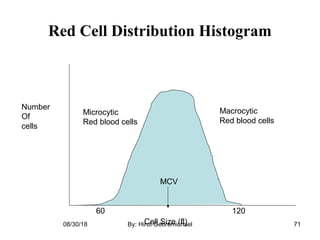
su
RBCs disorder and common laboratory tests
By:
Hirut G/Michael (DVM,Lecturer)
 
Appril, 2018
Bishoftu, Ethiopia
08/30/18 By: Hirut Gebremichael 1
RBCs disorder and
common laboratory tests
08/30/18 By: Hirut Gebremichael 2
Red Blood Cell Disorders
• Red blood cell (RBC) disorders are conditions 
that  affect  red  blood  cells,  the  cells  of  blood 
that carry oxygen from the lungs to all parts of 
the  body.   There  are  many  different  types  of 
red blood cell disorders, including:
08/30/18 By: Hirut Gebremichael 3
 Anemia 
• Red cell enzyme deficiencies (e.g. G6PD)
• Hemoglobinopathies (e.g. sickle cell 
disease and thalassemia) 
• Red cell membrane disorders among these;
08/30/18 By: Hirut Gebremichael 4
A  number  of  inherited  red  cell  disorders  due  to 
altered  membrane  function  have  been  identified. 
These include:
hereditary spherocytosis (HS), 
hereditary elliptocytosis (HE), 
hereditary ovalocytosis (HO), and 
hereditary stomatocytosis (HSt).
08/30/18 By: Hirut Gebremichael 5
• Most  RBC  diseases  are  lifelong,  chronic 
conditions that result in anemia, a reduction in 
the  ability  to  carry  oxygen  to  tissues  due  to 
reduced  numbers  of  RBCs  in  blood,  or  a 
reduction  in  the  amount  or  function  of 
hemoglobin in the RBCs….
08/30/18 By: Hirut Gebremichael 6
The  latter  diseases  are  also  known  as 
hemoglobinopathies,  and  include  sickle  cell 
disease (caused by a less functional hemoglobin 
beta,  selected  via  malaria  parasite),  as  well  as 
beta and alpha thalassemia (like sickle cell trait)
08/30/18 By: Hirut Gebremichael 7
• Thalassemia  is  an  inherited  blood disorder  in 
which  the  body  makes  an  abnormal  form  of 
hemoglobin
• The disorder results in excessive destruction of 
red blood cells, which leads to anemia
• It’s  caused  by  either  a  genetic  mutation  or  a 
deletion of certain key gene fragments.
08/30/18 By: Hirut Gebremichael 8
RED BLOOD CELL MORPHOLOGY
Abnormal  erythrocyte  morphology  is  found  in 
pathological states that may be : 
I. abnormalities in size (anisocytosis).  
II.In shape (poikilocytosis).  
III.In  hemoglobin  content  or  the  presence  of 
inclusion bodies in erythrocyte.
08/30/18 By: Hirut Gebremichael 9
I-Variation in erythrocyte size
(anisocytosis)
1-Microcytosis:
- Decrease in the red cell size. Red cells are smaller 
than ± 7µm in diameter. The nucleus of a small 
lymphocyte (± 8,µm) is a useful guide to the 
size of a red blood cell.
08/30/18 By: Hirut Gebremichael 10
Found in:
• Iron deficiency anemia.
- Thalassaemia.
- Lead poisoning.
- Anemia of chronic disease 
• Sideroblastic anemia
Sideroblastic anemia: is a form of anemia in which 
the bone marrow produces ringed sideroblasts 
rather than healthy red blood cells .08/30/18 By: Hirut Gebremichael 11
 
• In sideroblastic anemia, the body has iron available 
but cannot incorporate it into hemoglobin, which 
red blood cells need in order to transport oxygen 
efficiently. The disorder may be caused either by 
a genetic disorder or by hematological 
malignancies (especially acute myeloid leukemia)
08/30/18 By: Hirut Gebremichael 12
2-Macrocytosis:
Macrocytosis is a term used to describe red blood 
cells  that  are  larger  than  normal.  It  typically 
causes  no  signs  or  symptoms  and  is  usually 
detected incidentally on routine blood tests.
Macrocytosis  isn't  a  specific  disease,  but  it  may 
indicate  an  underlying  problem  that  requires 
medical evaluation. 
08/30/18 By: Hirut Gebremichael 13
08/30/18 By: Hirut Gebremichael 14
• Common causes of macrocytosis include:
• Vitamin B-12 deficiency
• Folate deficiency
• Liver disease
• Alcoholism(Human)
• Hypothyroidism08/30/18 By: Hirut Gebremichael 15
II-Variation in hemoglobin content
1-Hypochromasia:
Hypochromia  is  characterized  by  an  increase  in 
central  pallor.  It  is  said  to  be  present  when 
greater than 1/2 of the cell diameter has central 
pallor.
08/30/18 By: Hirut Gebremichael 16
• Hypochromia is caused by a deficiency in the 
production  of  hemoglobin,  which  is  usually 
the result of iron deficiency caused by chronic 
hemorrhage.
08/30/18 By: Hirut Gebremichael 17
2- Polychromasia:
• Polychromasia is a disorder where there is an 
abnormally  high  number  of  immature  red 
blood cells found in the bloodstream as a result 
of  being  prematurely  released  from  the  bone 
marrow during blood formation
08/30/18 By: Hirut Gebremichael 18
• Poly- refers to many, and -chromasia means color.
• Polychromasia  is  usually  a  sign  of  bone  marrow 
stress as well as immature red blood cells. 
• 3 types are recognized, with types (1) and (2) being 
referred to as 'young red blood cells' and type (3) as 
'old red blood cells'. 
• Giemsa stain is used to distinguish all three types in 
blood smears.08/30/18 By: Hirut Gebremichael 19
Found in:
Any  situation  with  reticulocytosis  -  for  example 
bleeding,  hemolysis  or  response  to  haemostatic 
factor replacement.
supravital Stain
08/30/18 By: Hirut Gebremichael 20
III- Variation of red cells shape
(Poikilocytosis)  
RBCs may have different shapes: 
1- Spherocytosis: 
Red cells are more spherical. Lack the central area 
of pallor on a stained blood film.
 Found in:
- Hereditary spherocytosis
- Immune haemolytic anemia08/30/18 By: Hirut Gebremichael 21
Spherocytosis
08/30/18 By: Hirut Gebremichael 22
2-Target Cells:
• Codocytes  also  known  as  target  cells  are  red 
blood  cells  that  have  the  appearance  of  a 
shooting target with a bullseye. 
08/30/18 By: Hirut Gebremichael 23
• In  optical  microscopy  these  cells  appear  to 
have a dark center (a central, hemoglobinized 
area)  surrounded  by  a  white  ring  (an  area  of 
relative  pallor),  followed  by  dark  outer 
(peripheral) second ring containing a band of 
hemoglobin. 
08/30/18 By: Hirut Gebremichael 24
Target cells or codocytes (black arrows) appear as a target 
with a bullseye. Dog, blood smear, Wright stain.
08/30/18 By: Hirut Gebremichael 25
Found in:
-Obstructive liver disease
- Severe iron deficiency
- Thalassaemia
- Haemoglobinopathies
- Post splenectomy
08/30/18 By: Hirut Gebremichael 26
3- Ovalocytes:
oval shape red blood cell
Found in:
- Thalassaemia major.
- Hereditary ovalocytosis.
- Sickle cell anemia
08/30/18 By: Hirut Gebremichael 27
08/30/18 By: Hirut Gebremichael 28
4- Elliptocytosis:
The red cells are oval or elliptical
in shape. Long axis is twice the
short axis.
Found in:
- Hereditary elliptocytosis
- Megaloblastic anemia
- Iron deficiency
08/30/18 By: Hirut Gebremichael 29
5- Tear Drop Cells:
Red cells shaped like a tear drop or pear
Found in:
- Bone marrow fibrosis
- Megaloblastic anemia
- Iron deficiency
- Thalassaemia
08/30/18 By: Hirut Gebremichael 30
08/30/18 By: Hirut Gebremichael 31
6- Blister cell:
A degmacyte (a.k.a. "bite cell") is an abnormally
shaped red blood cell with one or more
semicircular portions removed from the cell
margin.These "bites" result from the removal of
denatured hemoglobin by macrophages in the
spleen.
08/30/18 By: Hirut Gebremichael 32
Glucose-6-phosphate dehydrogenase deficiency,
in which uncontrolled oxidative stress causes
hemoglobin to denature and form Heinz
bodies, is a common disorder that leads to the
formation of bite cells.
Found in:
 Microangiopathic hemolytic anemia
 G6PD deficiency
08/30/18 By: Hirut Gebremichael 33
7- Schistocytosis:
Fragmentation of the red cells.
Found in:
- DIC
- Micro angiopathic haemolytic anemia
- Mechanical haemolytic anemia
08/30/18 By: Hirut Gebremichael 34
08/30/18 By: Hirut Gebremichael 35
8- Stomatocytosis:
Red cells with a central linear slit or stoma.
Seen as mouth-shaped form in peripheral
smear.
Found in:
- Hereditary stomatocytosis
- Hereditary spherocytosis
08/30/18 By: Hirut Gebremichael 36
08/30/18 By: Hirut Gebremichael 37
9- Burr (crenation ) cell:
Red cell with uniformly spaced, pointed
projections on their surface.
Found in:
- hemolytic anemia
- Uremia.
- Megaloblastic anemia
08/30/18 By: Hirut Gebremichael 38
10- Keratocytes (horn cell):
Part of the cell fuses back leaving two or
three horn-like projections. The
keratocyte is a fragile cell and remains
in circulation for only a few hours.
Found in:
- Uraemia
- Severe burns
- EDTA artifact
- Liver disease
08/30/18 By: Hirut Gebremichael 39
are red blood cells with irregularly
spaced projections, these
projections very in width but
usually contain a rounded end
Found in:
- Liver disease
- Post splenectomy
- Anorexia and starvation
11- Acanthocytosis:
08/30/18 By: Hirut Gebremichael 40
12- Sickle Cells:
Sickle shaped red cells
Found in:
Hb-S disease
08/30/18 By: Hirut Gebremichael 41
13- Rouleaux Formation:
Stacks of RBC's resembling a
stack of coins.
Found in:
- Hyperfibrinogenaemia
- Hyperglobulinaemia
08/30/18 By: Hirut Gebremichael 42
14- Red cell-agglutination:
Irregular clumps of red cells
Found in:
- Cold agglutinins
- Autoimmune hemolysis
08/30/18 By: Hirut Gebremichael 43
15- Nucleated red blood cells.
These red blood cells are released
from the bone marrow early into the
blood stream, due to the need for
oxygen. Normal red blood cells do
not contain a nucleus on a
peripheral smear.
08/30/18 By: Hirut Gebremichael 44
IV -Erythrocyte inclusion bodies
1- Howell-Jolly Bodies:
Small round cytoplasmic red cell
inclusion with same staining
characteristics as nuclei
Found in:
- Post splenectomy
- Megaloblastic anemia
08/30/18 By: Hirut Gebremichael 45
2- Siderotic Granules (Pappenheimer
Bodies)
RBCs which contain no hemoglobin iron granules. They
appear as dense blue, irregular granules which are
unevenly distributed in Wright stained
RBCs. Pappenheimer bodies can be increased in
hemolytic anemia, infections and post-splenectomy.
08/30/18 By: Hirut Gebremichael 46
3- Basophilic stippling:
Considerable numbers of small
basophilic inclusions in red cells.
Found in:
- Hemolytic anemia
- Liver disease
- Heavy metal poisoning.
08/30/18 By: Hirut Gebremichael 47
4- Heinz Bodies:
Represent denatured hemoglobin (methemoglobin - Fe+++)
within a cell. With a supravital stain like crystal violet,
Heinz bodies appear as round blue precipitates. Presence of
Heinz bodies indicates red cell injury and is usually
associated with G6PD-deficiency.
08/30/18 By: Hirut Gebremichael 48
5- Cabot Rings:
Reddish-blue threadlike rings in RBCs of severe
anemia's. These are remnants of the nuclear
membrane and appear as a ring or as the figure
pattern. Very rare; finding in patients with
Megaloblastic anemia, severe anemia's, lead
poisoning, and dyserythropoiesis.
08/30/18 By: Hirut Gebremichael 49
08/30/18 By: Hirut Gebremichael 50
6- Parasites of Red Cell:
are protozoan parasites which occur in many
species of birds and are the cause of avian
malaria. Transmitted by mosquitoes, infection
with Plasmodium can be a cause of hemolytic
anemia
08/30/18 By: Hirut Gebremichael 51
Common Laboratory Tests
08/30/18 By: Hirut Gebremichael 52
Let’s look at some nuances of 2 of most
commonly ordered lab tests
I. CBC (Complete Blood Count)
II. Blood chemistry test(Basic and Complete
metabolic panels)
08/30/18 By: Hirut Gebremichael 53
CBC
• This is the most common blood test performed
on pets and people. A CBC gives information
on hydration status, anaemia, infection, the
blood's clotting ability, and the ability of the
immune system to respond.
08/30/18 By: Hirut Gebremichael 54
• This test is essential for pets with fevers,
vomiting, diarrhoea, weakness, pale gums, or
loss of appetite. If your pet needs surgery, a
CBC can detect some bleeding disorders or
other unseen abnormalities.
08/30/18 By: Hirut Gebremichael 55
• Peripheral venous blood is collected in a
lavendar tube (contains the anticoagulant
EDTA) and should be thoroughly mixed.
08/30/18 By: Hirut Gebremichael 56
• Unacceptable specimen:
– Clotted or greater than 48 hours old
• Methodology of testing:
– Whole blood analyzer
08/30/18 By: Hirut Gebremichael 57
What is measured?
• Red blood cell data
– Total red blood cell count (RBC)
– Hemoglobin (Hgb)
– Hematocrit (Hct)
– Mean corpuscular volume (MCV)
– Red blood cell distribution width (RDW)08/30/18 By: Hirut Gebremichael 58
Total Red Blood Cell Count
• Count of the number of circulating red blood
cells in 1mm3
of peripheral venous blood
08/30/18 By: Hirut Gebremichael 59
Hemoglobin
• The hemoglobin concentration is a measure of
the amount of Hgb in the peripheral blood,
which reflects the number of red blood cells in
the blood
Result interpretation
– Hgb constitutes over 90% of the red blood
cells08/30/18 By: Hirut Gebremichael 60
• Decrease in Hgb concentration = anemia
• Increase in Hgb concentration = polycythemia
Hematocrit
Hematocrit is a measure of the percentage of the
total blood volume that is made up by the red
blood cells.
08/30/18 By: Hirut Gebremichael 61
• The hematocrit can be determined directly by
centrifugation (“spun hematocrit”)
– The height of the red blood cell column is
measured and compared to the column of
the whole blood
08/30/18 By: Hirut Gebremichael 62
Centrifuged blood (normal)
Red blood cells
Buffy coat (WBCs and Platelets)
Plasma
Normal Hct in adult males
40-54%
Normal Hct in adult females
34-51%
08/30/18 By: Hirut Gebremichael 63
Centrifuged blood
Anemia – there is a low percentage of RBCs
(low hematocrit)
RBCs
Buffy coat
Plasma
08/30/18 By: Hirut Gebremichael 64
Results
The percentage of the volume of blood occupied
by the red cells constitutes hematocrit or packed
cell volume.
Hct % = {Height of RBCs (mm) / Height of
RBCs and plasma (mm)} × 100
For example, if the height of packed red cells is
45 mm, then
= 45/ 100 × 100 = 45 percent.
08/30/18 By: Hirut Gebremichael 65
It also means that out of 100 volumes (or parts)
of blood 45 volumes (or parts) are red cells
and 55 volumes (or parts) are plasma. Thus,
out of 1 liter of blood, 450 ml are red cells and
550 ml are plasma.
08/30/18 By: Hirut Gebremichael 66
Mean Corpuscular Volume (MCV)
• It is defined as the average volume of red
cell
• It Provides information on red cell size
• It is measured in femtolitres.
• Normal range = 87±5fl
• 1fL= L
• It is determined by the distribution of the08/30/18 By: Hirut Gebremichael 67
08/30/18 By: Hirut Gebremichael 68
Cell Size (fl)
Number
Of
cells
60 120
MCV
Red Cell Distribution Histogram
08/30/18 By: Hirut Gebremichael 69
Use of MCV Result
• The MCV is important in classifying anemias
– Normal MCV = normocytic anemia
– Decreased MCV = microcytic anemia
– Increased MCV = macrocytic anemia
08/30/18 By: Hirut Gebremichael 70
Cell Size (fl)
Number
Of
cells
60 120
MCV
Red Cell Distribution Histogram
Microcytic
Red blood cells
Macrocytic
Red blood cells
08/30/18 By: Hirut Gebremichael 71
Red Blood Cell Distribution Width
• RDW is a measure of the range of variation of
red blood cell (RBC) volume that is reported as
part of a standard complete blood count.
• Usually red blood cells are a standard size of
about 6-8 μm in diameter. Certain disorders,
however, cause a significant variation in cell
size. 08/30/18 By: Hirut Gebremichael 72
• In general, an elevated RDW (indicating more
variation in the size of RBCs) has been
associated with anemias with various
deficiencies, such as iron, B12, or folate
• Thalassemia is a microcytic anemia that
characteristically has a normal RDW
08/30/18 By: Hirut Gebremichael 73
Mean Corpuscular Hemoglobin
It is defined as the amount of hemoglobin in
average red cell Or Average amount of Hb in
all the red cell
•It is directly proportional to the amount of
hemoglobin and the size of the erythrocyte
•Normal range = 27-32pg
•1pg = 08/30/18 By: Hirut Gebremichael 74
Formul
a
08/30/18 By: Hirut Gebremichael 75
Interpretation of MCH values
• Low MCH values: are found in microcytic
hypochromic anaemias and also when red
cells are microcytic and normochromic. In
thalassaemia the MCH is low even when
anaemia is mild (MCHC is often normal)
• Raised MCH values: found in macrocytic
normochromic anaemia
08/30/18 By: Hirut Gebremichael 76
Mean Corpuscular Hemoglobin
Concentration (MCHC)
• The MCHC expresses the average
concentration of hemoglobin per unit volume
of erythrocytes.
• It is expressed in percentage.
• Normal value = 32-36%
08/30/18 By: Hirut Gebremichael 77
B a s i c
formula
o
r
08/30/18 By: Hirut Gebremichael 78
Interpretation of MCHC values
• Low MCHC values are found in iron deficiency
anemia and other conditions in which the red
cells are microcytic and hypochromic.
• An increased MCHC can occur in marked
spherocytosis.
08/30/18 By: Hirut Gebremichael 79
Peripheral Blood Smear
• A small drop of the blood is placed on a slide
and spread out into a thin film. After drying, it
is stained with Wright's stain in an automatic
stainer.
08/30/18 By: Hirut Gebremichael 80
• The stained slide can the be viewed under a
microscope, and the hematologist can assess the
morphology and appearance of all the blood
elements: the red blood cells, the white blood cells
and the platelets.
08/30/18 By: Hirut Gebremichael 81
Erythrocyte Sedimentation Rate(ESR)
• Anticoagulated blood is placed in a thin tube
to stand for two hours. The red blood cells
(RBCs) settle to the bottom of the tube and a
top layer of plasma is left in the tube.
08/30/18 By: Hirut Gebremichael 82
• The result is seen through the number of
millimeters the RBCs fall in one to two hours.
The speed at which the RBCs fall is
determined by the concentration of a number
of plasma proteins which rise during acute
illnesses.
08/30/18 By: Hirut Gebremichael 83
• A high sedimentation rate is a nonspecific, but
highly sensitive, indicator for the presence of
diseases of all sorts.
08/30/18 By: Hirut Gebremichael 84
Westergren pipet array on StaRRsed automated
ESR analyzer
08/30/18 By: Hirut Gebremichael 85
Tests of the Coagulation System
• There are many types of tests of the coagulation
system, and these are categorized into routine
tests, bleeding diathesis (tendency to bleed),
• hypercoagulability tests (excess blood
clotting)
08/30/18 By: Hirut Gebremichael 86
• There is a whole array of indicators for tests
related to the coagulation system. Some of
these tests are performed routinely in a number
of medical situations, such as prior to major
surgical procedures.
08/30/18 By: Hirut Gebremichael 87
• They may also be performed if there is
evidence of a bleeding problem, such as easy
bruising. Alternatively, other tests are
performed if there is evidence of a disorder
leading to excessive blood clotting
(thrombosis).
08/30/18 By: Hirut Gebremichael 88
Blood chemistries
• These common blood serum tests evaluate organ
function, electrolyte status, hormone levels and
more. They are important in evaluating older pets,
pets with vomiting, diarrhoea or toxin exposure,
pets receiving long-term medications and health
before anaesthesia.08/30/18 By: Hirut Gebremichael 89
• A veterinary chemistry panel (also termed
'biochemical profile') includes tests for
multiple chemical constituents within one
sample. The quantities of these chemicals can
reveal many things about the various organs of
the body. Most veterinary chemistry panels
check blood electrolyte and for diseases of the
liver, kidneys, and pancreas
08/30/18 By: Hirut Gebremichael 90
• A small quantity of the serum from the original
collection is drawn into a single large machine.
Tiny samples from that serum are tested for
various chemical components. The results of each
test are compiled and printed out on a single
form. To make it easier for veterinarians and
physicians, the form lists the patient's results
along with the expected normal values for that
species08/30/18 By: Hirut Gebremichael 91
Types of panels
• Since the laboratory equipment has the ability
to run numerous different tests, there can be
many different chemistry panels (e.g., liver
panels, electrolyte panels, geriatric panels, pre-
surgical panels) produced depending upon
which individual tests are requested and
included.
08/30/18 By: Hirut Gebremichael 92
A typical veterinary chemistry panel will
measure the following
08/30/18 By: Hirut Gebremichael 93
08/30/18 By: Hirut Gebremichael 94
08/30/18 By: Hirut Gebremichael 95
08/30/18 By: Hirut Gebremichael 96
Thank You!
08/30/18 By: Hirut Gebremichael 97

Red Blood Cell disorder

  • 1.
    su RBCs disorder andcommon laboratory tests By: Hirut G/Michael (DVM,Lecturer)   Appril, 2018 Bishoftu, Ethiopia 08/30/18 By: Hirut Gebremichael 1
  • 2.
    RBCs disorder and commonlaboratory tests 08/30/18 By: Hirut Gebremichael 2
  • 3.
    Red Blood CellDisorders • Red blood cell (RBC) disorders are conditions  that  affect  red  blood  cells,  the  cells  of  blood  that carry oxygen from the lungs to all parts of  the  body.   There  are  many  different  types  of  red blood cell disorders, including: 08/30/18 By: Hirut Gebremichael 3
  • 4.
  • 5.
    A  number  of inherited  red  cell  disorders  due  to  altered  membrane  function  have  been  identified.  These include: hereditary spherocytosis (HS),  hereditary elliptocytosis (HE),  hereditary ovalocytosis (HO), and  hereditary stomatocytosis (HSt). 08/30/18 By: Hirut Gebremichael 5
  • 6.
    • Most  RBC diseases  are  lifelong,  chronic  conditions that result in anemia, a reduction in  the  ability  to  carry  oxygen  to  tissues  due  to  reduced  numbers  of  RBCs  in  blood,  or  a  reduction  in  the  amount  or  function  of  hemoglobin in the RBCs…. 08/30/18 By: Hirut Gebremichael 6
  • 7.
    The  latter  diseases are  also  known  as  hemoglobinopathies,  and  include  sickle  cell  disease (caused by a less functional hemoglobin  beta,  selected  via  malaria  parasite),  as  well  as  beta and alpha thalassemia (like sickle cell trait) 08/30/18 By: Hirut Gebremichael 7
  • 8.
    • Thalassemia  is an  inherited  blood disorder  in  which  the  body  makes  an  abnormal  form  of  hemoglobin • The disorder results in excessive destruction of  red blood cells, which leads to anemia • It’s  caused  by  either  a  genetic  mutation  or  a  deletion of certain key gene fragments. 08/30/18 By: Hirut Gebremichael 8
  • 9.
    RED BLOOD CELLMORPHOLOGY Abnormal  erythrocyte  morphology  is  found  in  pathological states that may be :  I. abnormalities in size (anisocytosis).   II.In shape (poikilocytosis).   III.In  hemoglobin  content  or  the  presence  of  inclusion bodies in erythrocyte. 08/30/18 By: Hirut Gebremichael 9
  • 10.
    I-Variation in erythrocytesize (anisocytosis) 1-Microcytosis: - Decrease in the red cell size. Red cells are smaller  than ± 7µm in diameter. The nucleus of a small  lymphocyte (± 8,µm) is a useful guide to the  size of a red blood cell. 08/30/18 By: Hirut Gebremichael 10
  • 11.
    Found in: • Iron deficiency anemia. - Thalassaemia. - Lead poisoning. - Anemia of chronic disease  •Sideroblastic anemia Sideroblastic anemia: is a form of anemia in which  the bone marrow produces ringed sideroblasts  rather than healthy red blood cells .08/30/18 By: Hirut Gebremichael 11
  • 12.
  • 13.
    2-Macrocytosis: Macrocytosis is a term used to describe red blood  cells  that  are larger  than  normal.  It  typically  causes  no  signs  or  symptoms  and  is  usually  detected incidentally on routine blood tests. Macrocytosis  isn't  a  specific  disease,  but  it  may  indicate  an  underlying  problem  that  requires  medical evaluation.  08/30/18 By: Hirut Gebremichael 13
  • 14.
    08/30/18 By: HirutGebremichael 14
  • 15.
    • Common causes of macrocytosis include: • Vitamin B-12 deficiency •Folate deficiency • Liver disease • Alcoholism(Human) • Hypothyroidism08/30/18 By: Hirut Gebremichael 15
  • 16.
    II-Variation in hemoglobincontent 1-Hypochromasia: Hypochromia  is  characterized  by  an  increase  in  central  pallor.  It  is  said  to  be  present  when  greater than 1/2 of the cell diameter has central  pallor. 08/30/18 By: Hirut Gebremichael 16
  • 17.
    • Hypochromia is caused by a deficiency in the  production  of hemoglobin,  which  is  usually  the result of iron deficiency caused by chronic  hemorrhage. 08/30/18 By: Hirut Gebremichael 17
  • 18.
    2- Polychromasia: • Polychromasia is a disorder where there is an  abnormally high  number  of  immature  red  blood cells found in the bloodstream as a result  of  being  prematurely  released  from  the  bone  marrow during blood formation 08/30/18 By: Hirut Gebremichael 18
  • 19.
    • Poly- refers to many, and -chromasia means color. • Polychromasia is  usually  a  sign  of  bone  marrow  stress as well as immature red blood cells.  • 3 types are recognized, with types (1) and (2) being  referred to as 'young red blood cells' and type (3) as  'old red blood cells'.  • Giemsa stain is used to distinguish all three types in  blood smears.08/30/18 By: Hirut Gebremichael 19
  • 20.
    Found in: Any  situation with  reticulocytosis  -  for  example  bleeding,  hemolysis  or  response  to  haemostatic  factor replacement. supravital Stain 08/30/18 By: Hirut Gebremichael 20
  • 21.
    III- Variation ofred cells shape (Poikilocytosis)   RBCs may have different shapes:  1- Spherocytosis:  Red cells are more spherical. Lack the central area  of pallor on a stained blood film.  Found in: - Hereditary spherocytosis - Immune haemolytic anemia08/30/18 By: Hirut Gebremichael 21
  • 22.
  • 23.
    2-Target Cells: • Codocytes also  known  as  target  cells  are  red  blood  cells  that  have  the  appearance  of  a  shooting target with a bullseye.  08/30/18 By: Hirut Gebremichael 23
  • 24.
    • In  optical microscopy  these  cells  appear  to  have a dark center (a central, hemoglobinized  area)  surrounded  by  a  white  ring  (an  area  of  relative  pallor),  followed  by  dark  outer  (peripheral) second ring containing a band of  hemoglobin.  08/30/18 By: Hirut Gebremichael 24
  • 25.
  • 26.
    Found in: -Obstructive liverdisease - Severe iron deficiency - Thalassaemia - Haemoglobinopathies - Post splenectomy 08/30/18 By: Hirut Gebremichael 26
  • 27.
    3- Ovalocytes: oval shapered blood cell Found in: - Thalassaemia major. - Hereditary ovalocytosis. - Sickle cell anemia 08/30/18 By: Hirut Gebremichael 27
  • 28.
    08/30/18 By: HirutGebremichael 28
  • 29.
    4- Elliptocytosis: The redcells are oval or elliptical in shape. Long axis is twice the short axis. Found in: - Hereditary elliptocytosis - Megaloblastic anemia - Iron deficiency 08/30/18 By: Hirut Gebremichael 29
  • 30.
    5- Tear DropCells: Red cells shaped like a tear drop or pear Found in: - Bone marrow fibrosis - Megaloblastic anemia - Iron deficiency - Thalassaemia 08/30/18 By: Hirut Gebremichael 30
  • 31.
    08/30/18 By: HirutGebremichael 31
  • 32.
    6- Blister cell: Adegmacyte (a.k.a. "bite cell") is an abnormally shaped red blood cell with one or more semicircular portions removed from the cell margin.These "bites" result from the removal of denatured hemoglobin by macrophages in the spleen. 08/30/18 By: Hirut Gebremichael 32
  • 33.
    Glucose-6-phosphate dehydrogenase deficiency, inwhich uncontrolled oxidative stress causes hemoglobin to denature and form Heinz bodies, is a common disorder that leads to the formation of bite cells. Found in:  Microangiopathic hemolytic anemia  G6PD deficiency 08/30/18 By: Hirut Gebremichael 33
  • 34.
    7- Schistocytosis: Fragmentation ofthe red cells. Found in: - DIC - Micro angiopathic haemolytic anemia - Mechanical haemolytic anemia 08/30/18 By: Hirut Gebremichael 34
  • 35.
    08/30/18 By: HirutGebremichael 35
  • 36.
    8- Stomatocytosis: Red cellswith a central linear slit or stoma. Seen as mouth-shaped form in peripheral smear. Found in: - Hereditary stomatocytosis - Hereditary spherocytosis 08/30/18 By: Hirut Gebremichael 36
  • 37.
    08/30/18 By: HirutGebremichael 37
  • 38.
    9- Burr (crenation) cell: Red cell with uniformly spaced, pointed projections on their surface. Found in: - hemolytic anemia - Uremia. - Megaloblastic anemia 08/30/18 By: Hirut Gebremichael 38
  • 39.
    10- Keratocytes (horncell): Part of the cell fuses back leaving two or three horn-like projections. The keratocyte is a fragile cell and remains in circulation for only a few hours. Found in: - Uraemia - Severe burns - EDTA artifact - Liver disease 08/30/18 By: Hirut Gebremichael 39
  • 40.
    are red bloodcells with irregularly spaced projections, these projections very in width but usually contain a rounded end Found in: - Liver disease - Post splenectomy - Anorexia and starvation 11- Acanthocytosis: 08/30/18 By: Hirut Gebremichael 40
  • 41.
    12- Sickle Cells: Sickleshaped red cells Found in: Hb-S disease 08/30/18 By: Hirut Gebremichael 41
  • 42.
    13- Rouleaux Formation: Stacksof RBC's resembling a stack of coins. Found in: - Hyperfibrinogenaemia - Hyperglobulinaemia 08/30/18 By: Hirut Gebremichael 42
  • 43.
    14- Red cell-agglutination: Irregularclumps of red cells Found in: - Cold agglutinins - Autoimmune hemolysis 08/30/18 By: Hirut Gebremichael 43
  • 44.
    15- Nucleated redblood cells. These red blood cells are released from the bone marrow early into the blood stream, due to the need for oxygen. Normal red blood cells do not contain a nucleus on a peripheral smear. 08/30/18 By: Hirut Gebremichael 44
  • 45.
    IV -Erythrocyte inclusionbodies 1- Howell-Jolly Bodies: Small round cytoplasmic red cell inclusion with same staining characteristics as nuclei Found in: - Post splenectomy - Megaloblastic anemia 08/30/18 By: Hirut Gebremichael 45
  • 46.
    2- Siderotic Granules(Pappenheimer Bodies) RBCs which contain no hemoglobin iron granules. They appear as dense blue, irregular granules which are unevenly distributed in Wright stained RBCs. Pappenheimer bodies can be increased in hemolytic anemia, infections and post-splenectomy. 08/30/18 By: Hirut Gebremichael 46
  • 47.
    3- Basophilic stippling: Considerablenumbers of small basophilic inclusions in red cells. Found in: - Hemolytic anemia - Liver disease - Heavy metal poisoning. 08/30/18 By: Hirut Gebremichael 47
  • 48.
    4- Heinz Bodies: Representdenatured hemoglobin (methemoglobin - Fe+++) within a cell. With a supravital stain like crystal violet, Heinz bodies appear as round blue precipitates. Presence of Heinz bodies indicates red cell injury and is usually associated with G6PD-deficiency. 08/30/18 By: Hirut Gebremichael 48
  • 49.
    5- Cabot Rings: Reddish-bluethreadlike rings in RBCs of severe anemia's. These are remnants of the nuclear membrane and appear as a ring or as the figure pattern. Very rare; finding in patients with Megaloblastic anemia, severe anemia's, lead poisoning, and dyserythropoiesis. 08/30/18 By: Hirut Gebremichael 49
  • 50.
    08/30/18 By: HirutGebremichael 50
  • 51.
    6- Parasites ofRed Cell: are protozoan parasites which occur in many species of birds and are the cause of avian malaria. Transmitted by mosquitoes, infection with Plasmodium can be a cause of hemolytic anemia 08/30/18 By: Hirut Gebremichael 51
  • 52.
    Common Laboratory Tests 08/30/18By: Hirut Gebremichael 52
  • 53.
    Let’s look atsome nuances of 2 of most commonly ordered lab tests I. CBC (Complete Blood Count) II. Blood chemistry test(Basic and Complete metabolic panels) 08/30/18 By: Hirut Gebremichael 53
  • 54.
    CBC • This isthe most common blood test performed on pets and people. A CBC gives information on hydration status, anaemia, infection, the blood's clotting ability, and the ability of the immune system to respond. 08/30/18 By: Hirut Gebremichael 54
  • 55.
    • This testis essential for pets with fevers, vomiting, diarrhoea, weakness, pale gums, or loss of appetite. If your pet needs surgery, a CBC can detect some bleeding disorders or other unseen abnormalities. 08/30/18 By: Hirut Gebremichael 55
  • 56.
    • Peripheral venousblood is collected in a lavendar tube (contains the anticoagulant EDTA) and should be thoroughly mixed. 08/30/18 By: Hirut Gebremichael 56
  • 57.
    • Unacceptable specimen: –Clotted or greater than 48 hours old • Methodology of testing: – Whole blood analyzer 08/30/18 By: Hirut Gebremichael 57
  • 58.
    What is measured? •Red blood cell data – Total red blood cell count (RBC) – Hemoglobin (Hgb) – Hematocrit (Hct) – Mean corpuscular volume (MCV) – Red blood cell distribution width (RDW)08/30/18 By: Hirut Gebremichael 58
  • 59.
    Total Red BloodCell Count • Count of the number of circulating red blood cells in 1mm3 of peripheral venous blood 08/30/18 By: Hirut Gebremichael 59
  • 60.
    Hemoglobin • The hemoglobinconcentration is a measure of the amount of Hgb in the peripheral blood, which reflects the number of red blood cells in the blood Result interpretation – Hgb constitutes over 90% of the red blood cells08/30/18 By: Hirut Gebremichael 60
  • 61.
    • Decrease inHgb concentration = anemia • Increase in Hgb concentration = polycythemia Hematocrit Hematocrit is a measure of the percentage of the total blood volume that is made up by the red blood cells. 08/30/18 By: Hirut Gebremichael 61
  • 62.
    • The hematocritcan be determined directly by centrifugation (“spun hematocrit”) – The height of the red blood cell column is measured and compared to the column of the whole blood 08/30/18 By: Hirut Gebremichael 62
  • 63.
    Centrifuged blood (normal) Redblood cells Buffy coat (WBCs and Platelets) Plasma Normal Hct in adult males 40-54% Normal Hct in adult females 34-51% 08/30/18 By: Hirut Gebremichael 63
  • 64.
    Centrifuged blood Anemia –there is a low percentage of RBCs (low hematocrit) RBCs Buffy coat Plasma 08/30/18 By: Hirut Gebremichael 64
  • 65.
    Results The percentage ofthe volume of blood occupied by the red cells constitutes hematocrit or packed cell volume. Hct % = {Height of RBCs (mm) / Height of RBCs and plasma (mm)} × 100 For example, if the height of packed red cells is 45 mm, then = 45/ 100 × 100 = 45 percent. 08/30/18 By: Hirut Gebremichael 65
  • 66.
    It also meansthat out of 100 volumes (or parts) of blood 45 volumes (or parts) are red cells and 55 volumes (or parts) are plasma. Thus, out of 1 liter of blood, 450 ml are red cells and 550 ml are plasma. 08/30/18 By: Hirut Gebremichael 66
  • 67.
    Mean Corpuscular Volume(MCV) • It is defined as the average volume of red cell • It Provides information on red cell size • It is measured in femtolitres. • Normal range = 87±5fl • 1fL= L • It is determined by the distribution of the08/30/18 By: Hirut Gebremichael 67
  • 68.
    08/30/18 By: HirutGebremichael 68
  • 69.
    Cell Size (fl) Number Of cells 60120 MCV Red Cell Distribution Histogram 08/30/18 By: Hirut Gebremichael 69
  • 70.
    Use of MCVResult • The MCV is important in classifying anemias – Normal MCV = normocytic anemia – Decreased MCV = microcytic anemia – Increased MCV = macrocytic anemia 08/30/18 By: Hirut Gebremichael 70
  • 71.
    Cell Size (fl) Number Of cells 60120 MCV Red Cell Distribution Histogram Microcytic Red blood cells Macrocytic Red blood cells 08/30/18 By: Hirut Gebremichael 71
  • 72.
    Red Blood CellDistribution Width • RDW is a measure of the range of variation of red blood cell (RBC) volume that is reported as part of a standard complete blood count. • Usually red blood cells are a standard size of about 6-8 μm in diameter. Certain disorders, however, cause a significant variation in cell size. 08/30/18 By: Hirut Gebremichael 72
  • 73.
    • In general,an elevated RDW (indicating more variation in the size of RBCs) has been associated with anemias with various deficiencies, such as iron, B12, or folate • Thalassemia is a microcytic anemia that characteristically has a normal RDW 08/30/18 By: Hirut Gebremichael 73
  • 74.
    Mean Corpuscular Hemoglobin Itis defined as the amount of hemoglobin in average red cell Or Average amount of Hb in all the red cell •It is directly proportional to the amount of hemoglobin and the size of the erythrocyte •Normal range = 27-32pg •1pg = 08/30/18 By: Hirut Gebremichael 74
  • 75.
  • 76.
    Interpretation of MCHvalues • Low MCH values: are found in microcytic hypochromic anaemias and also when red cells are microcytic and normochromic. In thalassaemia the MCH is low even when anaemia is mild (MCHC is often normal) • Raised MCH values: found in macrocytic normochromic anaemia 08/30/18 By: Hirut Gebremichael 76
  • 77.
    Mean Corpuscular Hemoglobin Concentration(MCHC) • The MCHC expresses the average concentration of hemoglobin per unit volume of erythrocytes. • It is expressed in percentage. • Normal value = 32-36% 08/30/18 By: Hirut Gebremichael 77
  • 78.
    B a si c formula o r 08/30/18 By: Hirut Gebremichael 78
  • 79.
    Interpretation of MCHCvalues • Low MCHC values are found in iron deficiency anemia and other conditions in which the red cells are microcytic and hypochromic. • An increased MCHC can occur in marked spherocytosis. 08/30/18 By: Hirut Gebremichael 79
  • 80.
    Peripheral Blood Smear •A small drop of the blood is placed on a slide and spread out into a thin film. After drying, it is stained with Wright's stain in an automatic stainer. 08/30/18 By: Hirut Gebremichael 80
  • 81.
    • The stainedslide can the be viewed under a microscope, and the hematologist can assess the morphology and appearance of all the blood elements: the red blood cells, the white blood cells and the platelets. 08/30/18 By: Hirut Gebremichael 81
  • 82.
    Erythrocyte Sedimentation Rate(ESR) •Anticoagulated blood is placed in a thin tube to stand for two hours. The red blood cells (RBCs) settle to the bottom of the tube and a top layer of plasma is left in the tube. 08/30/18 By: Hirut Gebremichael 82
  • 83.
    • The resultis seen through the number of millimeters the RBCs fall in one to two hours. The speed at which the RBCs fall is determined by the concentration of a number of plasma proteins which rise during acute illnesses. 08/30/18 By: Hirut Gebremichael 83
  • 84.
    • A highsedimentation rate is a nonspecific, but highly sensitive, indicator for the presence of diseases of all sorts. 08/30/18 By: Hirut Gebremichael 84
  • 85.
    Westergren pipet arrayon StaRRsed automated ESR analyzer 08/30/18 By: Hirut Gebremichael 85
  • 86.
    Tests of theCoagulation System • There are many types of tests of the coagulation system, and these are categorized into routine tests, bleeding diathesis (tendency to bleed), • hypercoagulability tests (excess blood clotting) 08/30/18 By: Hirut Gebremichael 86
  • 87.
    • There isa whole array of indicators for tests related to the coagulation system. Some of these tests are performed routinely in a number of medical situations, such as prior to major surgical procedures. 08/30/18 By: Hirut Gebremichael 87
  • 88.
    • They mayalso be performed if there is evidence of a bleeding problem, such as easy bruising. Alternatively, other tests are performed if there is evidence of a disorder leading to excessive blood clotting (thrombosis). 08/30/18 By: Hirut Gebremichael 88
  • 89.
    Blood chemistries • Thesecommon blood serum tests evaluate organ function, electrolyte status, hormone levels and more. They are important in evaluating older pets, pets with vomiting, diarrhoea or toxin exposure, pets receiving long-term medications and health before anaesthesia.08/30/18 By: Hirut Gebremichael 89
  • 90.
    • A veterinarychemistry panel (also termed 'biochemical profile') includes tests for multiple chemical constituents within one sample. The quantities of these chemicals can reveal many things about the various organs of the body. Most veterinary chemistry panels check blood electrolyte and for diseases of the liver, kidneys, and pancreas 08/30/18 By: Hirut Gebremichael 90
  • 91.
    • A smallquantity of the serum from the original collection is drawn into a single large machine. Tiny samples from that serum are tested for various chemical components. The results of each test are compiled and printed out on a single form. To make it easier for veterinarians and physicians, the form lists the patient's results along with the expected normal values for that species08/30/18 By: Hirut Gebremichael 91
  • 92.
    Types of panels •Since the laboratory equipment has the ability to run numerous different tests, there can be many different chemistry panels (e.g., liver panels, electrolyte panels, geriatric panels, pre- surgical panels) produced depending upon which individual tests are requested and included. 08/30/18 By: Hirut Gebremichael 92
  • 93.
    A typical veterinarychemistry panel will measure the following 08/30/18 By: Hirut Gebremichael 93
  • 94.
    08/30/18 By: HirutGebremichael 94
  • 95.
    08/30/18 By: HirutGebremichael 95
  • 96.
    08/30/18 By: HirutGebremichael 96
  • 97.
    Thank You! 08/30/18 By:Hirut Gebremichael 97