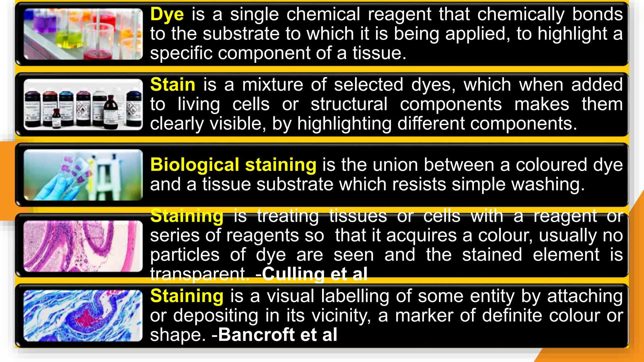
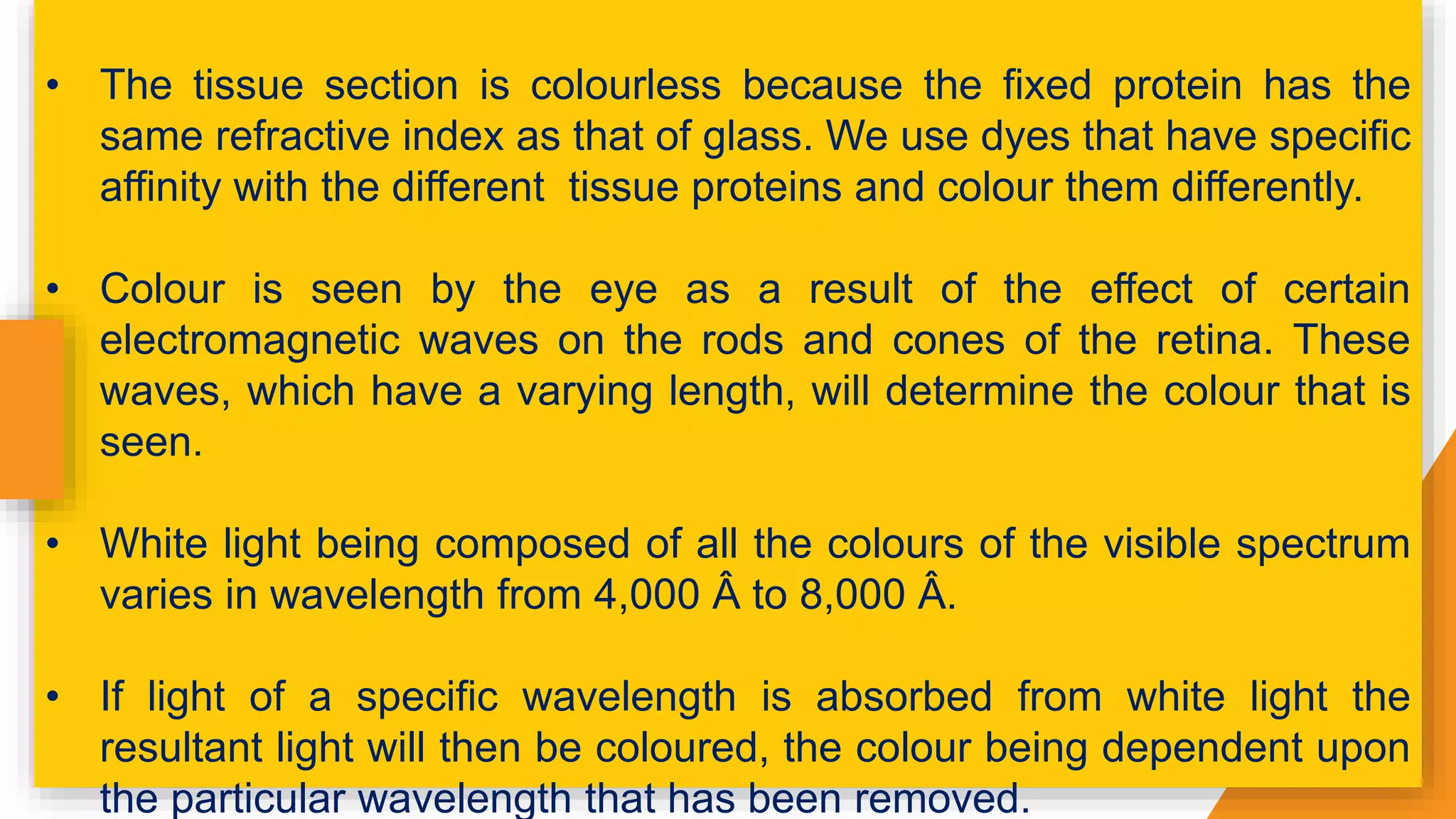
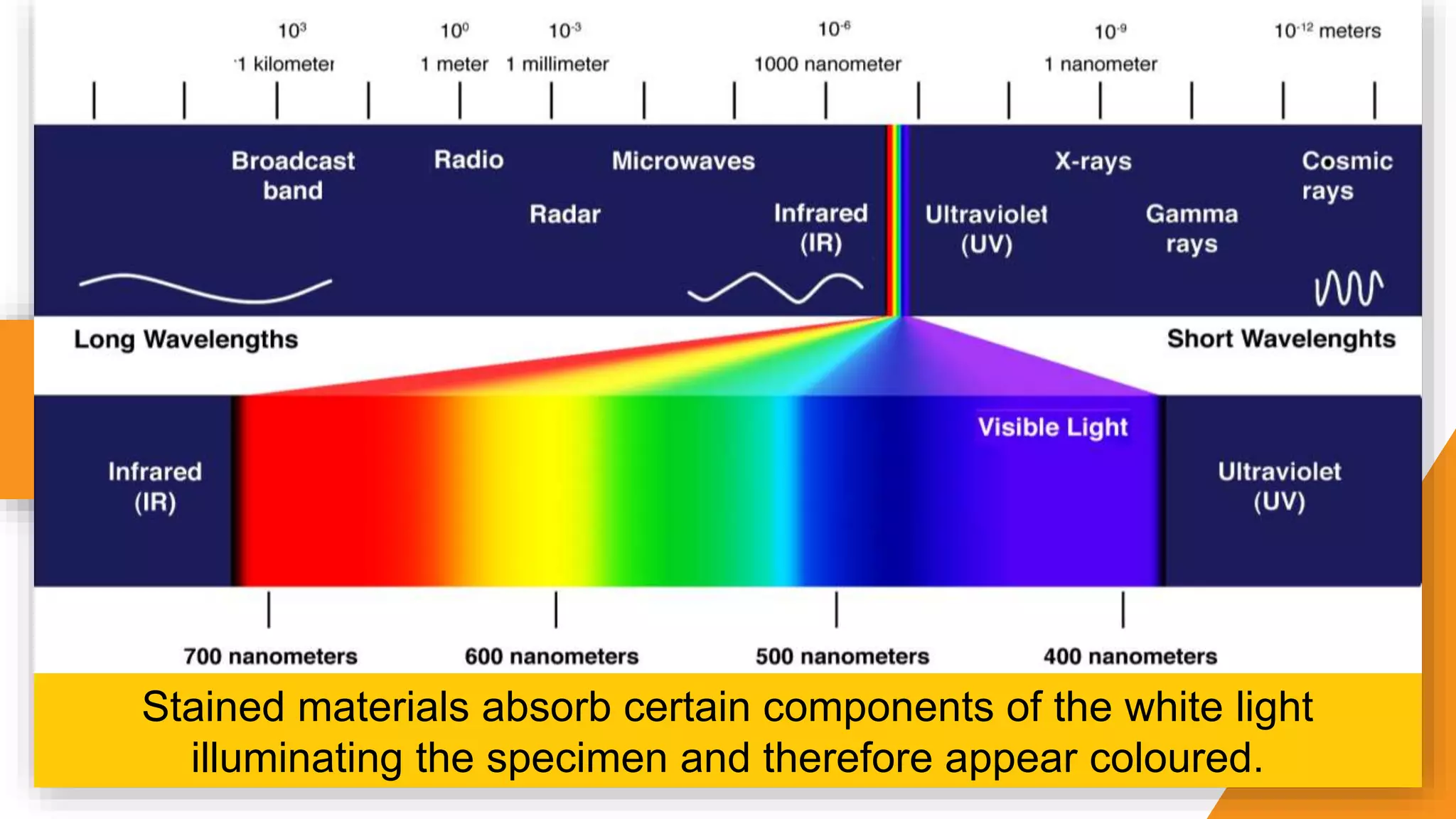
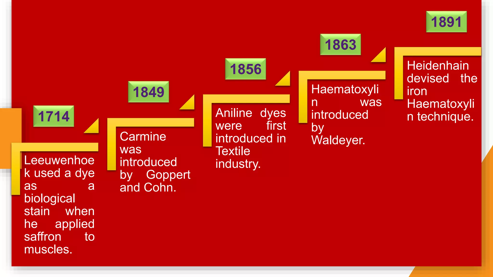
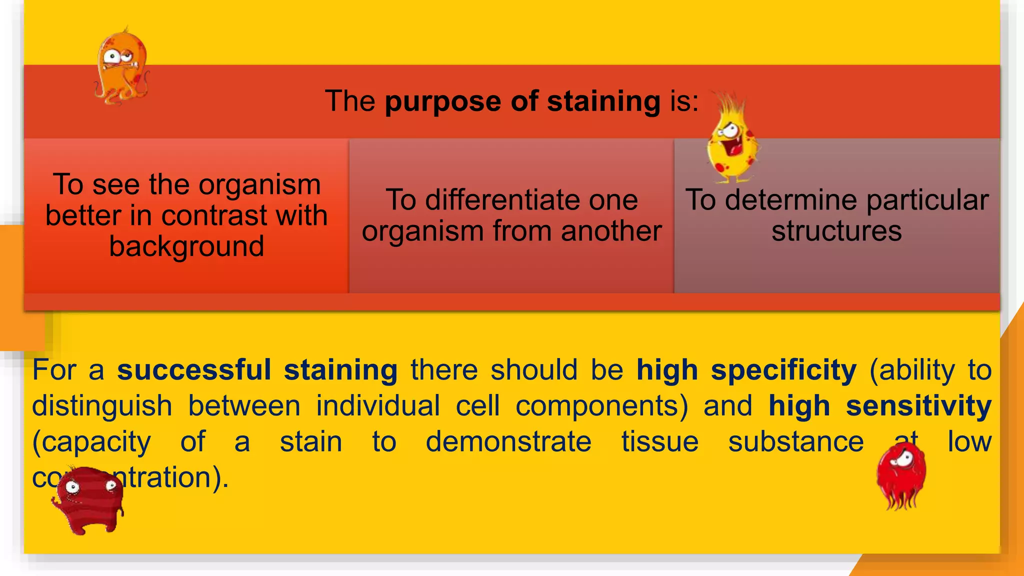
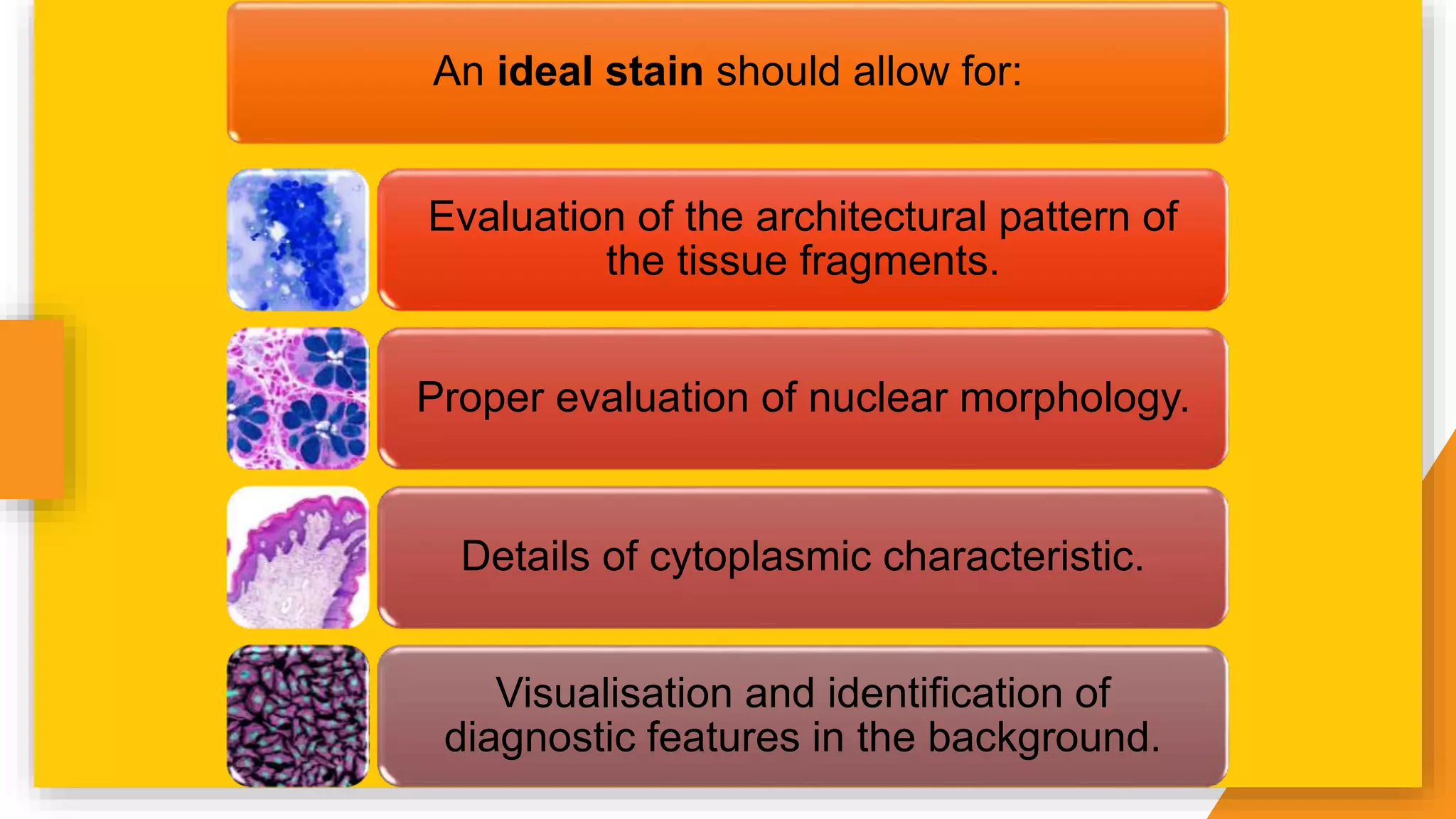
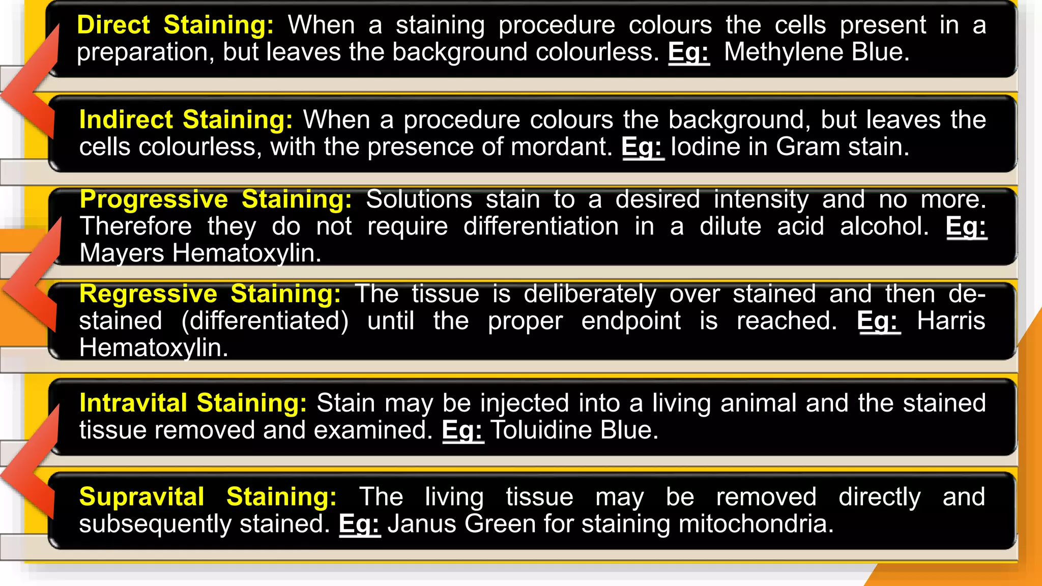
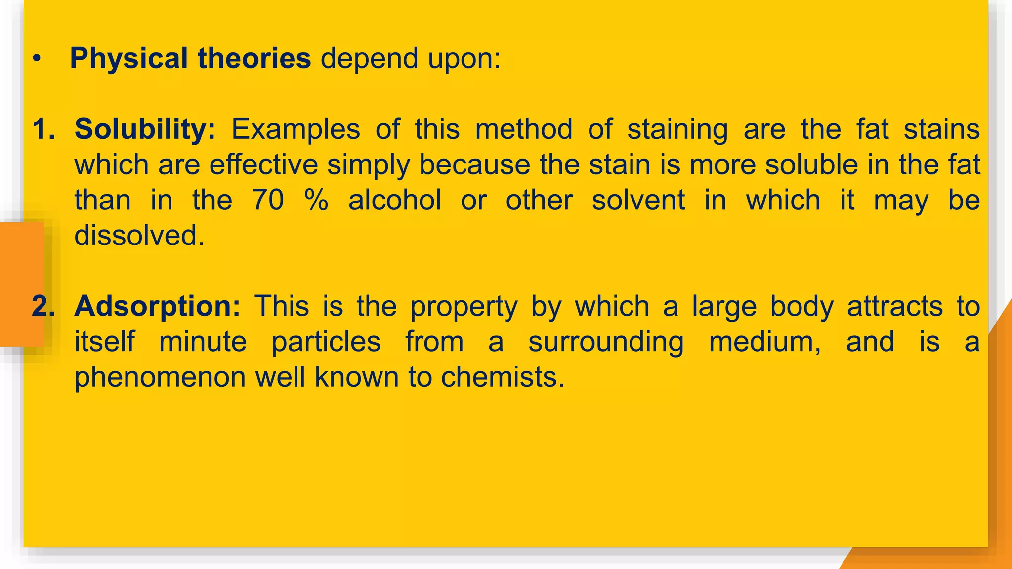
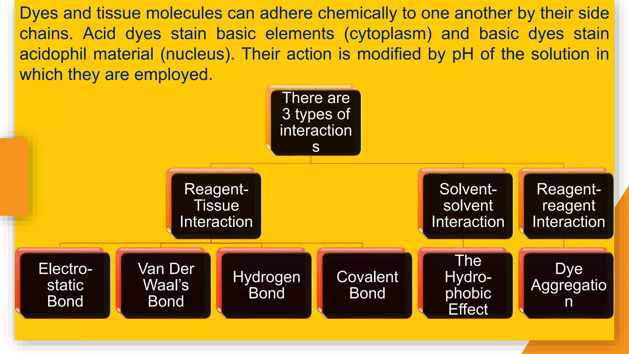
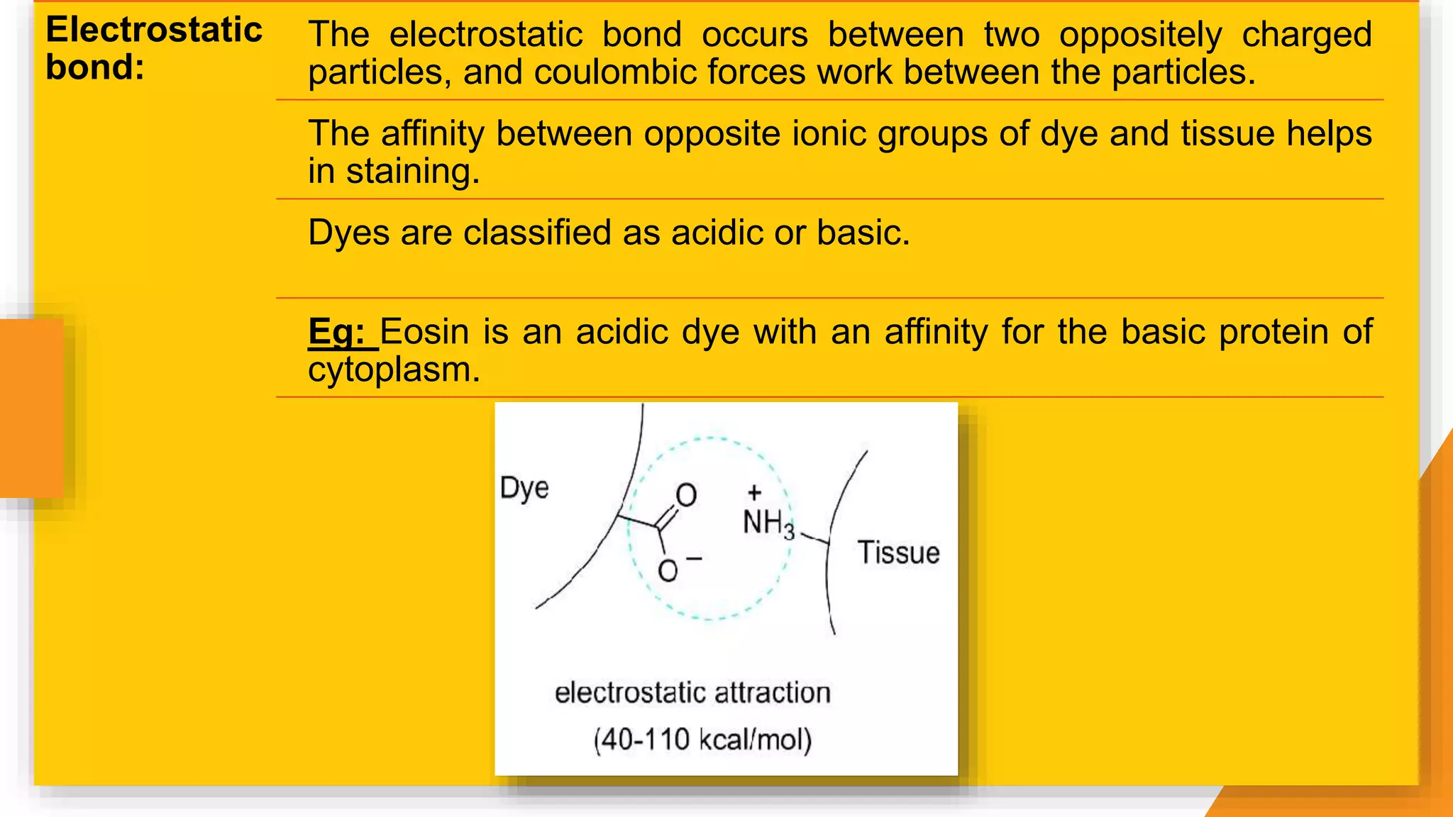
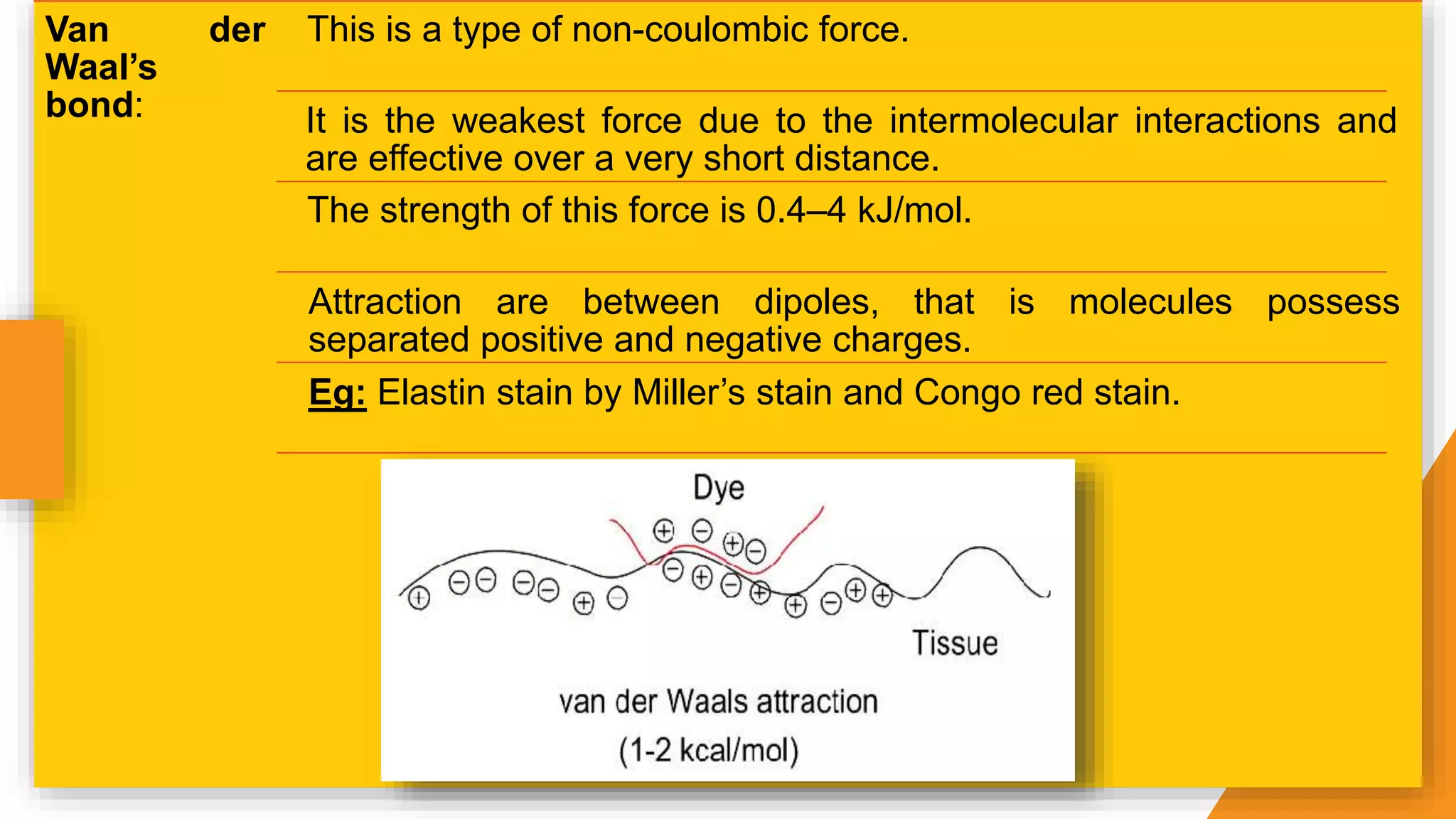
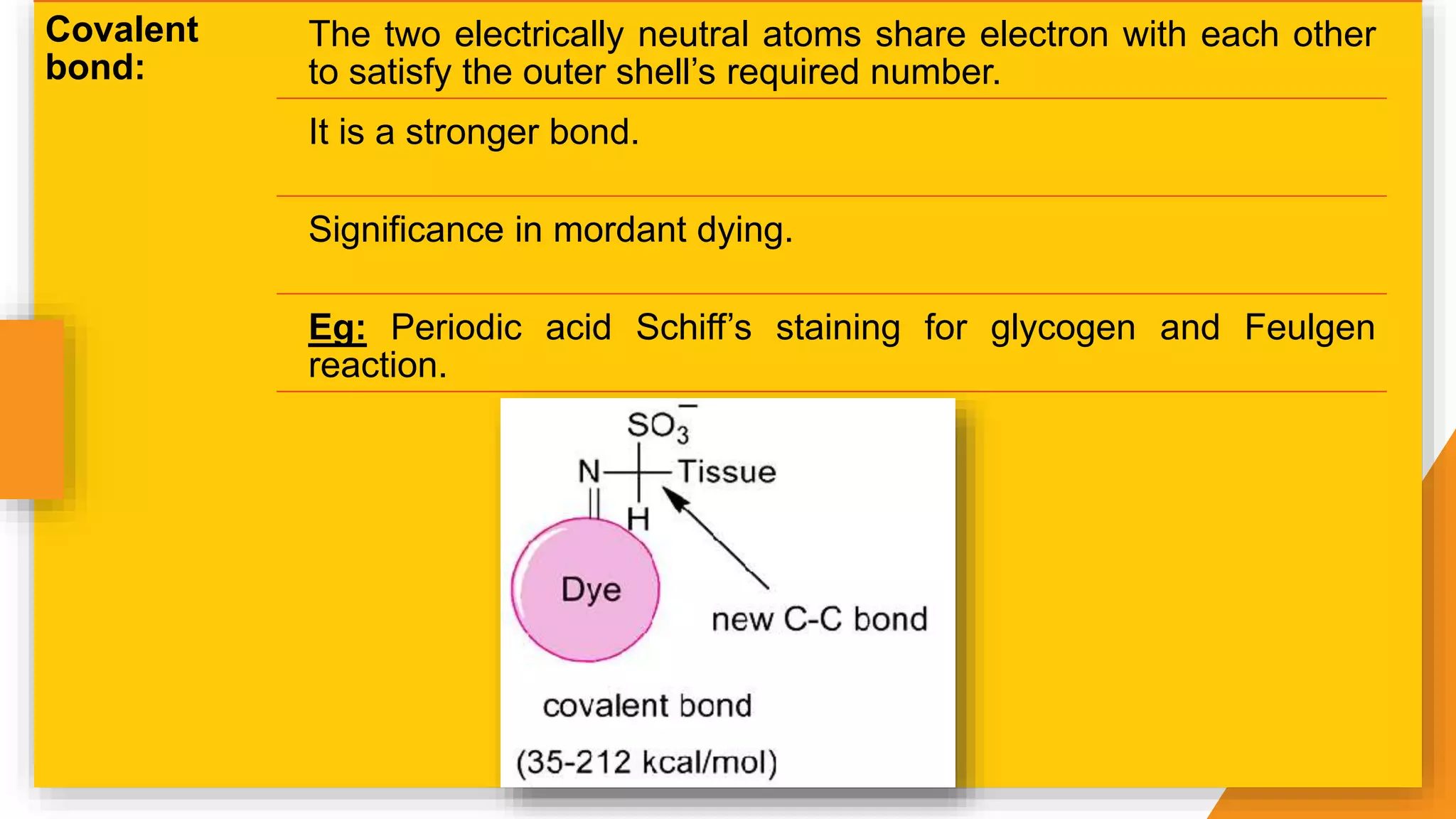
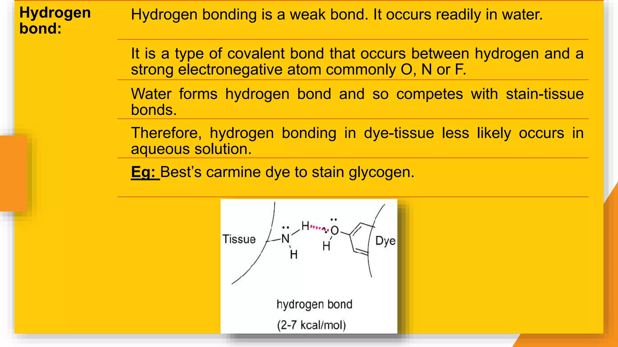
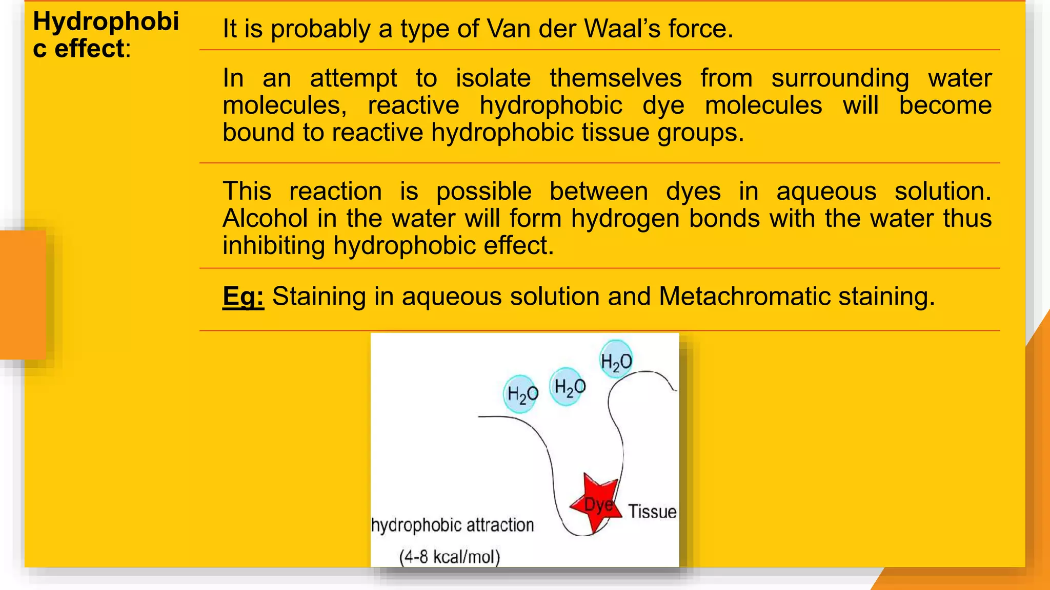
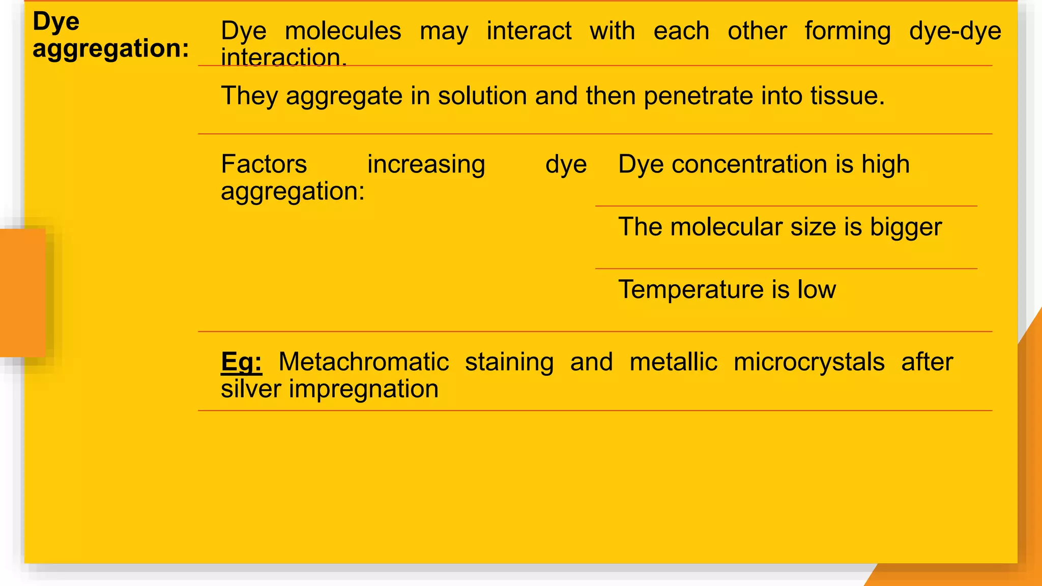
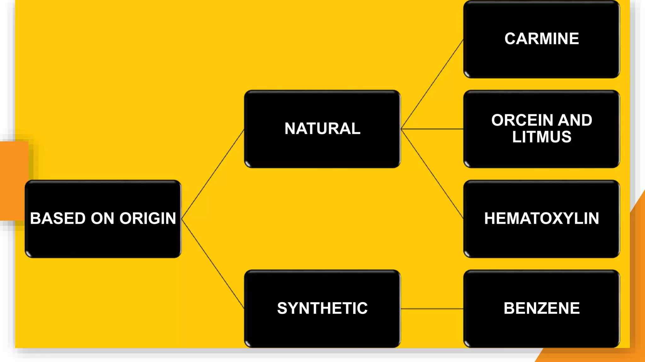
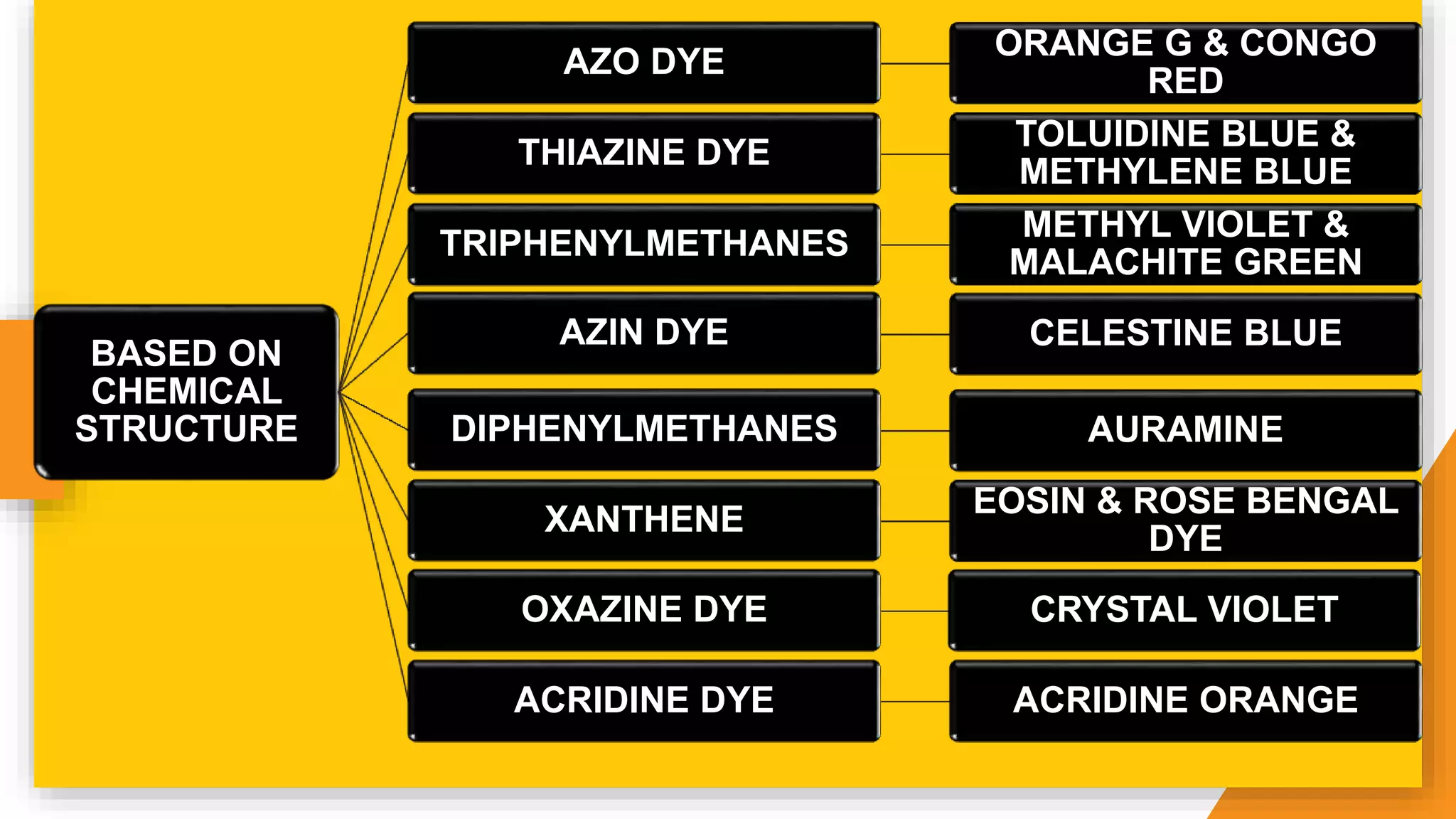
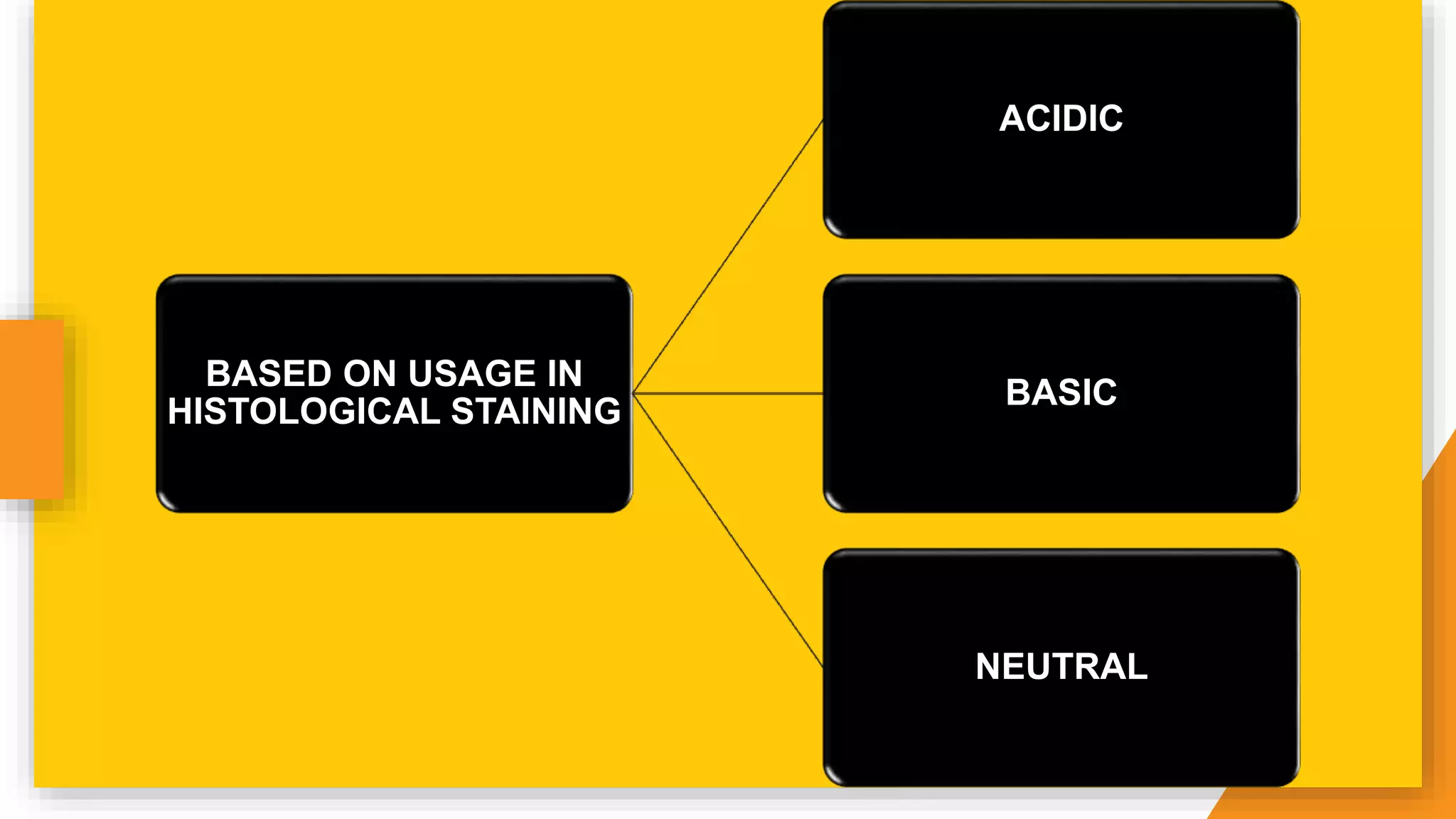
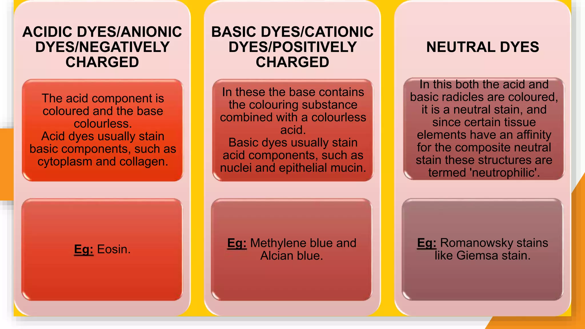
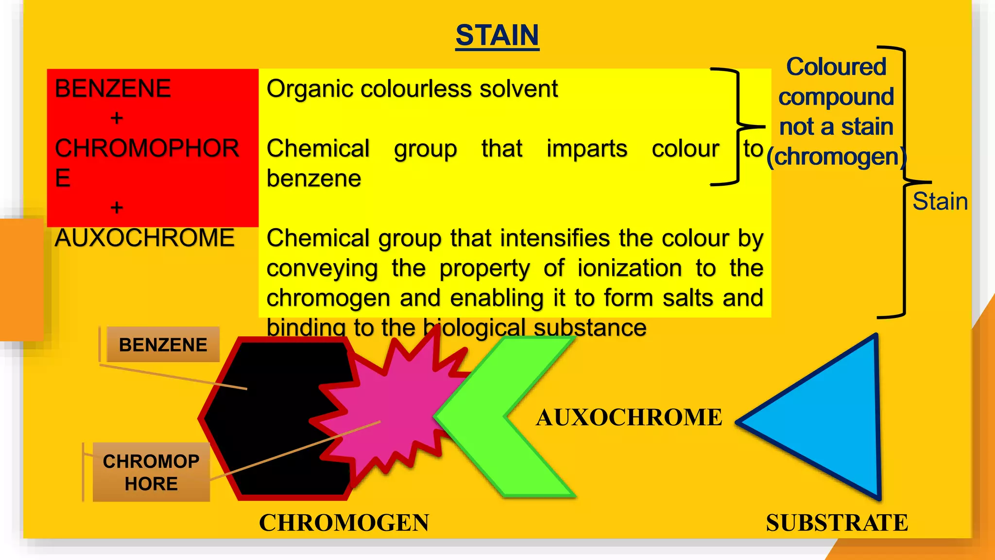
BY
DR. ISHITA SINGHAL
MDS SECOND YEAR
DEFINITIONS
INTRODUCTION
HISTORY
STAINING
TYPES OF STAINING
PHYSICAL THEORIES
CHEMICAL THEORIES
CLASSIFICATION OF DYES
PRINCIPLES OF SYNTHETIC DYE
CHEMISTRY
MORDANTS
ACCENTUATORS AND ACCELERATORS
METACHROMASY
REFERENCES
INDEX
DEFINITIONS
Dye is a single chemical reagent that chemically bonds
to the substrate to which it is being applied, to highlight a
specific component of a tissue.
Stain is a mixture of selected dyes, which when added
to living cells or structural components makes them
clearly visible, by highlighting different components.
Biological staining is the union between a coloured dye
and a tissue substrate which resists simple washing.
Staining is treating tissues or cells with a reagent or
series of reagents so that it acquires a colour, usually no
particles of dye are seen and the stained element is
transparent. -Culling et al
Staining is a visual labelling of some entity by attaching
or depositing in its vicinity, a marker of definite colour or
shape. -Bancroft et al
INTRODUCTION
• The tissue section is colourless because the fixed protein has the
same refractive index as that of glass. We use dyes that have specific
affinity with the different tissue proteins and colour them differently.
• Colour is seen by the eye as a result of the effect of certain
electromagnetic waves on the rods and cones of the retina. These
waves, which have a varying length, will determine the colour that is
seen.
• White light being composed of all the colours of the visible spectrum
varies in wavelength from 4,000 Â to 8,000 Â.
• If light of a specific wavelength is absorbed from white light the
resultant light will then be coloured, the colour being dependent upon
the particular wavelength that has been removed.
Stained materials absorb certain components of the white light
illuminating the specimen and therefore appear coloured.
HISTORY
Leeuwenhoe
k used a dye
as a
biological
stain when
he applied
saffron to
muscles.
Carmine
was
introduced
by Goppert
and Cohn.
Aniline dyes
were first
introduced in
Textile
industry.
Haematoxyli
n was
introduced
by
Waldeyer.
Heidenhain
devised the
iron
Haematoxyli
n technique.
1714
1849
1856
1863
1891
STAINING
The purpose of staining is:
To see the organism
better in contrast with
background
To differentiate one
organism from another
To determine particular
structures
For a successful staining there should be high specificity (ability to
distinguish between individual cell components) and high sensitivity
(capacity of a stain to demonstrate tissue substance at low
concentration).
An ideal stain should allow for:
Evaluation of the architectural pattern of
the tissue fragments.
Proper evaluation of nuclear morphology.
Details of cytoplasmic characteristic.
Visualisation and identification of
diagnostic features in the background.
Factors affecting staining:
Dye affinity to
the target
tissue
specimen:
The tendency to
bind a dye with
the target tissue
is known as dye
affinity. Affinity of
the dye to the
specific tissue is
influenced by the
pH and the
presence of
inorganic salt
concentration of
the solvent.
Specimen
geometry
• Thickness of
the tissue
• Surface
topography
• Inner geometry
of the tissue
Target
concentration:
The
concentration of
the target tissue
affects the
staining intensity
as the more the
amount of the
target tissue, the
more intense will
be the staining.
Rate of
reaction:
Rate of reaction
in the target
tissue also
influences the
staining pattern
such as in
Feulgen
reaction, the
short reaction
time exposes
only a few
aldehyde groups
producing weak
staining pattern.
Rate of stain
loss:
Staining pattern
is greatly
influenced by
rate of stain loss.
At times the stain
loss may be
intentional such
as differentiation
or de-staining.
The
differentiation
often removes
the excess stain
from the cell and
thus helps to
differentiate the
organisms.
TYPES
OF
STAINING
Direct Staining: When a staining procedure colours the cells present in a
preparation, but leaves the background colourless. Eg: Methylene Blue.
Indirect Staining: When a procedure colours the background, but leaves the
cells colourless, with the presence of mordant. Eg: Iodine in Gram stain.
Progressive Staining: Solutions stain to a desired intensity and no more.
Therefore they do not require differentiation in a dilute acid alcohol. Eg:
Mayers Hematoxylin.
Regressive Staining: The tissue is deliberately over stained and then de-
stained (differentiated) until the proper endpoint is reached. Eg: Harris
Hematoxylin.
Intravital Staining: Stain may be injected into a living animal and the stained
tissue removed and examined. Eg: Toluidine Blue.
Supravital Staining: The living tissue may be removed directly and
subsequently stained. Eg: Janus Green for staining mitochondria.
PHYSICAL
THEORIES
• Physical theories depend upon:
1. Solubility: Examples of this method of staining are the fat stains
which are effective simply because the stain is more soluble in the fat
than in the 70 % alcohol or other solvent in which it may be
dissolved.
2. Adsorption: This is the property by which a large body attracts to
itself minute particles from a surrounding medium, and is a
phenomenon well known to chemists.
CHEMICAL
THEORIES
There are
3 types of
interaction
s
Reagent-
Tissue
Interaction
Electro-
static
Bond
Van Der
Waal’s
Bond
Hydrogen
Bond
Covalent
Bond
Solvent-
solvent
Interaction
The
Hydro-
phobic
Effect
Reagent-
reagent
Interaction
Dye
Aggregatio
n
Dyes and tissue molecules can adhere chemically to one another by their side
chains. Acid dyes stain basic elements (cytoplasm) and basic dyes stain
acidophil material (nucleus). Their action is modified by pH of the solution in
which they are employed.
Electrostatic
bond:
The electrostatic bond occurs between two oppositely charged
particles, and coulombic forces work between the particles.
The affinity between opposite ionic groups of dye and tissue helps
in staining.
Dyes are classified as acidic or basic.
Eg: Eosin is an acidic dye with an affinity for the basic protein of
cytoplasm.
Schematic diagram showing
interaction of anionic and
cationic dye with the oppositely
charged tissue components.
Acid or anionic dye binds with
the cytoplasmic protein and
collagen, whereas the basic or
cationic dye binds with nucleic
acid of the nucleus.
Van der
Waal’s
bond:
This is a type of non-coulombic force.
It is the weakest force due to the intermolecular interactions and
are effective over a very short distance.
The strength of this force is 0.4–4 kJ/mol.
Attraction are between dipoles, that is molecules possess
separated positive and negative charges.
Eg: Elastin stain by Miller’s stain and Congo red stain.
Covalent
bond:
The two electrically neutral atoms share electron with each other
to satisfy the outer shell’s required number.
It is a stronger bond.
Significance in mordant dying.
Eg: Periodic acid Schiff’s staining for glycogen and Feulgen
reaction.
Hydrogen
bond:
Hydrogen bonding is a weak bond. It occurs readily in water.
It is a type of covalent bond that occurs between hydrogen and a
strong electronegative atom commonly O, N or F.
Water forms hydrogen bond and so competes with stain-tissue
bonds.
Therefore, hydrogen bonding in dye-tissue less likely occurs in
aqueous solution.
Eg: Best’s carmine dye to stain glycogen.
Hydrophobi
c effect:
It is probably a type of Van der Waal’s force.
In an attempt to isolate themselves from surrounding water
molecules, reactive hydrophobic dye molecules will become
bound to reactive hydrophobic tissue groups.
This reaction is possible between dyes in aqueous solution.
Alcohol in the water will form hydrogen bonds with the water thus
inhibiting hydrophobic effect.
Eg: Staining in aqueous solution and Metachromatic staining.
Dye
aggregation:
Dye molecules may interact with each other forming dye-dye
interaction.
They aggregate in solution and then penetrate into tissue.
Factors increasing dye
aggregation:
Dye concentration is high
The molecular size is bigger
Temperature is low
Eg: Metachromatic staining and metallic microcrystals after
silver impregnation
CLASSIFICATION
OF
DYE
BASED ON ORIGIN
NATURAL
CARMINE
ORCEIN AND
LITMUS
HEMATOXYLIN
SYNTHETIC BENZENE
BASED ON
CHEMICAL
STRUCTURE
AZO DYE
ORANGE G & CONGO
RED
THIAZINE DYE
TOLUIDINE BLUE &
METHYLENE BLUE
TRIPHENYLMETHANES
METHYL VIOLET &
MALACHITE GREEN
AZIN DYE CELESTINE BLUE
DIPHENYLMETHANES AURAMINE
XANTHENE
EOSIN & ROSE BENGAL
DYE
OXAZINE DYE CRYSTAL VIOLET
ACRIDINE DYE ACRIDINE ORANGE
BASED ON USAGE IN
HISTOLOGICAL STAINING
ACIDIC
BASIC
NEUTRAL
ACIDIC DYES/ANIONIC
DYES/NEGATIVELY
CHARGED
The acid component is
coloured and the base
colourless.
Acid dyes usually stain
basic components, such as
cytoplasm and collagen.
Eg: Eosin.
BASIC DYES/CATIONIC
DYES/POSITIVELY
CHARGED
In these the base contains
the colouring substance
combined with a colourless
acid.
Basic dyes usually stain
acid components, such as
nuclei and epithelial mucin.
Eg: Methylene blue and
Alcian blue.
NEUTRAL DYES
In this both the acid and
basic radicles are coloured,
it is a neutral stain, and
since certain tissue
elements have an affinity
for the composite neutral
stain these structures are
termed 'neutrophilic'.
Eg: Romanowsky stains
like Giemsa stain.
32
BENZENE
+
CHROMOPHOR
E
+
AUXOCHROME
Organic colourless solvent
Chemical group that imparts colour to
benzene
Chemical group that intensifies the colour by
conveying the property of ionization to the
chromogen and enabling it to form salts and
binding to the biological substance
Coloured
compound
not a stain
(chromogen)
Stain
STAIN
CHROMOGEN
AUXOCHROME
SUBSTRATE
CHROMOP
HORE
Coloured
compound
not a stain
(chromogen)
Coloured
compound
not a stain
(chromogen)
BENZENE
BENZENE
• When 2 of the hydrogen atoms are replaced by oxygen, a
readjustment of the double bonds takes place and a new compound,
quinone (C6H402), is formed, compounds from which are called
quinoid compounds.
• The change from benzene to quinone is important, because benzene
derivatives absorb light only in the UV band and are colourless,
whereas quinoid compounds have absorption bands in the visible
spectrum and are coloured.
Formed by 6
carbon atoms
and 6 hydrogen
atoms to form a
ring
Each carbon
atom has a
double bond on
one side and a
single bond on
the other
CHROMOPHORES
• Certain chemical group
introduced into benzene
ring by substitution
induce colour to the
compound. This is known
as chromophore, and the
chemical compound
formed by the grouping is
known as chromogen.
• These groups have many
free electrons that absorb
the ultraviolet rays of light
which are not in the
visible range.
• Eg: Active chromophore
group, nitro grouping and
azo coupling.
AUXOCHROMES
• It helps to intensify the
light. It is an ionizing
group that also helps to
stick the stain with the
tissue.
• The auxochrome group
augments more free
electrons in the
chromophore groups.
• The increased number of
electrons in the system
helps to absorb light of
longer wavelength in the
visible range.
• Eg: Acidic, such as the
hydroxyl group or basic,
such as the amino group
(NH2).
SULPHONIC GROUP
• The sulphonic group is
important in dye
chemistry because it
forms acid salts.
• It is only slightly
auxochromic and is used
principally either to make
an insoluble dye soluble
in water, or to convert an
otherwise basic dye into
an acid dye.
• Eg: Basic fuchsin is
converted to acid fuchsin
by the introduction of a
sulphonic group.
Schematic
diagram showing
colour generation
by dye. The
chromophore
group absorbs
light and imparts
colour to the
stain. The
auxochrome
group donates
more electrons to
the chromophore
group and helps
to absorb light of
longer
MORDANTS
• Mordant is a metallic substance that forms a link between the tissue
and the dye.
• It is a substance that forms an insoluble compound with a stain and
helps to fix the colour to the cell components.
• + MORDANT LAKE
• Some lakes are unstable, and with such compounds tissue may be
soaked in the mordant immediately before treatment with the stain, or
even afterwards. Eg: Heidenhain's iron haematoxylin stain and
Gram’s stain respectively.
• The term mordant is only strictly applicable to salts and hydroxides of
di-valent and tri-valent metals.
DYE
ACCENTUATORS
AND
ACCELERATORS
• Accentuators are chemical substances which increase the selectivity
or staining power of the dyes. Eg: Potassium hydroxide in methylene
blue, and phenol in carbol thionin and carbol fuchsin.
• The effect on staining of this group is due to the change in pH of the
staining solution.
• Accentuators when used in the impregnation of the nervous system
with metallic salts have been called accelerators. Eg: Veronal in
Cajal's method for axis cylinders, and chloral hydrate in Cajal's
method for motor end-plates.
METACHROMASY
• A dye which has the ability to change its colour without changing its
chemical structure is said to be metachromatic.
• Staining reaction is known as metachromasy, the tissue is said to
exhibit metachromasia, and the dye is known as a metachromatic
dye.
• Eg:
1. Cationic dye/ Positively charged/ Basic dye: Toluidine blue,
methylene blue, azure A and B, methyl violet, brilliant cresyl blue,
etc.
2. Anionic dye/ Negatively charged/ Acidic dye: Biebrich scarlet and
bromophenol blue.
• Among the principal tissue components which exhibit metachromasia
Mechanism of dye
aggregation in
metachromasia.
The cationic (positively
charged) dye in aqueous
solution is neutralized
with the polyanions of
the tissues.
Subsequently the
nonpolar aromatic ring
of the dye binds with the
other dye by van der
Waals force. The dye
absorbs shorter
wavelength of light, and
the visible colour
changes.
The different types of
metachromasia are
highlighted in this
schematic diagram.
Factors enhancing
metachromasia:
1. Higher dye concentration
2. Low pH
3. Decreased temperature
4. Aqueous solution
REFERENCES
1. Culling CF. Handbook of histopathological and histochemical
techniques: including museum techniques. Butterworth-Heinemann;
2013 Oct 22.
2. Suvarna KS, Layton C, Bancroft JD, editors. Bancroft's theory and
practice of histological techniques E-Book. Elsevier Health Sciences;
2018 Feb 27.
3. Dey P. Basic and advanced laboratory techniques in histopathology
and cytology. Springer Singapore; 2018 Jun 8.
4. https://www.biologydiscussion.com/micro-biology/staining/stains-
meaning-purpose-and-components-microorganisms/55080
5. Veuthey, T., Herrera, G., & Dodero, V. Dyes and stains: from
Theory of staining

Theory of staining

  • 1.
  • 2.
    DEFINITIONS INTRODUCTION HISTORY STAINING TYPES OF STAINING PHYSICALTHEORIES CHEMICAL THEORIES CLASSIFICATION OF DYES PRINCIPLES OF SYNTHETIC DYE CHEMISTRY MORDANTS ACCENTUATORS AND ACCELERATORS METACHROMASY REFERENCES INDEX
  • 3.
  • 4.
    Dye is asingle chemical reagent that chemically bonds to the substrate to which it is being applied, to highlight a specific component of a tissue. Stain is a mixture of selected dyes, which when added to living cells or structural components makes them clearly visible, by highlighting different components. Biological staining is the union between a coloured dye and a tissue substrate which resists simple washing. Staining is treating tissues or cells with a reagent or series of reagents so that it acquires a colour, usually no particles of dye are seen and the stained element is transparent. -Culling et al Staining is a visual labelling of some entity by attaching or depositing in its vicinity, a marker of definite colour or shape. -Bancroft et al
  • 5.
  • 6.
    • The tissuesection is colourless because the fixed protein has the same refractive index as that of glass. We use dyes that have specific affinity with the different tissue proteins and colour them differently. • Colour is seen by the eye as a result of the effect of certain electromagnetic waves on the rods and cones of the retina. These waves, which have a varying length, will determine the colour that is seen. • White light being composed of all the colours of the visible spectrum varies in wavelength from 4,000 Â to 8,000 Â. • If light of a specific wavelength is absorbed from white light the resultant light will then be coloured, the colour being dependent upon the particular wavelength that has been removed.
  • 7.
    Stained materials absorbcertain components of the white light illuminating the specimen and therefore appear coloured.
  • 8.
  • 9.
    Leeuwenhoe k used adye as a biological stain when he applied saffron to muscles. Carmine was introduced by Goppert and Cohn. Aniline dyes were first introduced in Textile industry. Haematoxyli n was introduced by Waldeyer. Heidenhain devised the iron Haematoxyli n technique. 1714 1849 1856 1863 1891
  • 10.
  • 11.
    The purpose ofstaining is: To see the organism better in contrast with background To differentiate one organism from another To determine particular structures For a successful staining there should be high specificity (ability to distinguish between individual cell components) and high sensitivity (capacity of a stain to demonstrate tissue substance at low concentration).
  • 12.
    An ideal stainshould allow for: Evaluation of the architectural pattern of the tissue fragments. Proper evaluation of nuclear morphology. Details of cytoplasmic characteristic. Visualisation and identification of diagnostic features in the background.
  • 13.
    Factors affecting staining: Dyeaffinity to the target tissue specimen: The tendency to bind a dye with the target tissue is known as dye affinity. Affinity of the dye to the specific tissue is influenced by the pH and the presence of inorganic salt concentration of the solvent. Specimen geometry • Thickness of the tissue • Surface topography • Inner geometry of the tissue Target concentration: The concentration of the target tissue affects the staining intensity as the more the amount of the target tissue, the more intense will be the staining. Rate of reaction: Rate of reaction in the target tissue also influences the staining pattern such as in Feulgen reaction, the short reaction time exposes only a few aldehyde groups producing weak staining pattern. Rate of stain loss: Staining pattern is greatly influenced by rate of stain loss. At times the stain loss may be intentional such as differentiation or de-staining. The differentiation often removes the excess stain from the cell and thus helps to differentiate the organisms.
  • 14.
  • 15.
    Direct Staining: Whena staining procedure colours the cells present in a preparation, but leaves the background colourless. Eg: Methylene Blue. Indirect Staining: When a procedure colours the background, but leaves the cells colourless, with the presence of mordant. Eg: Iodine in Gram stain. Progressive Staining: Solutions stain to a desired intensity and no more. Therefore they do not require differentiation in a dilute acid alcohol. Eg: Mayers Hematoxylin. Regressive Staining: The tissue is deliberately over stained and then de- stained (differentiated) until the proper endpoint is reached. Eg: Harris Hematoxylin. Intravital Staining: Stain may be injected into a living animal and the stained tissue removed and examined. Eg: Toluidine Blue. Supravital Staining: The living tissue may be removed directly and subsequently stained. Eg: Janus Green for staining mitochondria.
  • 16.
  • 17.
    • Physical theoriesdepend upon: 1. Solubility: Examples of this method of staining are the fat stains which are effective simply because the stain is more soluble in the fat than in the 70 % alcohol or other solvent in which it may be dissolved. 2. Adsorption: This is the property by which a large body attracts to itself minute particles from a surrounding medium, and is a phenomenon well known to chemists.
  • 18.
  • 19.
    There are 3 typesof interaction s Reagent- Tissue Interaction Electro- static Bond Van Der Waal’s Bond Hydrogen Bond Covalent Bond Solvent- solvent Interaction The Hydro- phobic Effect Reagent- reagent Interaction Dye Aggregatio n Dyes and tissue molecules can adhere chemically to one another by their side chains. Acid dyes stain basic elements (cytoplasm) and basic dyes stain acidophil material (nucleus). Their action is modified by pH of the solution in which they are employed.
  • 20.
    Electrostatic bond: The electrostatic bondoccurs between two oppositely charged particles, and coulombic forces work between the particles. The affinity between opposite ionic groups of dye and tissue helps in staining. Dyes are classified as acidic or basic. Eg: Eosin is an acidic dye with an affinity for the basic protein of cytoplasm.
  • 21.
    Schematic diagram showing interactionof anionic and cationic dye with the oppositely charged tissue components. Acid or anionic dye binds with the cytoplasmic protein and collagen, whereas the basic or cationic dye binds with nucleic acid of the nucleus.
  • 22.
    Van der Waal’s bond: This isa type of non-coulombic force. It is the weakest force due to the intermolecular interactions and are effective over a very short distance. The strength of this force is 0.4–4 kJ/mol. Attraction are between dipoles, that is molecules possess separated positive and negative charges. Eg: Elastin stain by Miller’s stain and Congo red stain.
  • 23.
    Covalent bond: The two electricallyneutral atoms share electron with each other to satisfy the outer shell’s required number. It is a stronger bond. Significance in mordant dying. Eg: Periodic acid Schiff’s staining for glycogen and Feulgen reaction.
  • 24.
    Hydrogen bond: Hydrogen bonding isa weak bond. It occurs readily in water. It is a type of covalent bond that occurs between hydrogen and a strong electronegative atom commonly O, N or F. Water forms hydrogen bond and so competes with stain-tissue bonds. Therefore, hydrogen bonding in dye-tissue less likely occurs in aqueous solution. Eg: Best’s carmine dye to stain glycogen.
  • 25.
    Hydrophobi c effect: It isprobably a type of Van der Waal’s force. In an attempt to isolate themselves from surrounding water molecules, reactive hydrophobic dye molecules will become bound to reactive hydrophobic tissue groups. This reaction is possible between dyes in aqueous solution. Alcohol in the water will form hydrogen bonds with the water thus inhibiting hydrophobic effect. Eg: Staining in aqueous solution and Metachromatic staining.
  • 26.
    Dye aggregation: Dye molecules mayinteract with each other forming dye-dye interaction. They aggregate in solution and then penetrate into tissue. Factors increasing dye aggregation: Dye concentration is high The molecular size is bigger Temperature is low Eg: Metachromatic staining and metallic microcrystals after silver impregnation
  • 27.
  • 28.
    BASED ON ORIGIN NATURAL CARMINE ORCEINAND LITMUS HEMATOXYLIN SYNTHETIC BENZENE
  • 29.
    BASED ON CHEMICAL STRUCTURE AZO DYE ORANGEG & CONGO RED THIAZINE DYE TOLUIDINE BLUE & METHYLENE BLUE TRIPHENYLMETHANES METHYL VIOLET & MALACHITE GREEN AZIN DYE CELESTINE BLUE DIPHENYLMETHANES AURAMINE XANTHENE EOSIN & ROSE BENGAL DYE OXAZINE DYE CRYSTAL VIOLET ACRIDINE DYE ACRIDINE ORANGE
  • 30.
    BASED ON USAGEIN HISTOLOGICAL STAINING ACIDIC BASIC NEUTRAL
  • 31.
    ACIDIC DYES/ANIONIC DYES/NEGATIVELY CHARGED The acidcomponent is coloured and the base colourless. Acid dyes usually stain basic components, such as cytoplasm and collagen. Eg: Eosin. BASIC DYES/CATIONIC DYES/POSITIVELY CHARGED In these the base contains the colouring substance combined with a colourless acid. Basic dyes usually stain acid components, such as nuclei and epithelial mucin. Eg: Methylene blue and Alcian blue. NEUTRAL DYES In this both the acid and basic radicles are coloured, it is a neutral stain, and since certain tissue elements have an affinity for the composite neutral stain these structures are termed 'neutrophilic'. Eg: Romanowsky stains like Giemsa stain.
  • 32.
  • 33.
    BENZENE + CHROMOPHOR E + AUXOCHROME Organic colourless solvent Chemicalgroup that imparts colour to benzene Chemical group that intensifies the colour by conveying the property of ionization to the chromogen and enabling it to form salts and binding to the biological substance Coloured compound not a stain (chromogen) Stain STAIN CHROMOGEN AUXOCHROME SUBSTRATE CHROMOP HORE Coloured compound not a stain (chromogen) Coloured compound not a stain (chromogen) BENZENE
  • 34.
    BENZENE • When 2of the hydrogen atoms are replaced by oxygen, a readjustment of the double bonds takes place and a new compound, quinone (C6H402), is formed, compounds from which are called quinoid compounds. • The change from benzene to quinone is important, because benzene derivatives absorb light only in the UV band and are colourless, whereas quinoid compounds have absorption bands in the visible spectrum and are coloured. Formed by 6 carbon atoms and 6 hydrogen atoms to form a ring Each carbon atom has a double bond on one side and a single bond on the other
  • 36.
    CHROMOPHORES • Certain chemicalgroup introduced into benzene ring by substitution induce colour to the compound. This is known as chromophore, and the chemical compound formed by the grouping is known as chromogen. • These groups have many free electrons that absorb the ultraviolet rays of light which are not in the visible range. • Eg: Active chromophore group, nitro grouping and azo coupling. AUXOCHROMES • It helps to intensify the light. It is an ionizing group that also helps to stick the stain with the tissue. • The auxochrome group augments more free electrons in the chromophore groups. • The increased number of electrons in the system helps to absorb light of longer wavelength in the visible range. • Eg: Acidic, such as the hydroxyl group or basic, such as the amino group (NH2). SULPHONIC GROUP • The sulphonic group is important in dye chemistry because it forms acid salts. • It is only slightly auxochromic and is used principally either to make an insoluble dye soluble in water, or to convert an otherwise basic dye into an acid dye. • Eg: Basic fuchsin is converted to acid fuchsin by the introduction of a sulphonic group.
  • 37.
    Schematic diagram showing colour generation bydye. The chromophore group absorbs light and imparts colour to the stain. The auxochrome group donates more electrons to the chromophore group and helps to absorb light of longer
  • 39.
  • 40.
    • Mordant isa metallic substance that forms a link between the tissue and the dye. • It is a substance that forms an insoluble compound with a stain and helps to fix the colour to the cell components. • + MORDANT LAKE • Some lakes are unstable, and with such compounds tissue may be soaked in the mordant immediately before treatment with the stain, or even afterwards. Eg: Heidenhain's iron haematoxylin stain and Gram’s stain respectively. • The term mordant is only strictly applicable to salts and hydroxides of di-valent and tri-valent metals. DYE
  • 43.
  • 44.
    • Accentuators arechemical substances which increase the selectivity or staining power of the dyes. Eg: Potassium hydroxide in methylene blue, and phenol in carbol thionin and carbol fuchsin. • The effect on staining of this group is due to the change in pH of the staining solution. • Accentuators when used in the impregnation of the nervous system with metallic salts have been called accelerators. Eg: Veronal in Cajal's method for axis cylinders, and chloral hydrate in Cajal's method for motor end-plates.
  • 45.
  • 46.
    • A dyewhich has the ability to change its colour without changing its chemical structure is said to be metachromatic. • Staining reaction is known as metachromasy, the tissue is said to exhibit metachromasia, and the dye is known as a metachromatic dye. • Eg: 1. Cationic dye/ Positively charged/ Basic dye: Toluidine blue, methylene blue, azure A and B, methyl violet, brilliant cresyl blue, etc. 2. Anionic dye/ Negatively charged/ Acidic dye: Biebrich scarlet and bromophenol blue. • Among the principal tissue components which exhibit metachromasia
  • 47.
    Mechanism of dye aggregationin metachromasia. The cationic (positively charged) dye in aqueous solution is neutralized with the polyanions of the tissues. Subsequently the nonpolar aromatic ring of the dye binds with the other dye by van der Waals force. The dye absorbs shorter wavelength of light, and the visible colour changes.
  • 48.
    The different typesof metachromasia are highlighted in this schematic diagram. Factors enhancing metachromasia: 1. Higher dye concentration 2. Low pH 3. Decreased temperature 4. Aqueous solution
  • 49.
  • 50.
    1. Culling CF.Handbook of histopathological and histochemical techniques: including museum techniques. Butterworth-Heinemann; 2013 Oct 22. 2. Suvarna KS, Layton C, Bancroft JD, editors. Bancroft's theory and practice of histological techniques E-Book. Elsevier Health Sciences; 2018 Feb 27. 3. Dey P. Basic and advanced laboratory techniques in histopathology and cytology. Springer Singapore; 2018 Jun 8. 4. https://www.biologydiscussion.com/micro-biology/staining/stains- meaning-purpose-and-components-microorganisms/55080 5. Veuthey, T., Herrera, G., & Dodero, V. Dyes and stains: from