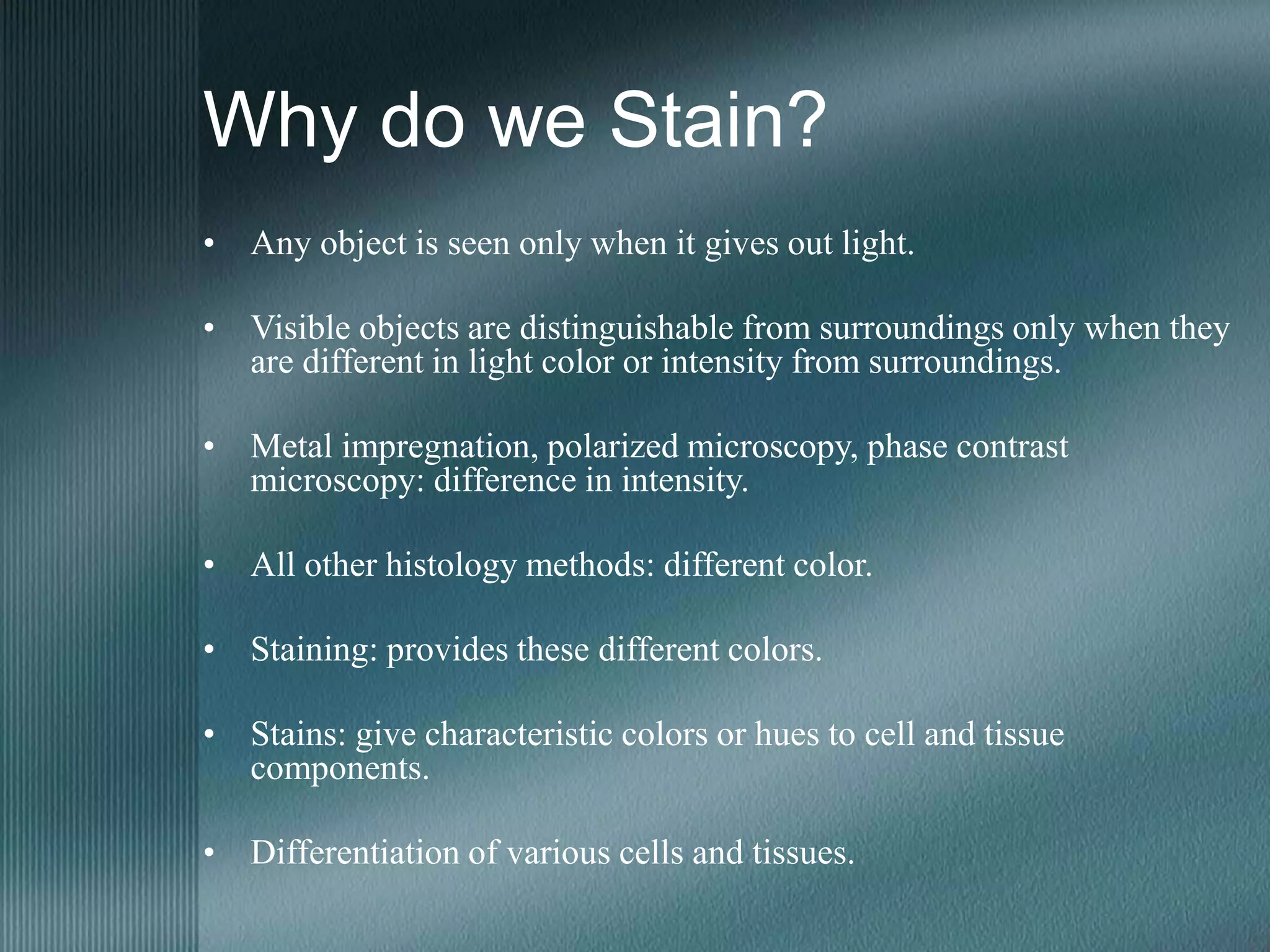
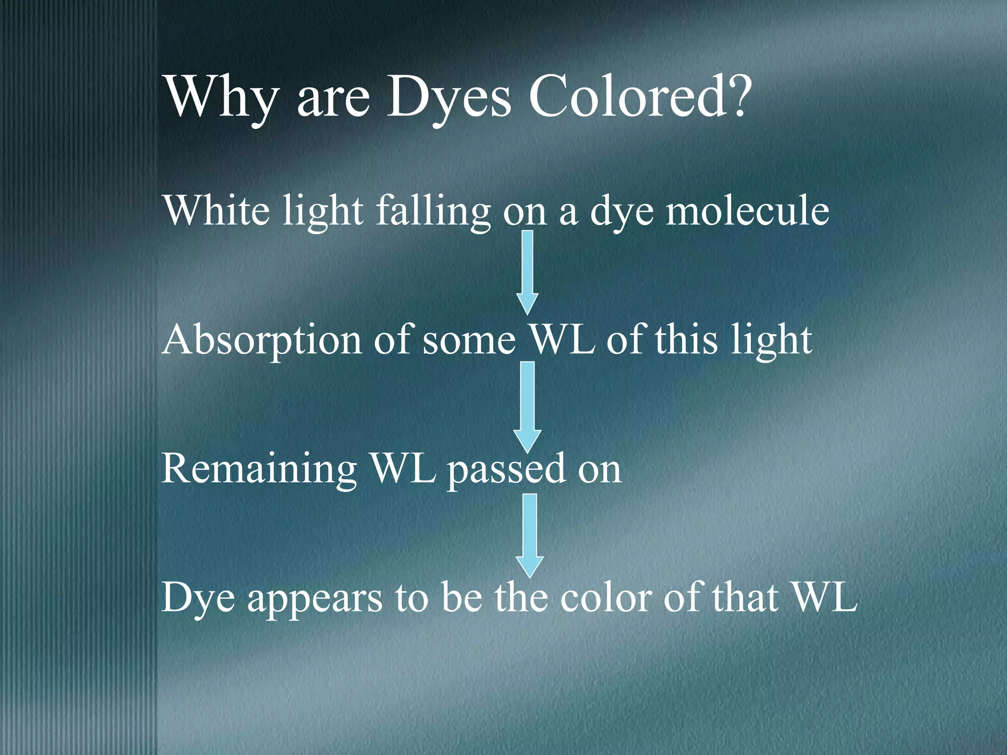
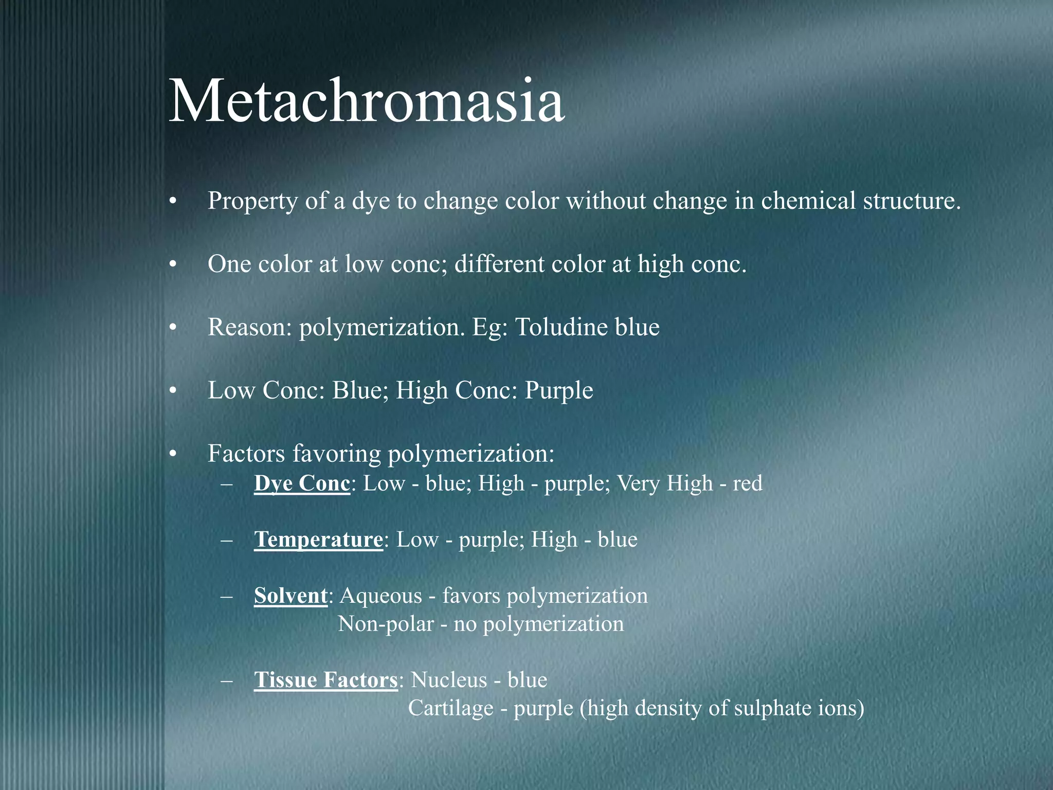
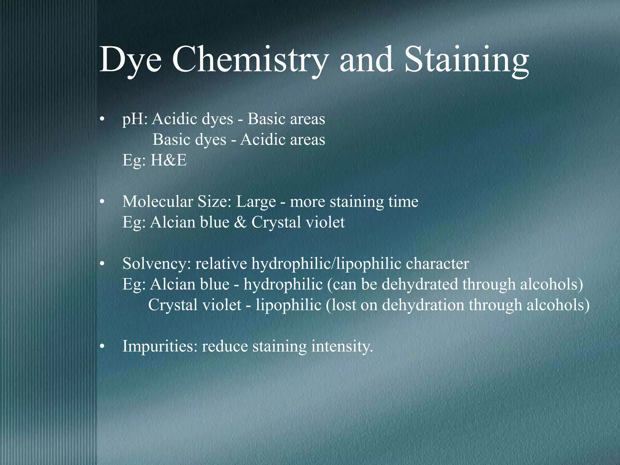
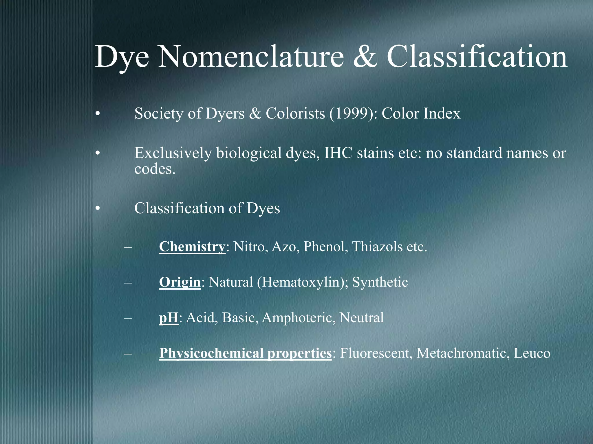
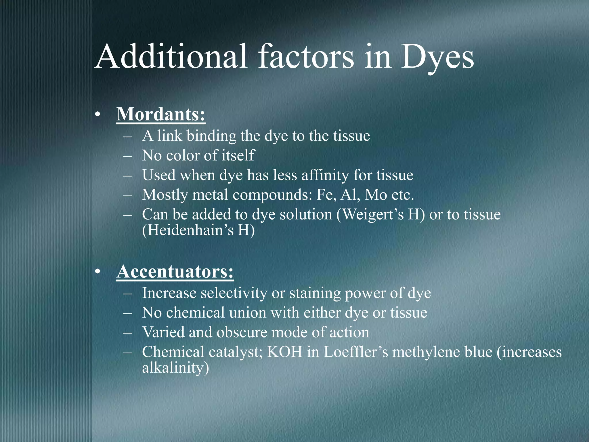
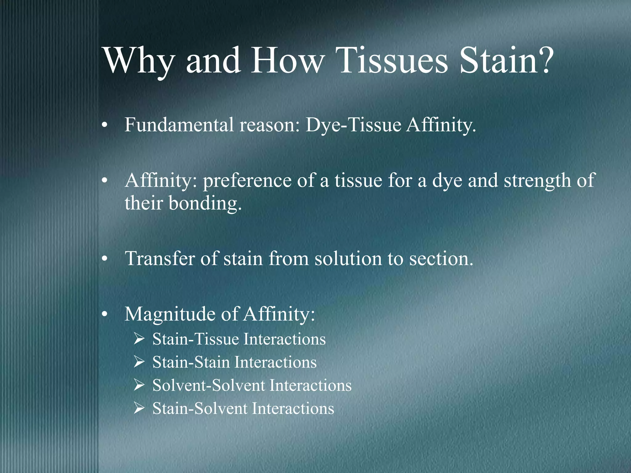
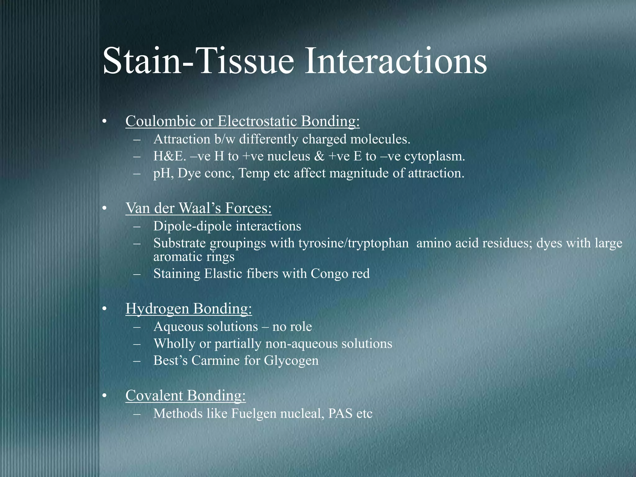
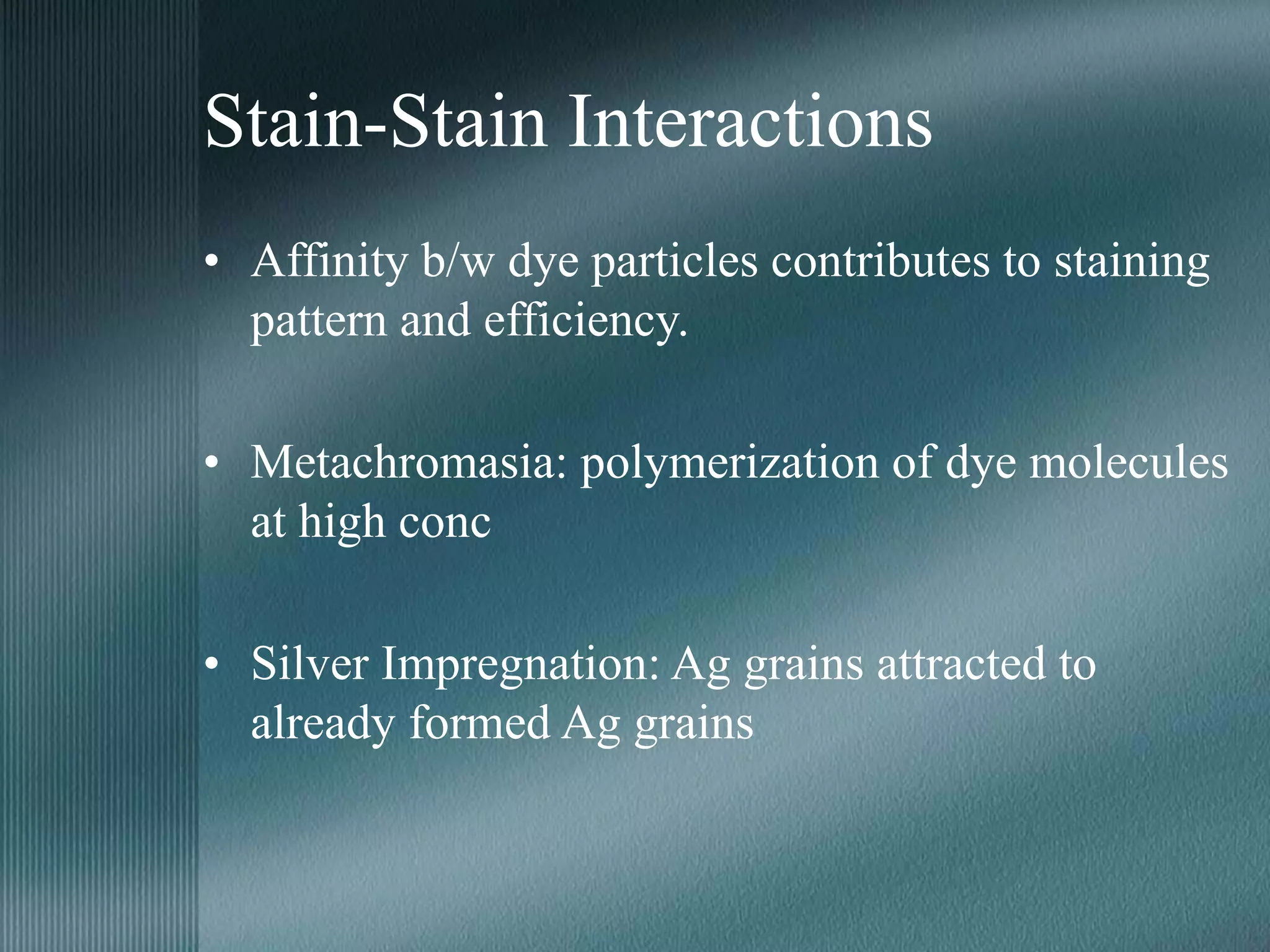
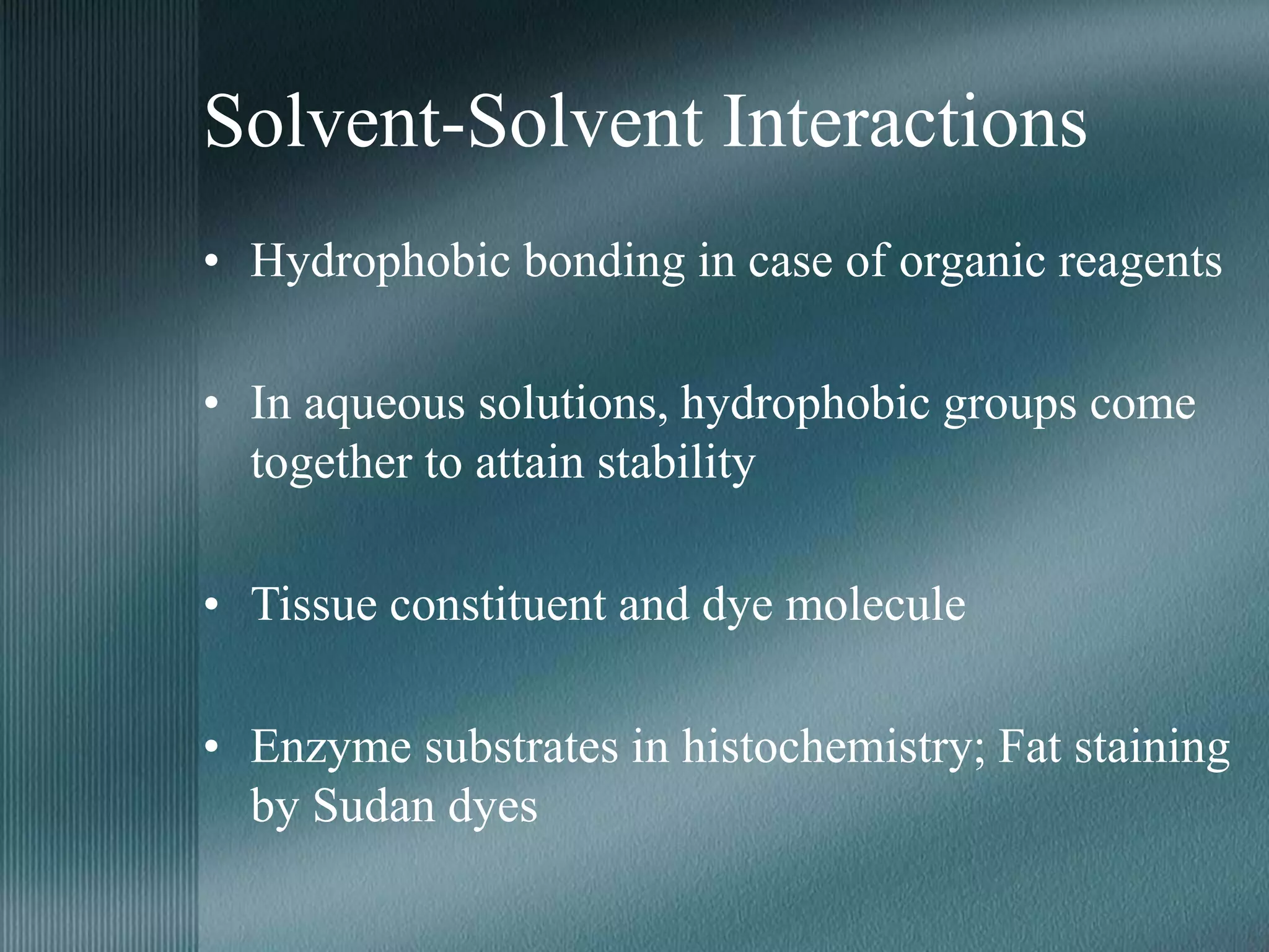
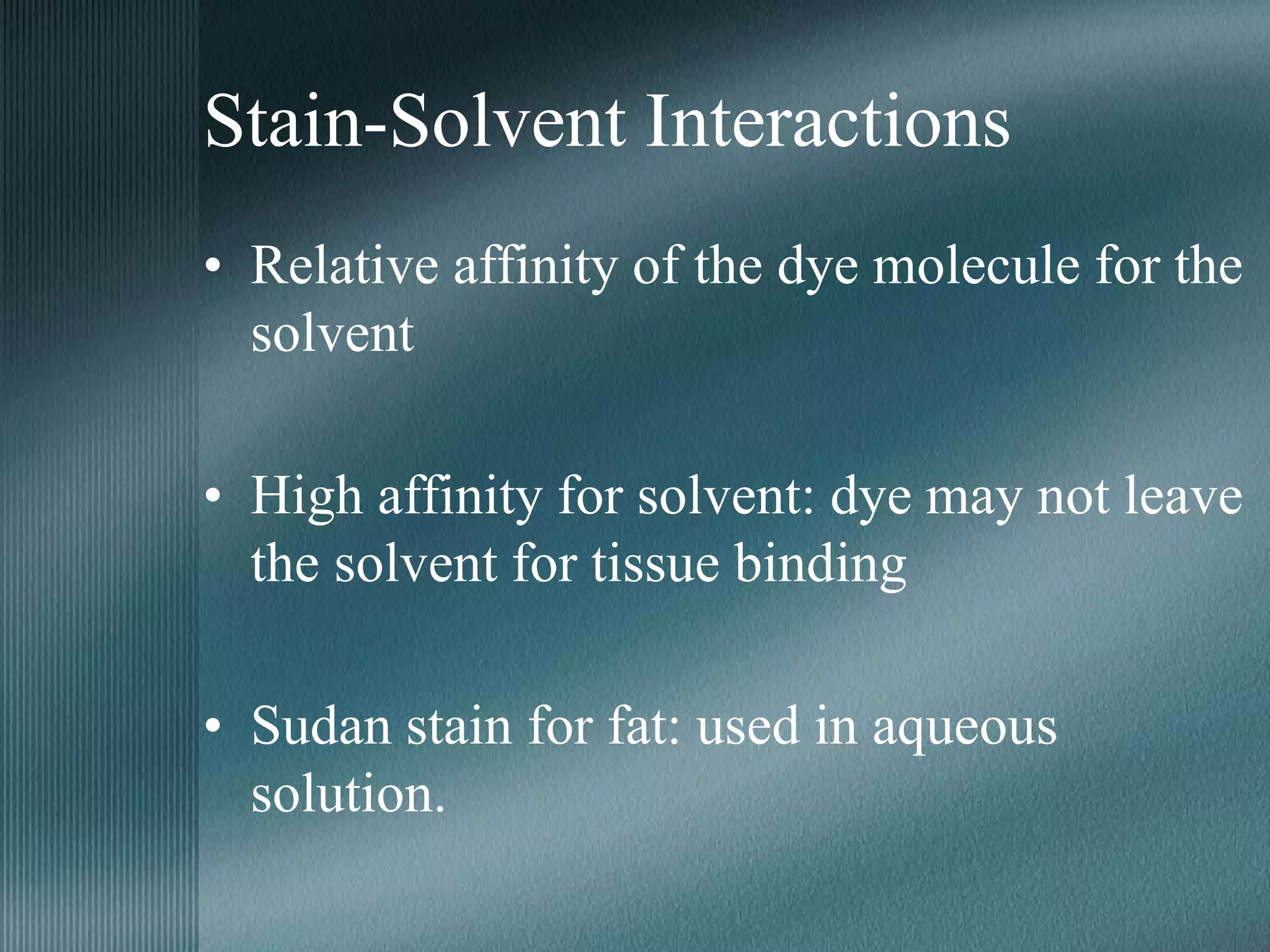
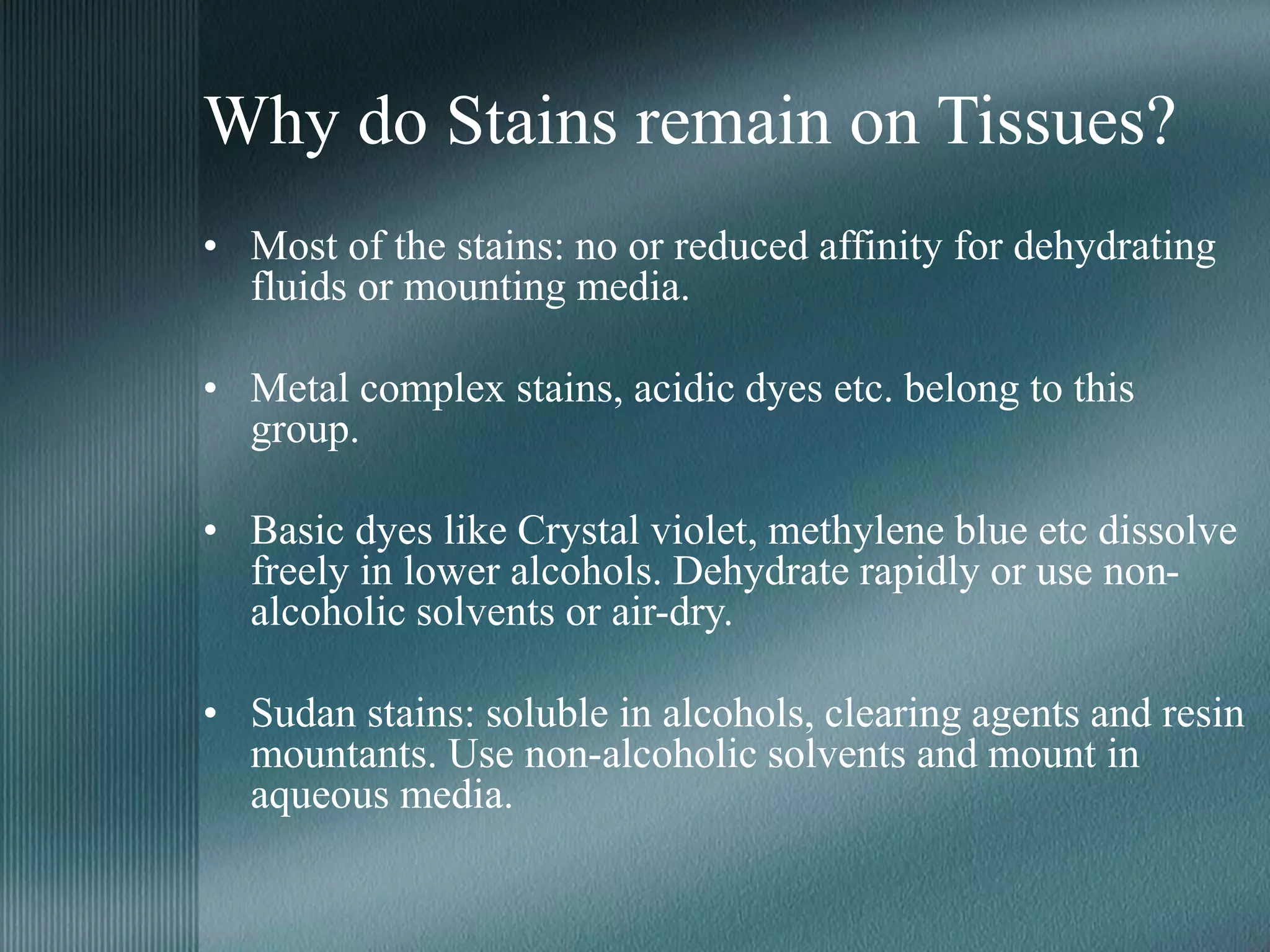
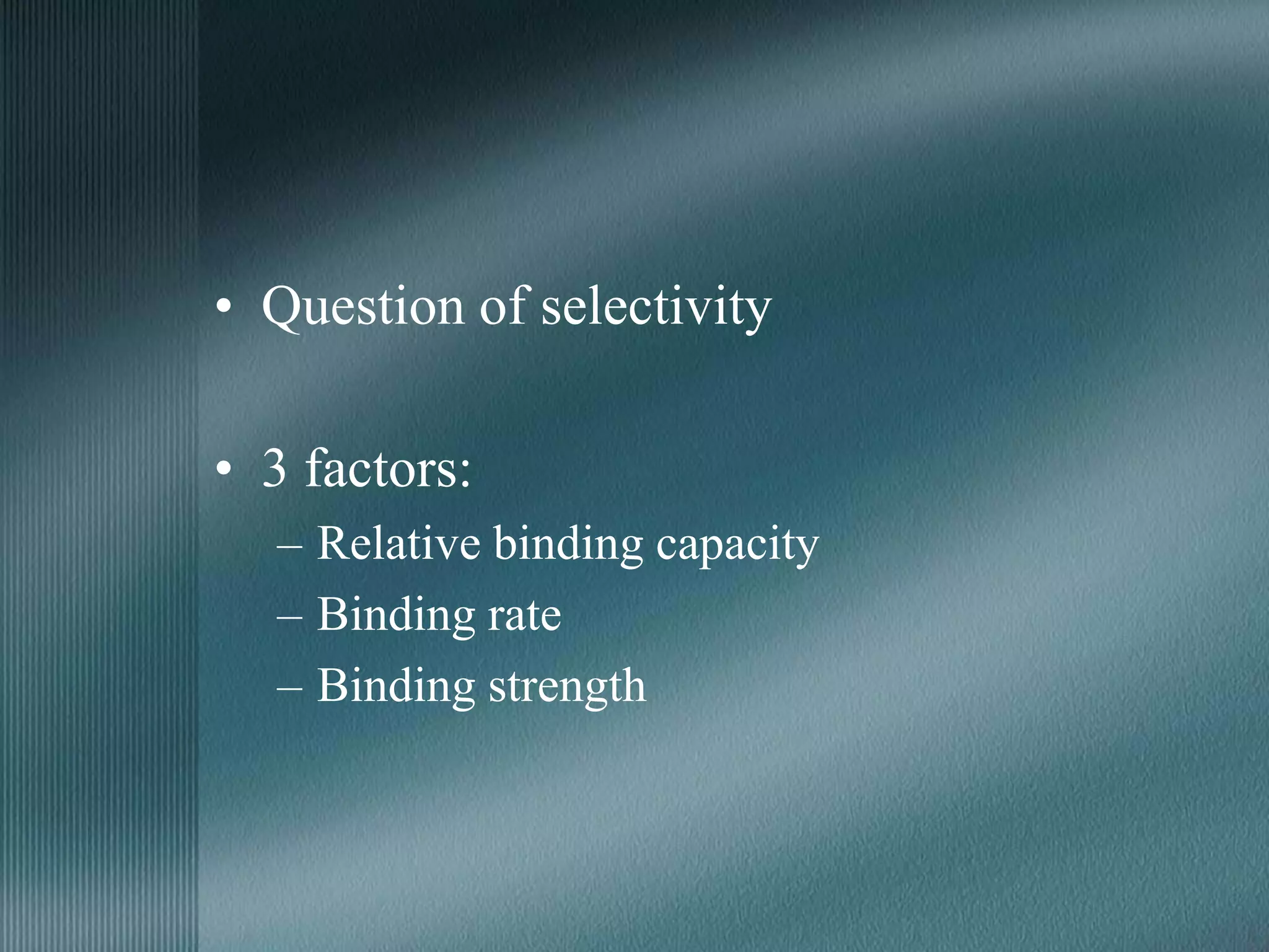
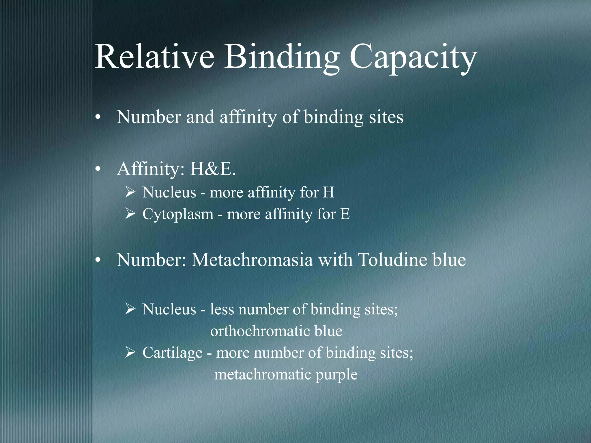
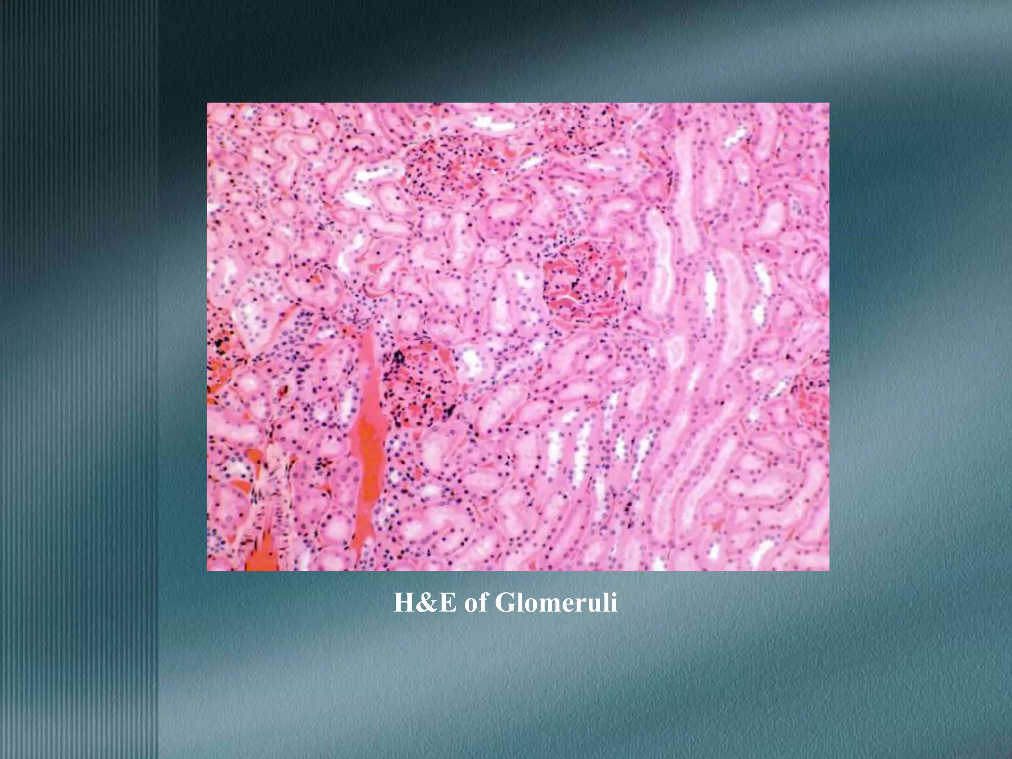
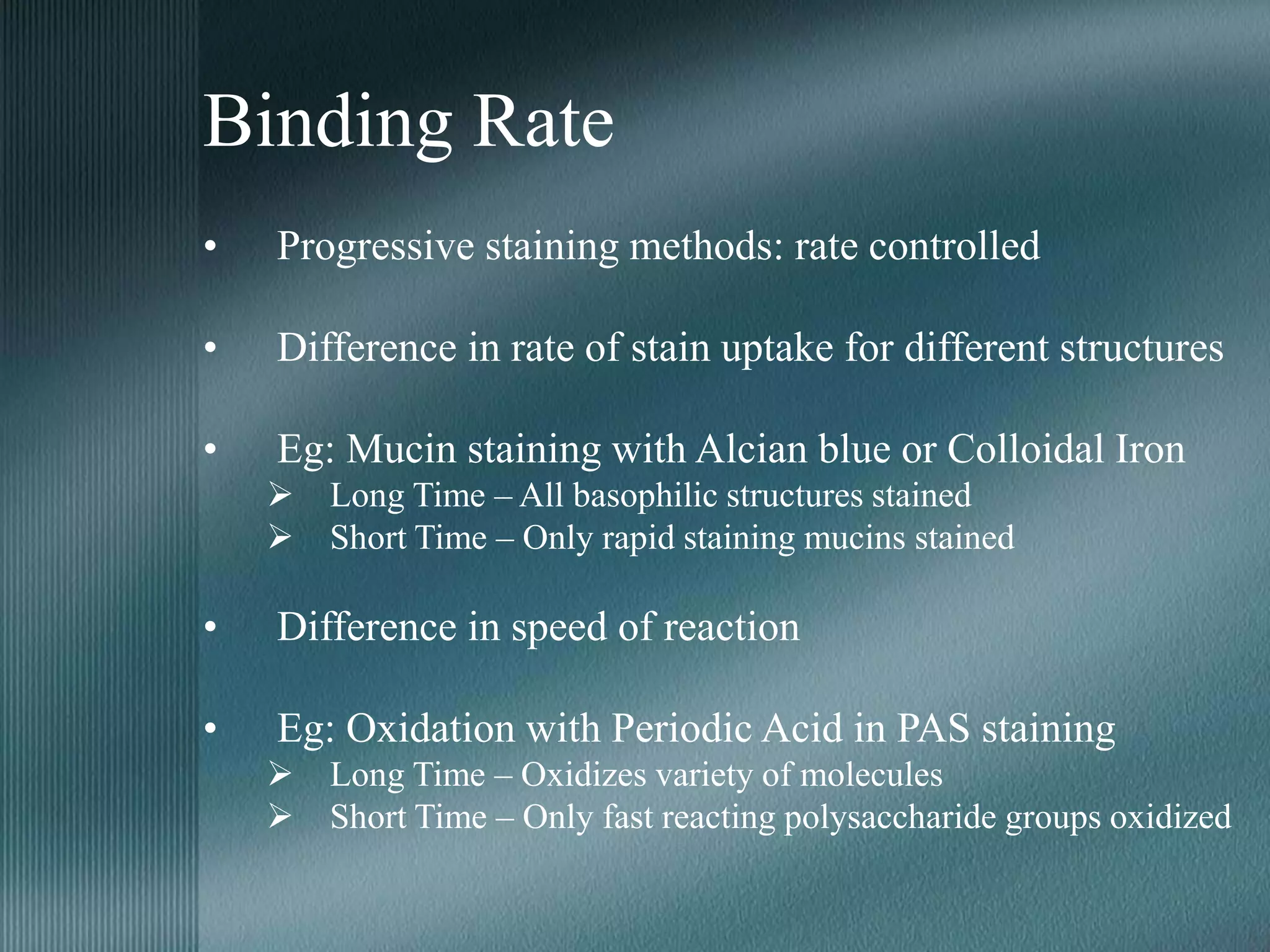
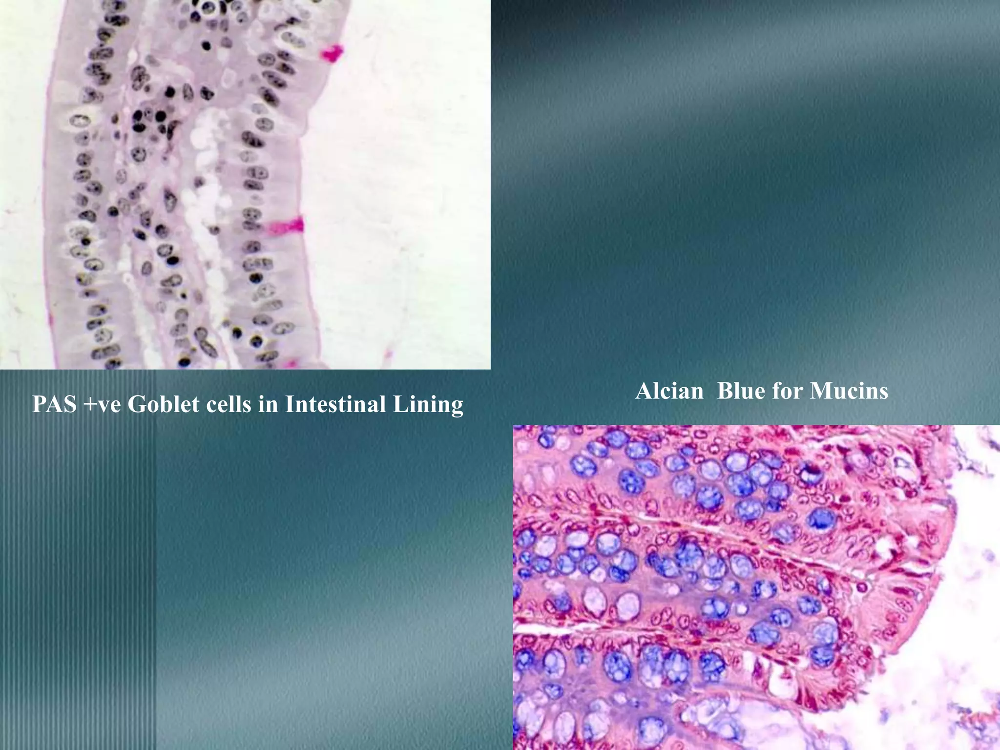
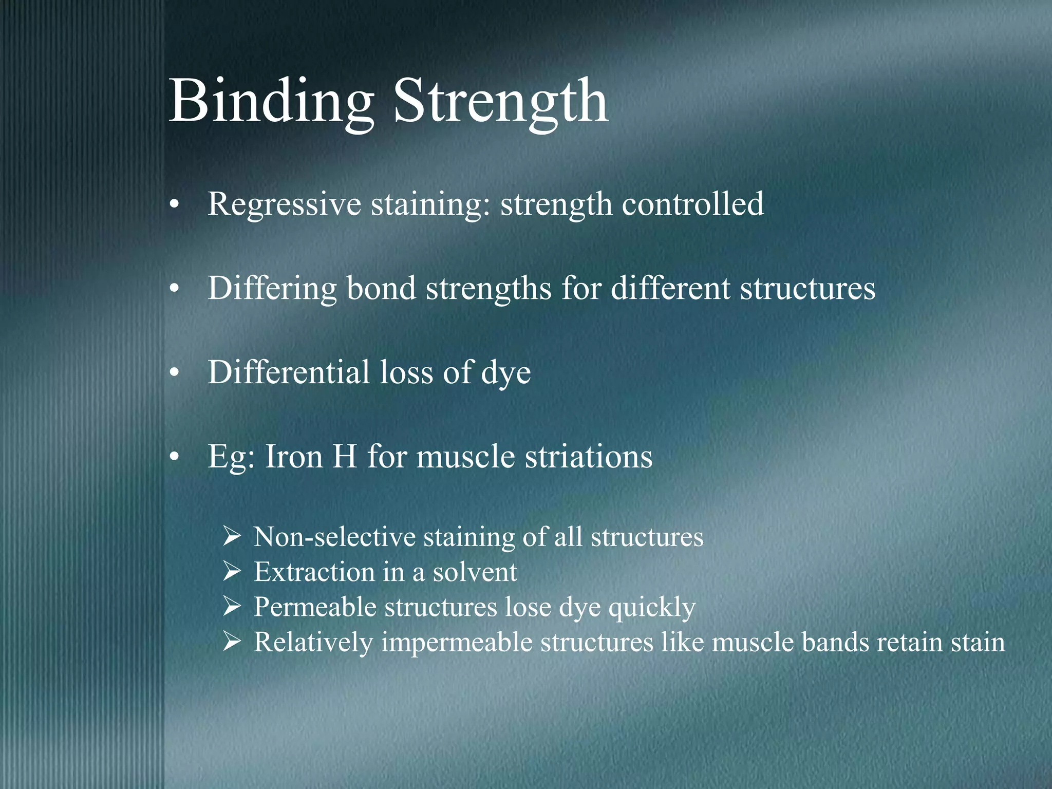
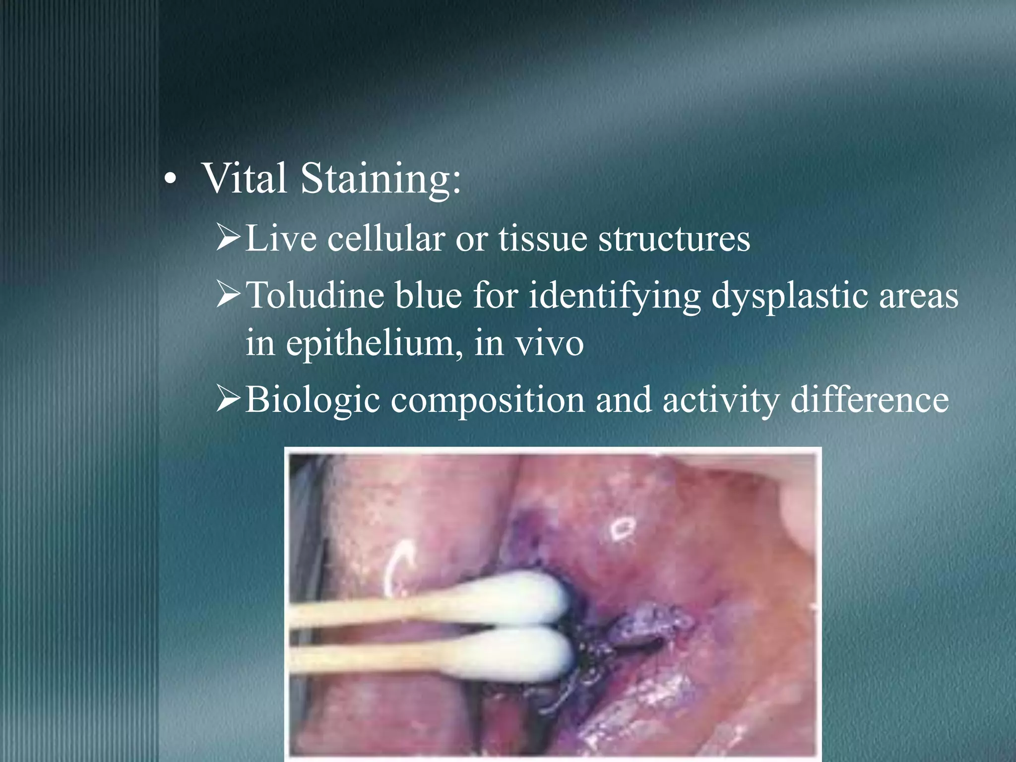
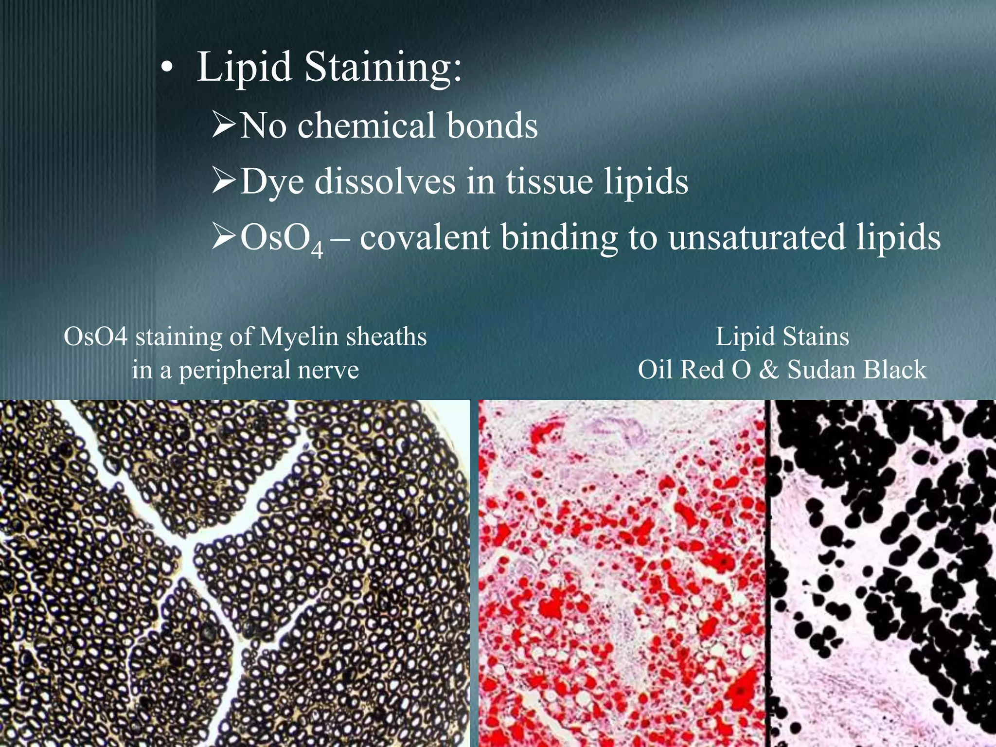
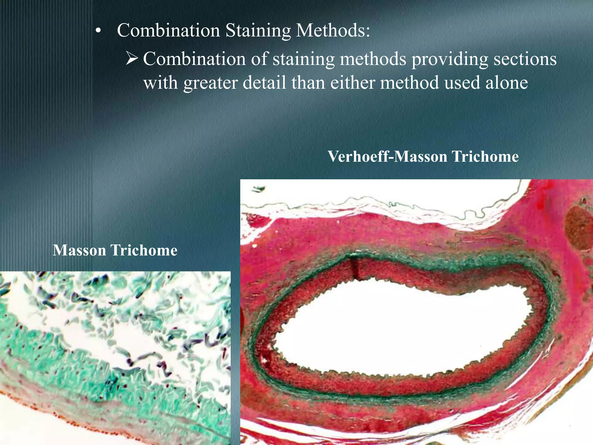
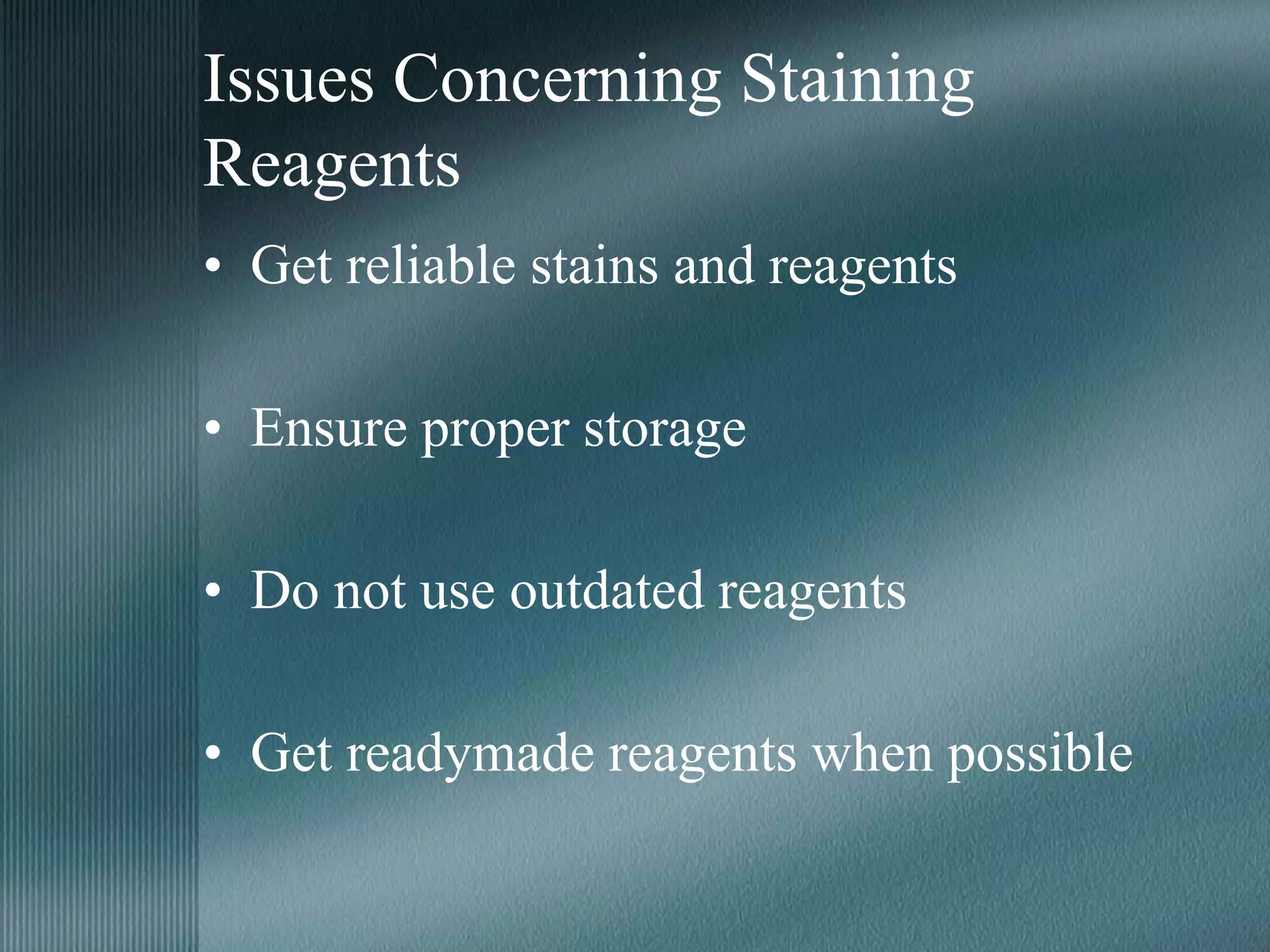
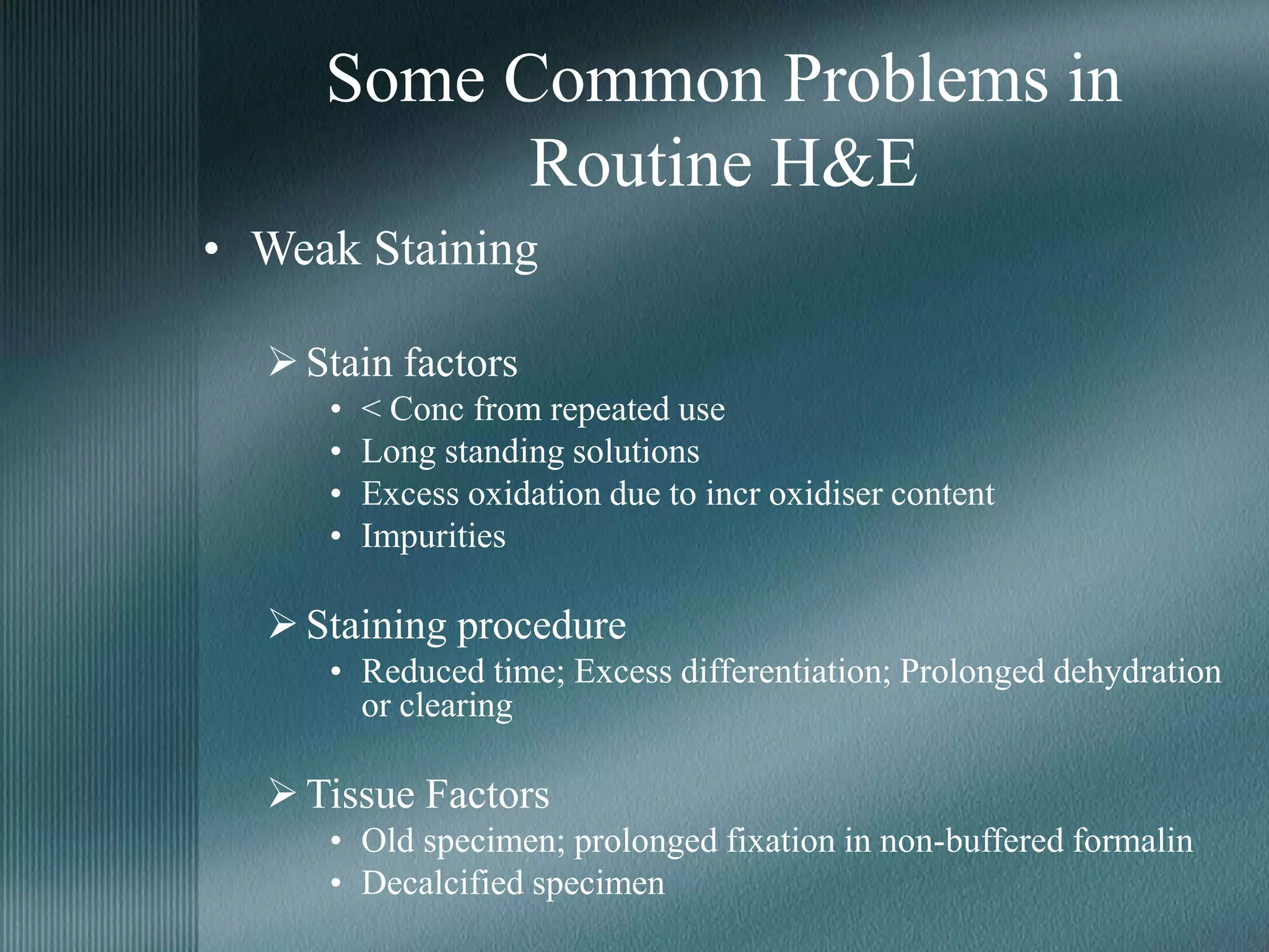
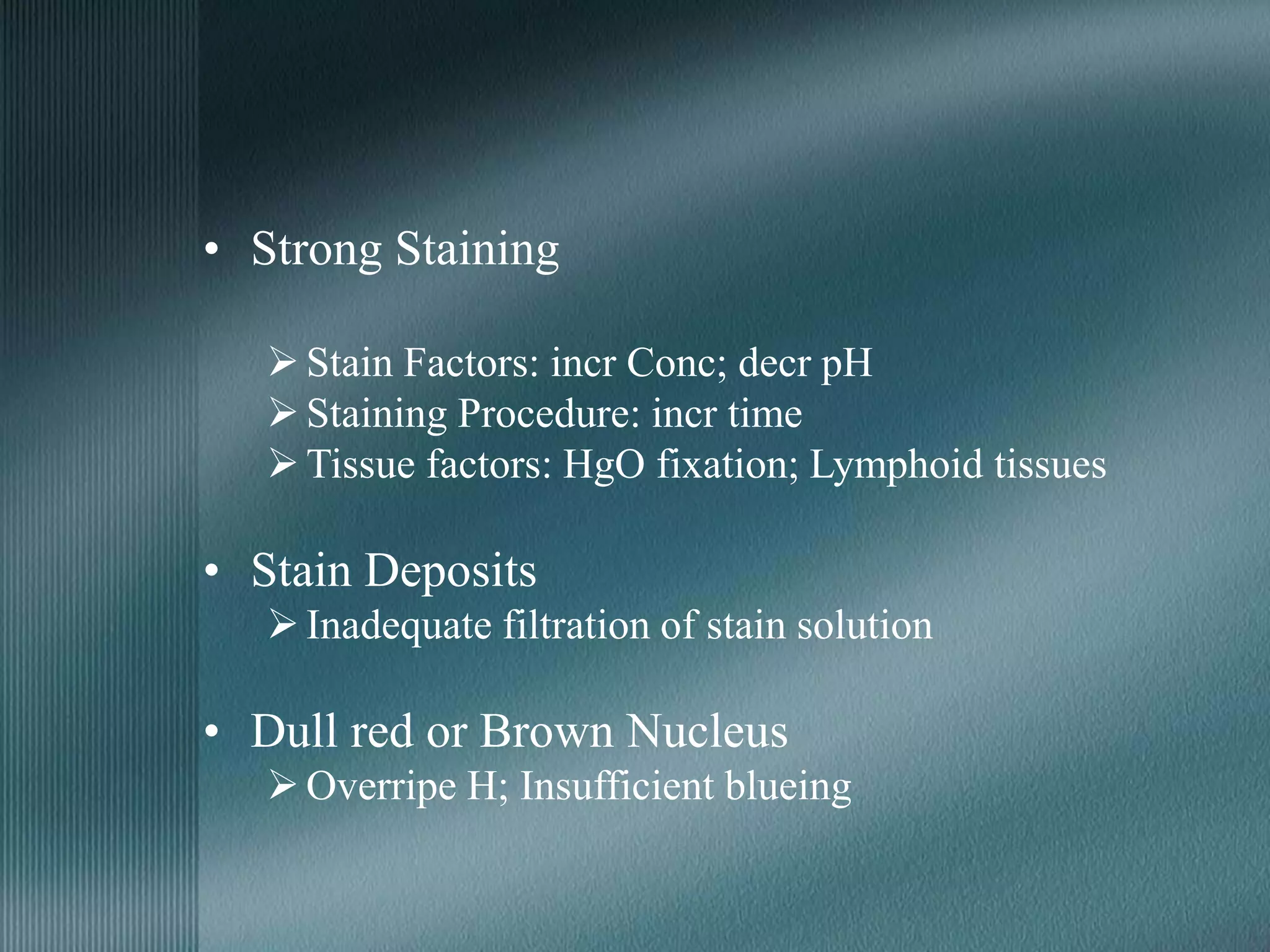
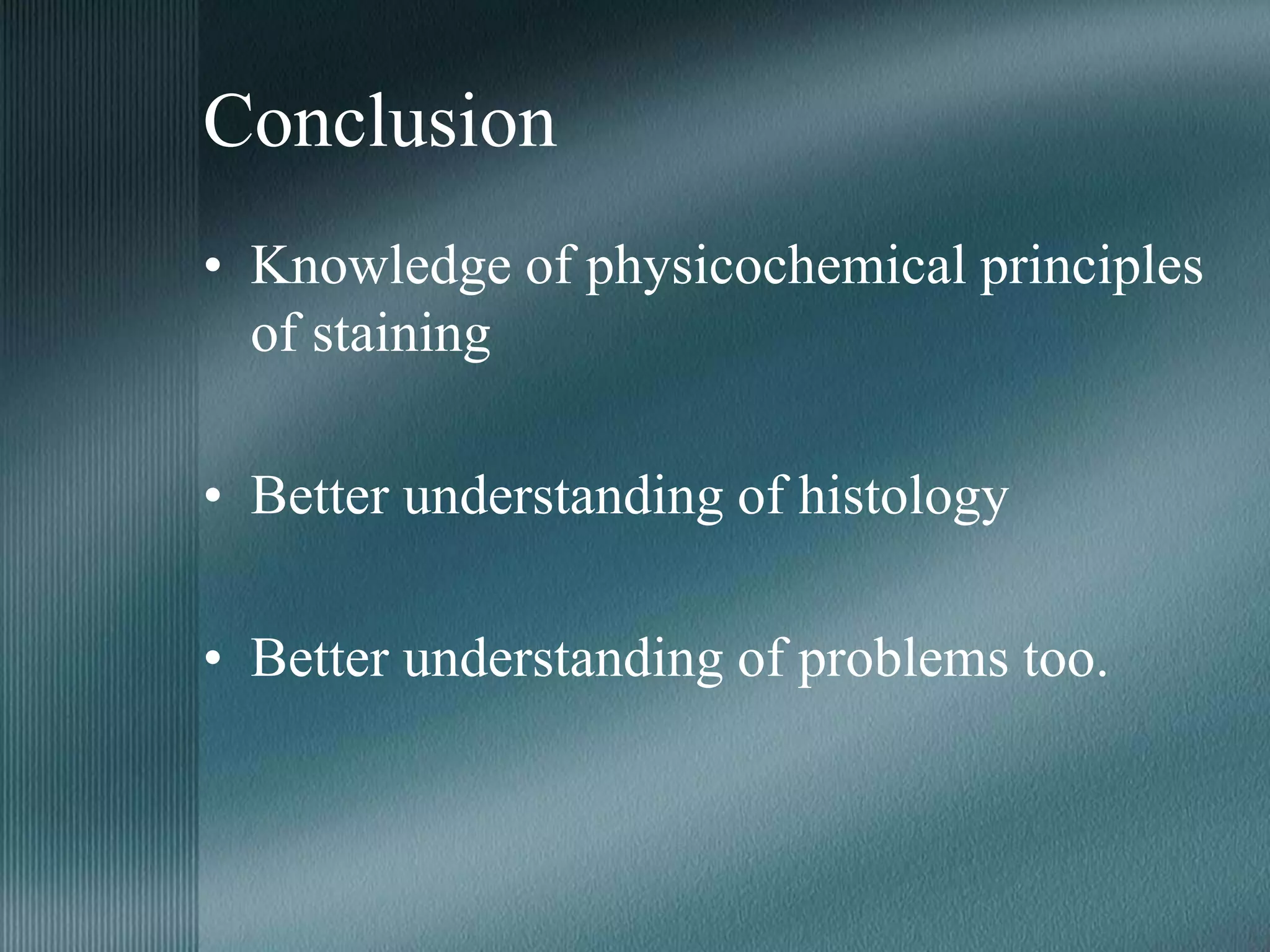
Theory of Staining
Practical Implications
• Biological stains have been used for centuries in
histology.
• Ignorance of the mechanisms involved (except in
histochemistry).
• Understanding of the principles involved –
judicious approach to staining.
• Investigation of failures and problems with
wisdom.
Overview
• The need for staining?
• Basic Principle involved?
• Properties of Stains?
• Problems & Troubleshooting?
1. How and why tissues are stained?
2. Properties of Dyes
3. Basis of Differential staining
4. Factors affecting
5. Problems and solutions
Why do we Stain?
• Any object is seen only when it gives out light.
• Visible objects are distinguishable from surroundings only when they
are different in light color or intensity from surroundings.
• Metal impregnation, polarized microscopy, phase contrast
microscopy: difference in intensity.
• All other histology methods: different color.
• Staining: provides these different colors.
• Stains: give characteristic colors or hues to cell and tissue
components.
• Differentiation of various cells and tissues.
Properties of Dyes
Why are Dyes Colored?
White light falling on a dye molecule
Absorption of some WL of this light
Remaining WL passed on
Dye appears to be the color of that WL
Metachromasia
• Property of a dye to change color without change in chemical structure.
• One color at low conc; different color at high conc.
• Reason: polymerization. Eg: Toludine blue
• Low Conc: Blue; High Conc: Purple
• Factors favoring polymerization:
– Dye Conc: Low - blue; High - purple; Very High - red
– Temperature: Low - purple; High - blue
– Solvent: Aqueous - favors polymerization
Non-polar - no polymerization
– Tissue Factors: Nucleus - blue
Cartilage - purple (high density of sulphate ions)
T.Blue Staining of Muscle fibers
T.Blue Staining of Kidney
Dye Chemistry and Staining
• pH: Acidic dyes - Basic areas
Basic dyes - Acidic areas
Eg: H&E
• Molecular Size: Large - more staining time
Eg: Alcian blue & Crystal violet
• Solvency: relative hydrophilic/lipophilic character
Eg: Alcian blue - hydrophilic (can be dehydrated through alcohols)
Crystal violet - lipophilic (lost on dehydration through alcohols)
• Impurities: reduce staining intensity.
Dye Nomenclature & Classification
• Society of Dyers & Colorists (1999): Color Index
• Exclusively biological dyes, IHC stains etc: no standard names or
codes.
• Classification of Dyes
– Chemistry: Nitro, Azo, Phenol, Thiazols etc.
– Origin: Natural (Hematoxylin); Synthetic
– pH: Acid, Basic, Amphoteric, Neutral
– Physicochemical properties: Fluorescent, Metachromatic, Leuco
Additional factors in Dyes
• Mordants:
– A link binding the dye to the tissue
– No color of itself
– Used when dye has less affinity for tissue
– Mostly metal compounds: Fe, Al, Mo etc.
– Can be added to dye solution (Weigert’s H) or to tissue
(Heidenhain’s H)
• Accentuators:
– Increase selectivity or staining power of dye
– No chemical union with either dye or tissue
– Varied and obscure mode of action
– Chemical catalyst; KOH in Loeffler’s methylene blue (increases
alkalinity)
Basic Theory of Staining
Why and How Tissues Stain?
• Fundamental reason: Dye-Tissue Affinity.
• Affinity: preference of a tissue for a dye and strength of
their bonding.
• Transfer of stain from solution to section.
• Magnitude of Affinity:
 Stain-Tissue Interactions
 Stain-Stain Interactions
 Solvent-Solvent Interactions
 Stain-Solvent Interactions
Stain-Tissue Interactions
• Coulombic or Electrostatic Bonding:
– Attraction b/w differently charged molecules.
– H&E. –ve H to +ve nucleus & +ve E to –ve cytoplasm.
– pH, Dye conc, Temp etc affect magnitude of attraction.
• Van der Waal’s Forces:
– Dipole-dipole interactions
– Substrate groupings with tyrosine/tryptophan amino acid residues; dyes with large
aromatic rings
– Staining Elastic fibers with Congo red
• Hydrogen Bonding:
– Aqueous solutions – no role
– Wholly or partially non-aqueous solutions
– Best’s Carmine for Glycogen
• Covalent Bonding:
– Methods like Fuelgen nucleal, PAS etc
Stain-Stain Interactions
• Affinity b/w dye particles contributes to staining
pattern and efficiency.
• Metachromasia: polymerization of dye molecules
at high conc
• Silver Impregnation: Ag grains attracted to
already formed Ag grains
Solvent-Solvent Interactions
• Hydrophobic bonding in case of organic reagents
• In aqueous solutions, hydrophobic groups come
together to attain stability
• Tissue constituent and dye molecule
• Enzyme substrates in histochemistry; Fat staining
by Sudan dyes
Stain-Solvent Interactions
• Relative affinity of the dye molecule for the
solvent
• High affinity for solvent: dye may not leave
the solvent for tissue binding
• Sudan stain for fat: used in aqueous
solution.
Why do Stains remain on Tissues?
• Most of the stains: no or reduced affinity for dehydrating
fluids or mounting media.
• Metal complex stains, acidic dyes etc. belong to this
group.
• Basic dyes like Crystal violet, methylene blue etc dissolve
freely in lower alcohols. Dehydrate rapidly or use non-
alcoholic solvents or air-dry.
• Sudan stains: soluble in alcohols, clearing agents and resin
mountants. Use non-alcoholic solvents and mount in
aqueous media.
Basis of Differential Staining
• Question of selectivity
• 3 factors:
– Relative binding capacity
– Binding rate
– Binding strength
Relative Binding Capacity
• Number and affinity of binding sites
• Affinity: H&E.
 Nucleus - more affinity for H
 Cytoplasm - more affinity for E
• Number: Metachromasia with Toludine blue
 Nucleus - less number of binding sites;
orthochromatic blue
 Cartilage - more number of binding sites;
metachromatic purple
H&E of Glomeruli
Binding Rate
• Progressive staining methods: rate controlled
• Difference in rate of stain uptake for different structures
• Eg: Mucin staining with Alcian blue or Colloidal Iron
 Long Time – All basophilic structures stained
 Short Time – Only rapid staining mucins stained
• Difference in speed of reaction
• Eg: Oxidation with Periodic Acid in PAS staining
 Long Time – Oxidizes variety of molecules
 Short Time – Only fast reacting polysaccharide groups oxidized
PAS +ve Goblet cells in Intestinal Lining
Alcian Blue for Mucins
Binding Strength
• Regressive staining: strength controlled
• Differing bond strengths for different structures
• Differential loss of dye
• Eg: Iron H for muscle striations
 Non-selective staining of all structures
 Extraction in a solvent
 Permeable structures lose dye quickly
 Relatively impermeable structures like muscle bands retain stain
Cardiac Muscle Iron Hematoxylin Stain
Other Factors Influencing
Staining
Fixation & Staining
• Fixation: to prevent tissue autolysis and breakdown by
bacteria
• Ability of fixative to retain a tissue substance
• Eg: Lipids
 Well preserved with OsO4
 Poorly retained in formalin or alcohol fixed tissues
• Eg: Proteins in IHC
 Glutaraldehyde – retains proteins better but Ag sites are blocked;
so useless in IHC
 Acetone – poorer fixative of proteins but retains antigenicity of
tissue
Specimen Geometry & Staining
• Microscopy: 2-D image of 3-D structure
• Small difference in specimen thickness: alter staining
pattern
• Simple Geometrical Influences
 Thin sections stain faster
 Specimens with irregular surfaces stain faster (Cryosections)
 Dispersed specimens (smears) stain faster
• Complex Geometrical Influences
 Artefactual: Chatter – alternate dark and light stained areas
 Biological: Large secretory granules v/s Small granules
Some Terminologies &
Special Methods in Staining
• Progressive Staining:
 Tissue immersed in dye until only desired structures are stained
 Difficult process to control
 Dyes with good degree of selectivity
• Regressive Staining:
 Overstaining all tissue structures
 Removing from unwanted groups
 Depends on dye-tissue bond strength
• Negative Staining:
• Structures disclosed by outlining them with stain
• Nigrosine for microbes; Picro-thionine for bone
matrix
-ve Staining of Sperm
-ve Stained Phage virus
• Vital Staining:
Live cellular or tissue structures
Toludine blue for identifying dysplastic areas
in epithelium, in vivo
Biologic composition and activity difference
• Lipid Staining:
No chemical bonds
Dye dissolves in tissue lipids
OsO4 – covalent binding to unsaturated lipids
OsO4 staining of Myelin sheaths
in a peripheral nerve
Lipid Stains
Oil Red O & Sudan Black
• Combination Staining Methods:
Combination of staining methods providing sections
with greater detail than either method used alone
Verhoeff-Masson Trichome
Masson Trichome
Problem Avoidance &
Troubleshooting
Issues Concerning Staining
Procedures
• Choose fixative and embedding media suitable for
proposed stain
• Use a routine, standardized protocol
• Use controls proactively
• Keep samples of effective batches of stains for cross-
checking
• Difficult procedures: monitoring by experienced
personnel.
Issues Concerning Staining
Reagents
• Get reliable stains and reagents
• Ensure proper storage
• Do not use outdated reagents
• Get readymade reagents when possible
Some Common Problems in
Routine H&E
• Weak Staining
Stain factors
• < Conc from repeated use
• Long standing solutions
• Excess oxidation due to incr oxidiser content
• Impurities
Staining procedure
• Reduced time; Excess differentiation; Prolonged dehydration
or clearing
Tissue Factors
• Old specimen; prolonged fixation in non-buffered formalin
• Decalcified specimen
• Strong Staining
Stain Factors: incr Conc; decr pH
Staining Procedure: incr time
Tissue factors: HgO fixation; Lymphoid tissues
• Stain Deposits
Inadequate filtration of stain solution
• Dull red or Brown Nucleus
Overripe H; Insufficient blueing
Conclusion
• Knowledge of physicochemical principles
of staining
• Better understanding of histology
• Better understanding of problems too.
References
• Theory & Practice of Histological
Techniques. Bancroft
• Cellular Pathology techniques. Culling
• Histological staining methods. Disbrey
Theory_of_Staining.ppt

Theory_of_Staining.ppt

  • 1.
  • 2.
    • Biological stainshave been used for centuries in histology. • Ignorance of the mechanisms involved (except in histochemistry). • Understanding of the principles involved – judicious approach to staining. • Investigation of failures and problems with wisdom.
  • 3.
    Overview • The needfor staining? • Basic Principle involved? • Properties of Stains? • Problems & Troubleshooting? 1. How and why tissues are stained? 2. Properties of Dyes 3. Basis of Differential staining 4. Factors affecting 5. Problems and solutions
  • 4.
    Why do weStain? • Any object is seen only when it gives out light. • Visible objects are distinguishable from surroundings only when they are different in light color or intensity from surroundings. • Metal impregnation, polarized microscopy, phase contrast microscopy: difference in intensity. • All other histology methods: different color. • Staining: provides these different colors. • Stains: give characteristic colors or hues to cell and tissue components. • Differentiation of various cells and tissues.
  • 5.
  • 6.
    Why are DyesColored? White light falling on a dye molecule Absorption of some WL of this light Remaining WL passed on Dye appears to be the color of that WL
  • 7.
    Metachromasia • Property ofa dye to change color without change in chemical structure. • One color at low conc; different color at high conc. • Reason: polymerization. Eg: Toludine blue • Low Conc: Blue; High Conc: Purple • Factors favoring polymerization: – Dye Conc: Low - blue; High - purple; Very High - red – Temperature: Low - purple; High - blue – Solvent: Aqueous - favors polymerization Non-polar - no polymerization – Tissue Factors: Nucleus - blue Cartilage - purple (high density of sulphate ions)
  • 8.
    T.Blue Staining ofMuscle fibers T.Blue Staining of Kidney
  • 9.
    Dye Chemistry andStaining • pH: Acidic dyes - Basic areas Basic dyes - Acidic areas Eg: H&E • Molecular Size: Large - more staining time Eg: Alcian blue & Crystal violet • Solvency: relative hydrophilic/lipophilic character Eg: Alcian blue - hydrophilic (can be dehydrated through alcohols) Crystal violet - lipophilic (lost on dehydration through alcohols) • Impurities: reduce staining intensity.
  • 10.
    Dye Nomenclature &Classification • Society of Dyers & Colorists (1999): Color Index • Exclusively biological dyes, IHC stains etc: no standard names or codes. • Classification of Dyes – Chemistry: Nitro, Azo, Phenol, Thiazols etc. – Origin: Natural (Hematoxylin); Synthetic – pH: Acid, Basic, Amphoteric, Neutral – Physicochemical properties: Fluorescent, Metachromatic, Leuco
  • 11.
    Additional factors inDyes • Mordants: – A link binding the dye to the tissue – No color of itself – Used when dye has less affinity for tissue – Mostly metal compounds: Fe, Al, Mo etc. – Can be added to dye solution (Weigert’s H) or to tissue (Heidenhain’s H) • Accentuators: – Increase selectivity or staining power of dye – No chemical union with either dye or tissue – Varied and obscure mode of action – Chemical catalyst; KOH in Loeffler’s methylene blue (increases alkalinity)
  • 12.
  • 13.
    Why and HowTissues Stain? • Fundamental reason: Dye-Tissue Affinity. • Affinity: preference of a tissue for a dye and strength of their bonding. • Transfer of stain from solution to section. • Magnitude of Affinity:  Stain-Tissue Interactions  Stain-Stain Interactions  Solvent-Solvent Interactions  Stain-Solvent Interactions
  • 14.
    Stain-Tissue Interactions • Coulombicor Electrostatic Bonding: – Attraction b/w differently charged molecules. – H&E. –ve H to +ve nucleus & +ve E to –ve cytoplasm. – pH, Dye conc, Temp etc affect magnitude of attraction. • Van der Waal’s Forces: – Dipole-dipole interactions – Substrate groupings with tyrosine/tryptophan amino acid residues; dyes with large aromatic rings – Staining Elastic fibers with Congo red • Hydrogen Bonding: – Aqueous solutions – no role – Wholly or partially non-aqueous solutions – Best’s Carmine for Glycogen • Covalent Bonding: – Methods like Fuelgen nucleal, PAS etc
  • 15.
    Stain-Stain Interactions • Affinityb/w dye particles contributes to staining pattern and efficiency. • Metachromasia: polymerization of dye molecules at high conc • Silver Impregnation: Ag grains attracted to already formed Ag grains
  • 16.
    Solvent-Solvent Interactions • Hydrophobicbonding in case of organic reagents • In aqueous solutions, hydrophobic groups come together to attain stability • Tissue constituent and dye molecule • Enzyme substrates in histochemistry; Fat staining by Sudan dyes
  • 17.
    Stain-Solvent Interactions • Relativeaffinity of the dye molecule for the solvent • High affinity for solvent: dye may not leave the solvent for tissue binding • Sudan stain for fat: used in aqueous solution.
  • 18.
    Why do Stainsremain on Tissues? • Most of the stains: no or reduced affinity for dehydrating fluids or mounting media. • Metal complex stains, acidic dyes etc. belong to this group. • Basic dyes like Crystal violet, methylene blue etc dissolve freely in lower alcohols. Dehydrate rapidly or use non- alcoholic solvents or air-dry. • Sudan stains: soluble in alcohols, clearing agents and resin mountants. Use non-alcoholic solvents and mount in aqueous media.
  • 19.
  • 20.
    • Question ofselectivity • 3 factors: – Relative binding capacity – Binding rate – Binding strength
  • 21.
    Relative Binding Capacity •Number and affinity of binding sites • Affinity: H&E.  Nucleus - more affinity for H  Cytoplasm - more affinity for E • Number: Metachromasia with Toludine blue  Nucleus - less number of binding sites; orthochromatic blue  Cartilage - more number of binding sites; metachromatic purple
  • 22.
  • 23.
    Binding Rate • Progressivestaining methods: rate controlled • Difference in rate of stain uptake for different structures • Eg: Mucin staining with Alcian blue or Colloidal Iron  Long Time – All basophilic structures stained  Short Time – Only rapid staining mucins stained • Difference in speed of reaction • Eg: Oxidation with Periodic Acid in PAS staining  Long Time – Oxidizes variety of molecules  Short Time – Only fast reacting polysaccharide groups oxidized
  • 24.
    PAS +ve Gobletcells in Intestinal Lining Alcian Blue for Mucins
  • 25.
    Binding Strength • Regressivestaining: strength controlled • Differing bond strengths for different structures • Differential loss of dye • Eg: Iron H for muscle striations  Non-selective staining of all structures  Extraction in a solvent  Permeable structures lose dye quickly  Relatively impermeable structures like muscle bands retain stain
  • 26.
    Cardiac Muscle IronHematoxylin Stain
  • 27.
  • 28.
    Fixation & Staining •Fixation: to prevent tissue autolysis and breakdown by bacteria • Ability of fixative to retain a tissue substance • Eg: Lipids  Well preserved with OsO4  Poorly retained in formalin or alcohol fixed tissues • Eg: Proteins in IHC  Glutaraldehyde – retains proteins better but Ag sites are blocked; so useless in IHC  Acetone – poorer fixative of proteins but retains antigenicity of tissue
  • 29.
    Specimen Geometry &Staining • Microscopy: 2-D image of 3-D structure • Small difference in specimen thickness: alter staining pattern • Simple Geometrical Influences  Thin sections stain faster  Specimens with irregular surfaces stain faster (Cryosections)  Dispersed specimens (smears) stain faster • Complex Geometrical Influences  Artefactual: Chatter – alternate dark and light stained areas  Biological: Large secretory granules v/s Small granules
  • 30.
    Some Terminologies & SpecialMethods in Staining
  • 31.
    • Progressive Staining: Tissue immersed in dye until only desired structures are stained  Difficult process to control  Dyes with good degree of selectivity • Regressive Staining:  Overstaining all tissue structures  Removing from unwanted groups  Depends on dye-tissue bond strength
  • 32.
    • Negative Staining: •Structures disclosed by outlining them with stain • Nigrosine for microbes; Picro-thionine for bone matrix -ve Staining of Sperm -ve Stained Phage virus
  • 33.
    • Vital Staining: Livecellular or tissue structures Toludine blue for identifying dysplastic areas in epithelium, in vivo Biologic composition and activity difference
  • 34.
    • Lipid Staining: Nochemical bonds Dye dissolves in tissue lipids OsO4 – covalent binding to unsaturated lipids OsO4 staining of Myelin sheaths in a peripheral nerve Lipid Stains Oil Red O & Sudan Black
  • 35.
    • Combination StainingMethods: Combination of staining methods providing sections with greater detail than either method used alone Verhoeff-Masson Trichome Masson Trichome
  • 36.
  • 37.
    Issues Concerning Staining Procedures •Choose fixative and embedding media suitable for proposed stain • Use a routine, standardized protocol • Use controls proactively • Keep samples of effective batches of stains for cross- checking • Difficult procedures: monitoring by experienced personnel.
  • 38.
    Issues Concerning Staining Reagents •Get reliable stains and reagents • Ensure proper storage • Do not use outdated reagents • Get readymade reagents when possible
  • 39.
    Some Common Problemsin Routine H&E • Weak Staining Stain factors • < Conc from repeated use • Long standing solutions • Excess oxidation due to incr oxidiser content • Impurities Staining procedure • Reduced time; Excess differentiation; Prolonged dehydration or clearing Tissue Factors • Old specimen; prolonged fixation in non-buffered formalin • Decalcified specimen
  • 40.
    • Strong Staining StainFactors: incr Conc; decr pH Staining Procedure: incr time Tissue factors: HgO fixation; Lymphoid tissues • Stain Deposits Inadequate filtration of stain solution • Dull red or Brown Nucleus Overripe H; Insufficient blueing
  • 41.
    Conclusion • Knowledge ofphysicochemical principles of staining • Better understanding of histology • Better understanding of problems too.
  • 42.
    References • Theory &Practice of Histological Techniques. Bancroft • Cellular Pathology techniques. Culling • Histological staining methods. Disbrey