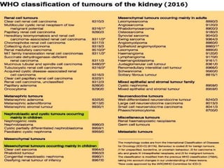
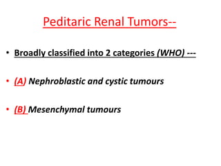
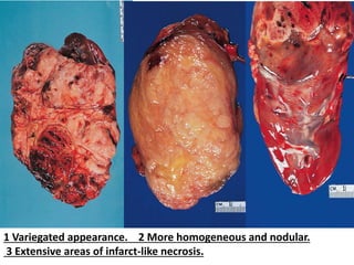
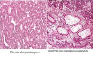
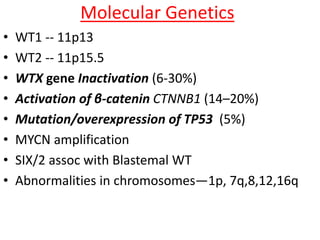
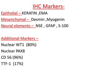
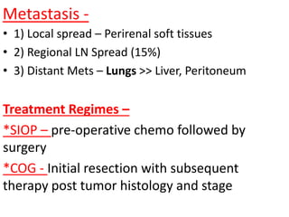
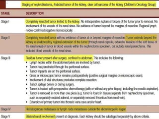
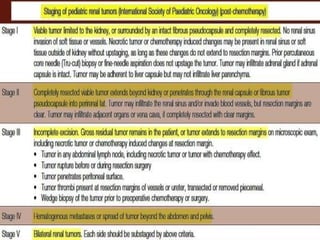
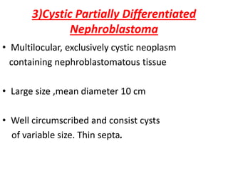
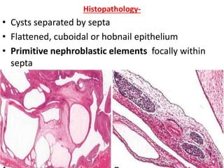
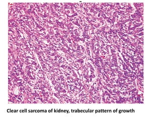
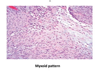
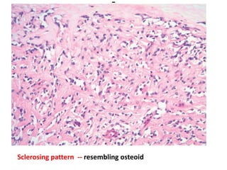
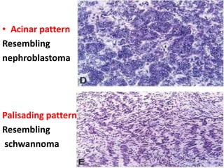
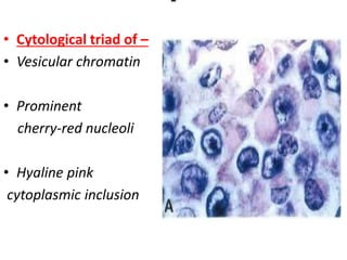
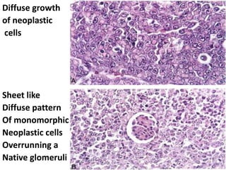
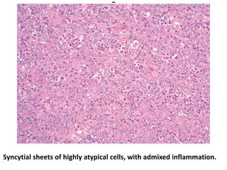
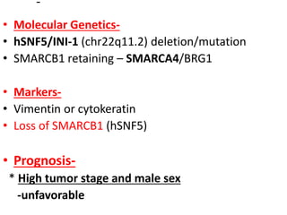
Pediatric Renal Tumors
Presenter –
Dr Utsab Das
Dept of Pathology ,Calcutta National Medical
College
Moderator –
Dr Piyali Ghosh
Demonstrator ,Dept of Pathology ,Calcutta
National Medical College
Peditaric Renal Tumors--
• Broadly classified into 2 categories (WHO) ---
• (A) Nephroblastic and cystic tumours
• (B) Mesenchymal tumours
(A) Nephroblastic and cystic tumours
occurring mainly in children
• 1) Nephrogenic Rest
• 2) Nephroblastoma / Wilm’s Tumour
• 3) Cystic partially differentiated nephroblastoma
• 4) Paediatric cystic nephroma
1) Nephrogenic Rest
• Defn – Persistent foci of embryonal cells
(> 36 wk POG)
• Epidemiology— 40% cases of Wilms Tumor
• Histopathology- Perilobar and Intralobar type-
• Each type subclassified into-
• Dormant/incipient
• Regressing /sclerosing
• Hyperplastic
Perilobar Nephrogenic Rest-
.
Intralobar Nephrogenic Rests
.
2) Nephroblastoma / Wilm’s Tumour
• Defn- Malignant embryonal neoplasm derived from
nephrogenic blastemal cells,mimics developing
kidney &shows divergent patterns of differentiation
• Epidemiology- 1 in 8000
• 90% cases <6yrs age
• Signs and symptoms-
• Abdominal mass
• Pain ,haematuria, proteinuria
• Hypertension
• Secondary to traumatic rupture
• Conditions associated with highest risk of Wilms tumor-
• WAGR syndrome
• Beckwith–Wiedemann syndrome
• Hemihypertrophy
• Denys–Drash syndrome
• Familial nephroblastoma
• Other conditions -
• Cutaneous nevi and angiomas
• Trisomy 18
• Klippel–Trénaunay syndrome
• Neurofibromatosis
• Bloom syndrome
• Frasier syndrome
• Perlman syndrome
• Cerebral gigantism (Sotos syndrome).
-
• Macroscopy-
• Gross- Solitary, well circumscribed, soft
• cut section -Pale gray ,cystic change, necrosis,
hemorrhage .
1 Variegated appearance. 2 More homogeneous and nodular.
3 Extensive areas of infarct-like necrosis.
• Histopathology-
• Triphasic pattern-
• 1) Undifferentiated blastema
• 2) Mesenchymal (stromal) tissue
• 3) Epithelial tissue.
• Blastemal- Highly cellular, overlapping nuclei .
• Diffuse, nodular ,serpentine, basaloid growth
• Mesenchymal- Spindle cells with smooth muscle
features
• Epithelial- Small round tubular structures
resembling rosettes
1 combination of blastema, stroma,epithelial tubular formations,immature glomeruli
2 blastema, stroma, and immature tubular formations.
-
Diffuse
blastemal
Pattern
Serpentine
blastemal
pattern
Molecular Genetics
• WT1 -- 11p13
• WT2 -- 11p15.5
• WTX gene Inactivation (6-30%)
• Activation of β-catenin CTNNB1 (14–20%)
• Mutation/overexpression of TP53 (5%)
• MYCN amplification
• SIX/2 assoc with Blastemal WT
• Abnormalities in chromosomes—1p, 7q,8,12,16q
IHC Markers-
Epithelial – KERATIN ,EMA
Mesenchymal – Desmin ,Myogenin
Neural elements – NSE , GFAP , S-100
Additional Markers –
Nuclear WT1 (80%)
Nuclear PAX8
CD 56 (96%)
TTF-1 (17%)
Metastasis -
• 1) Local spread – Perirenal soft tissues
• 2) Regional LN Spread (15%)
• 3) Distant Mets – Lungs >> Liver, Peritoneum
Treatment Regimes –
*SIOP – pre-operative chemo followed by
surgery
*COG - Initial resection with subsequent
therapy post tumor histology and stage
Prognosis -
• 1. Age
• 2. Stage
• 3. Anaplasia*** (4% of the cases )
***criteria for anaplasia-
(A)marked enlargement of nuclei
(B) obvious hyperchromasia
(C) multipolar mitotic figures
• Prognosis – (contd..)
• 4. Size
• 5. Extensive tubular differentiation***
• 6. Skeletal muscle differentiation***
• 7. Post-chemotherapy morphology
• 8. TP53 mutation
• 9. Loss Of Heterozygosity at 1p and 16q
Anaplastic (“Unfavorable Histology”) Wilms Tumor. Marked
pleomorphism with giant hyperchromatic nuclei and atypical mitoses.
Anaplastic (“Unfavorable Histology”) Wilms Tumor.
Strong nuclear immunoreactivity for p53.
3)Cystic Partially Differentiated
Nephroblastoma
• Multilocular, exclusively cystic neoplasm
containing nephroblastomatous tissue
• Large size ,mean diameter 10 cm
• Well circumscribed and consist cysts
of variable size. Thin septa.
Histopathology-
• Cysts separated by septa
• Flattened, cuboidal or hobnail epithelium
• Primitive nephroblastic elements focally within
septa
4) Paediatric cystic nephroma
• Multilocular, cystic
• Septa - fibrous tissue & differentiated tubules
• Histopathology- Cysts separated by septa
• D/D- Cystic partially differentiated nephroblastoma
• Genetic profile- DICER 1 mutations.
Gross appearance of pediatric cystic nephroma involving
most of the kidney
• .
Low-power microscopic
appearance -
Multiple cysts
lined by flattened epithelium
separated by
cellular spindle cell stroma
The epithelial lining of the cyst has a hobnail quality
stroma is loose and hypocellular
Pediatric Cystic Nephroma-- stroma shows subtle
cellularity adjacent to the epithelium, which is more
atrophic.
Type- B) Mesenchymal tumours
occurring
mainly in children
1) Clear Cell Sarcoma
2) Rhabdoid Tumor
3) Congenital mesoblastic nephroma
4) Ossifying renal tumour of infancy
1) Clear Cell Sarcoma
Synonym –
Bone-metastasizing renal tumour
Features-
• 4% of malignant renal tumours in childhood
• Male:female 2: 1
• Genetics-
• Somatic internal tandem duplication in BCOR
sequence (85%)
• YWHAE-NUTM2 fusion (10%).
Gross Appearance –
Well circumscribed
and whitish, bulges on
the cut surface
• Histology-
• Nests or cords of cells separated by
regularly spaced, arborizing fibrovascular septa
Histologic Patterns-
1)Classical
2)Trabecular
3)Myxoid
4)Sclerosing
5)Acinar
6)Palisading
7)Cellular
8)Storiform
9)Anaplastic
Clear cell sarcoma of kidney, prototypical appearance-branching
capillary Vasculature and open chromatin of epithelioid cord cells
Clear cell sarcoma of kidney, trabecular pattern of growth
Myxoid pattern
Sclerosing pattern -- resembling osteoid
-
• Acinar pattern
Resembling
nephroblastoma
Palisading pattern
Resembling
schwannoma
-
Cellular pattern
Resembling
blastemal
nephroblastoma
Storiform pattern
Resembling
Fibrohistiocytic
Neoplasia
• Metastasis-
Skeletal metastases,particularly skull
Regional lymph nodes, brain, lung, liver
*Prognosis-
• Treatment with doxorubicin
• Stage
• Age at diagnosis
• Tumor necrosis -- all are independent prognostic
factors
2) Rhabdoid Tumor
• Highly Aggressive
• Part of SMACRB1-associated neoplasms
• Epidemiology- 2% of renal neoplasms in children
• Clinical features-
• Haematuria , abdominal mass
• CNS counterpart : Atypical Teratoid/ Rhabdoid
tumour(posterior fossa) (15%)
Gross-
• Solid ,soft ,well circumscribed
• Microscopy-
• Large eosinophilic hyaline globule
displace nucleus laterally (Plasmacytoid)
-
• Cytological triad of –
• Vesicular chromatin
• Prominent
cherry-red nucleoli
• Hyaline pink
cytoplasmic inclusion
Rhabdoid Tumor of Kidney. The nuclear grade is high.
An eosinophilic amorphous (“hyaline”) material fills the scanty
cytoplasm and pushes the nucleus aside
-
Diffuse growth
of neoplastic
cells
Sheet like
Diffuse pattern
Of monomorphic
Neoplastic cells
Overrunning a
Native glomeruli
Syncytial sheets of highly atypical cells, with admixed inflammation.
• Molecular Genetics-
• hSNF5/INI-1 (chr22q11.2) deletion/mutation
• SMARCB1 retaining – SMARCA4/BRG1
• Markers-
• Vimentin or cytokeratin
• Loss of SMARCB1 (hSNF5)
• Prognosis-
* High tumor stage and male sex
-unfavorable
3)Congenital mesoblastic nephroma
*Low-grade fibroblastic neoplasm
*Most common renal tumor
of newborns(2-4%)
• Clinical features
* 90% <1 yr age
• Abdominal mass
-
• Macroscopy-
• Solid
• Yellow-gray
• Well-circumscribed
and its fibrous
cut surface
well illustrated
Classic CMN- monotonous proliferation of spindle
cells with bland nuclei, resembling “Infantile Fibromatosis”
Cellular CMN- Accompanied by Mitotic Activity.
Resembles “Infantile Fibrosarcoma”
• Genetic profile
• Polysomy of chromosomes 8, 11, 17,20
*Cellular CMN – t(12;15)(p13;q25)
results in ETV6–NTRK3 gene fusion
Treatment & Prognosis-
*Nephrectomy
*7% recurrence with local invasion
*Age at diagnosis & adequacy of excision -more
important than morphology
4)Ossifying renal tumour of infancy
• Intracalyceal mass composed of -
1) Osteoid trabeculae
2) Osteoblast-like cells
3) Spindle cell component
* Arise from and attached to Medullary Pyramid.
• Clinical features-
• Male predominance,
• Age <2 years
• Gross haematuria **
• Macroscopy-
• Well circumscribed , 1 -6 cm diameter
• Histopathology
• Osteoid core
• Osteoblastic cells within and periphery
• Bland spindle cells
• Genetic profile
• Relation with other paediatric renal neoplasms
uncertain
• A small number of cases shown trisomy 4
Ossifying renal tumour of infancy
My references-
• 1) WHO Classification of Tumours of
Urogenital system -2016
• 2) ROSAI AND ACKERMAN’S Surgical Pathology
11th Ed
• 3) Sternberg's Diagnostic Surgical Pathology
6th Ed
-

Pediatric Renal Tumors

  • 1.
    Pediatric Renal Tumors Presenter– Dr Utsab Das Dept of Pathology ,Calcutta National Medical College Moderator – Dr Piyali Ghosh Demonstrator ,Dept of Pathology ,Calcutta National Medical College
  • 3.
    Peditaric Renal Tumors-- •Broadly classified into 2 categories (WHO) --- • (A) Nephroblastic and cystic tumours • (B) Mesenchymal tumours
  • 4.
    (A) Nephroblastic andcystic tumours occurring mainly in children • 1) Nephrogenic Rest • 2) Nephroblastoma / Wilm’s Tumour • 3) Cystic partially differentiated nephroblastoma • 4) Paediatric cystic nephroma
  • 5.
    1) Nephrogenic Rest •Defn – Persistent foci of embryonal cells (> 36 wk POG) • Epidemiology— 40% cases of Wilms Tumor • Histopathology- Perilobar and Intralobar type- • Each type subclassified into- • Dormant/incipient • Regressing /sclerosing • Hyperplastic
  • 7.
  • 8.
  • 9.
    2) Nephroblastoma /Wilm’s Tumour • Defn- Malignant embryonal neoplasm derived from nephrogenic blastemal cells,mimics developing kidney &shows divergent patterns of differentiation • Epidemiology- 1 in 8000 • 90% cases <6yrs age • Signs and symptoms- • Abdominal mass • Pain ,haematuria, proteinuria • Hypertension • Secondary to traumatic rupture
  • 10.
    • Conditions associatedwith highest risk of Wilms tumor- • WAGR syndrome • Beckwith–Wiedemann syndrome • Hemihypertrophy • Denys–Drash syndrome • Familial nephroblastoma • Other conditions - • Cutaneous nevi and angiomas • Trisomy 18 • Klippel–Trénaunay syndrome • Neurofibromatosis • Bloom syndrome • Frasier syndrome • Perlman syndrome • Cerebral gigantism (Sotos syndrome).
  • 11.
    - • Macroscopy- • Gross-Solitary, well circumscribed, soft • cut section -Pale gray ,cystic change, necrosis, hemorrhage .
  • 12.
    1 Variegated appearance.2 More homogeneous and nodular. 3 Extensive areas of infarct-like necrosis.
  • 13.
    • Histopathology- • Triphasicpattern- • 1) Undifferentiated blastema • 2) Mesenchymal (stromal) tissue • 3) Epithelial tissue. • Blastemal- Highly cellular, overlapping nuclei . • Diffuse, nodular ,serpentine, basaloid growth • Mesenchymal- Spindle cells with smooth muscle features • Epithelial- Small round tubular structures resembling rosettes
  • 14.
    1 combination ofblastema, stroma,epithelial tubular formations,immature glomeruli 2 blastema, stroma, and immature tubular formations.
  • 15.
  • 17.
    Molecular Genetics • WT1-- 11p13 • WT2 -- 11p15.5 • WTX gene Inactivation (6-30%) • Activation of β-catenin CTNNB1 (14–20%) • Mutation/overexpression of TP53 (5%) • MYCN amplification • SIX/2 assoc with Blastemal WT • Abnormalities in chromosomes—1p, 7q,8,12,16q
  • 18.
    IHC Markers- Epithelial –KERATIN ,EMA Mesenchymal – Desmin ,Myogenin Neural elements – NSE , GFAP , S-100 Additional Markers – Nuclear WT1 (80%) Nuclear PAX8 CD 56 (96%) TTF-1 (17%)
  • 19.
    Metastasis - • 1)Local spread – Perirenal soft tissues • 2) Regional LN Spread (15%) • 3) Distant Mets – Lungs >> Liver, Peritoneum Treatment Regimes – *SIOP – pre-operative chemo followed by surgery *COG - Initial resection with subsequent therapy post tumor histology and stage
  • 20.
    Prognosis - • 1.Age • 2. Stage • 3. Anaplasia*** (4% of the cases ) ***criteria for anaplasia- (A)marked enlargement of nuclei (B) obvious hyperchromasia (C) multipolar mitotic figures
  • 21.
    • Prognosis –(contd..) • 4. Size • 5. Extensive tubular differentiation*** • 6. Skeletal muscle differentiation*** • 7. Post-chemotherapy morphology • 8. TP53 mutation • 9. Loss Of Heterozygosity at 1p and 16q
  • 22.
    Anaplastic (“Unfavorable Histology”)Wilms Tumor. Marked pleomorphism with giant hyperchromatic nuclei and atypical mitoses.
  • 23.
    Anaplastic (“Unfavorable Histology”)Wilms Tumor. Strong nuclear immunoreactivity for p53.
  • 26.
    3)Cystic Partially Differentiated Nephroblastoma •Multilocular, exclusively cystic neoplasm containing nephroblastomatous tissue • Large size ,mean diameter 10 cm • Well circumscribed and consist cysts of variable size. Thin septa.
  • 27.
    Histopathology- • Cysts separatedby septa • Flattened, cuboidal or hobnail epithelium • Primitive nephroblastic elements focally within septa
  • 28.
    4) Paediatric cysticnephroma • Multilocular, cystic • Septa - fibrous tissue & differentiated tubules • Histopathology- Cysts separated by septa • D/D- Cystic partially differentiated nephroblastoma • Genetic profile- DICER 1 mutations.
  • 29.
    Gross appearance ofpediatric cystic nephroma involving most of the kidney
  • 30.
    • . Low-power microscopic appearance- Multiple cysts lined by flattened epithelium separated by cellular spindle cell stroma
  • 31.
    The epithelial liningof the cyst has a hobnail quality stroma is loose and hypocellular
  • 32.
    Pediatric Cystic Nephroma--stroma shows subtle cellularity adjacent to the epithelium, which is more atrophic.
  • 33.
    Type- B) Mesenchymaltumours occurring mainly in children 1) Clear Cell Sarcoma 2) Rhabdoid Tumor 3) Congenital mesoblastic nephroma 4) Ossifying renal tumour of infancy
  • 34.
    1) Clear CellSarcoma Synonym – Bone-metastasizing renal tumour Features- • 4% of malignant renal tumours in childhood • Male:female 2: 1 • Genetics- • Somatic internal tandem duplication in BCOR sequence (85%) • YWHAE-NUTM2 fusion (10%).
  • 35.
    Gross Appearance – Wellcircumscribed and whitish, bulges on the cut surface
  • 36.
    • Histology- • Nestsor cords of cells separated by regularly spaced, arborizing fibrovascular septa Histologic Patterns- 1)Classical 2)Trabecular 3)Myxoid 4)Sclerosing 5)Acinar 6)Palisading 7)Cellular 8)Storiform 9)Anaplastic
  • 37.
    Clear cell sarcomaof kidney, prototypical appearance-branching capillary Vasculature and open chromatin of epithelioid cord cells
  • 38.
    Clear cell sarcomaof kidney, trabecular pattern of growth
  • 39.
  • 40.
    Sclerosing pattern --resembling osteoid
  • 41.
  • 42.
  • 43.
    • Metastasis- Skeletal metastases,particularlyskull Regional lymph nodes, brain, lung, liver *Prognosis- • Treatment with doxorubicin • Stage • Age at diagnosis • Tumor necrosis -- all are independent prognostic factors
  • 44.
    2) Rhabdoid Tumor •Highly Aggressive • Part of SMACRB1-associated neoplasms • Epidemiology- 2% of renal neoplasms in children • Clinical features- • Haematuria , abdominal mass • CNS counterpart : Atypical Teratoid/ Rhabdoid tumour(posterior fossa) (15%)
  • 45.
    Gross- • Solid ,soft,well circumscribed • Microscopy- • Large eosinophilic hyaline globule displace nucleus laterally (Plasmacytoid)
  • 46.
    - • Cytological triadof – • Vesicular chromatin • Prominent cherry-red nucleoli • Hyaline pink cytoplasmic inclusion
  • 47.
    Rhabdoid Tumor ofKidney. The nuclear grade is high. An eosinophilic amorphous (“hyaline”) material fills the scanty cytoplasm and pushes the nucleus aside
  • 48.
    - Diffuse growth of neoplastic cells Sheetlike Diffuse pattern Of monomorphic Neoplastic cells Overrunning a Native glomeruli
  • 49.
    Syncytial sheets ofhighly atypical cells, with admixed inflammation.
  • 50.
    • Molecular Genetics- •hSNF5/INI-1 (chr22q11.2) deletion/mutation • SMARCB1 retaining – SMARCA4/BRG1 • Markers- • Vimentin or cytokeratin • Loss of SMARCB1 (hSNF5) • Prognosis- * High tumor stage and male sex -unfavorable
  • 51.
    3)Congenital mesoblastic nephroma *Low-gradefibroblastic neoplasm *Most common renal tumor of newborns(2-4%) • Clinical features * 90% <1 yr age • Abdominal mass
  • 52.
    - • Macroscopy- • Solid •Yellow-gray • Well-circumscribed and its fibrous cut surface well illustrated
  • 53.
    Classic CMN- monotonousproliferation of spindle cells with bland nuclei, resembling “Infantile Fibromatosis”
  • 54.
    Cellular CMN- Accompaniedby Mitotic Activity. Resembles “Infantile Fibrosarcoma”
  • 55.
    • Genetic profile •Polysomy of chromosomes 8, 11, 17,20 *Cellular CMN – t(12;15)(p13;q25) results in ETV6–NTRK3 gene fusion Treatment & Prognosis- *Nephrectomy *7% recurrence with local invasion *Age at diagnosis & adequacy of excision -more important than morphology
  • 56.
    4)Ossifying renal tumourof infancy • Intracalyceal mass composed of - 1) Osteoid trabeculae 2) Osteoblast-like cells 3) Spindle cell component * Arise from and attached to Medullary Pyramid. • Clinical features- • Male predominance, • Age <2 years • Gross haematuria **
  • 57.
    • Macroscopy- • Wellcircumscribed , 1 -6 cm diameter • Histopathology • Osteoid core • Osteoblastic cells within and periphery • Bland spindle cells • Genetic profile • Relation with other paediatric renal neoplasms uncertain • A small number of cases shown trisomy 4
  • 58.
  • 59.
    My references- • 1)WHO Classification of Tumours of Urogenital system -2016 • 2) ROSAI AND ACKERMAN’S Surgical Pathology 11th Ed • 3) Sternberg's Diagnostic Surgical Pathology 6th Ed
  • 60.