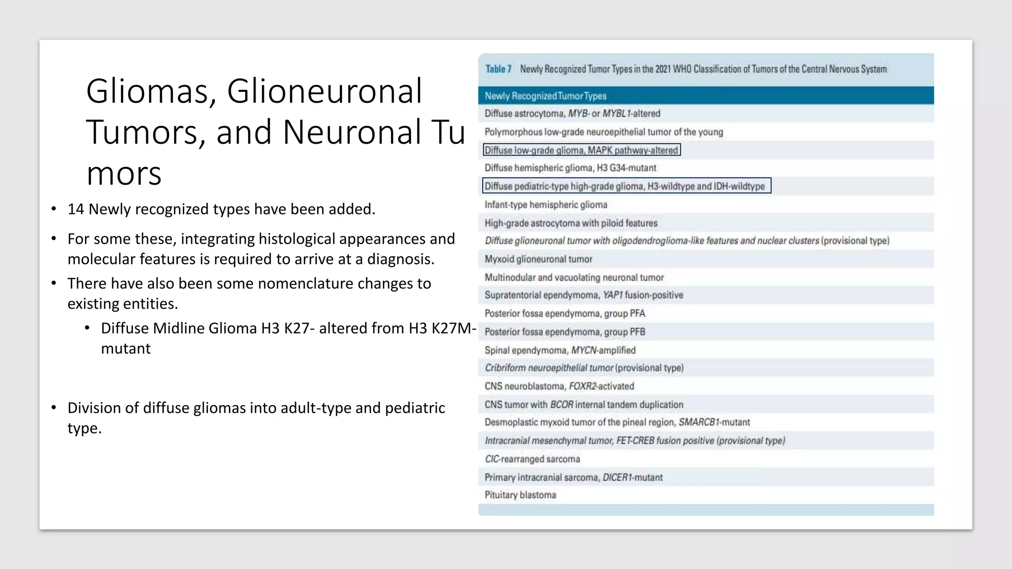
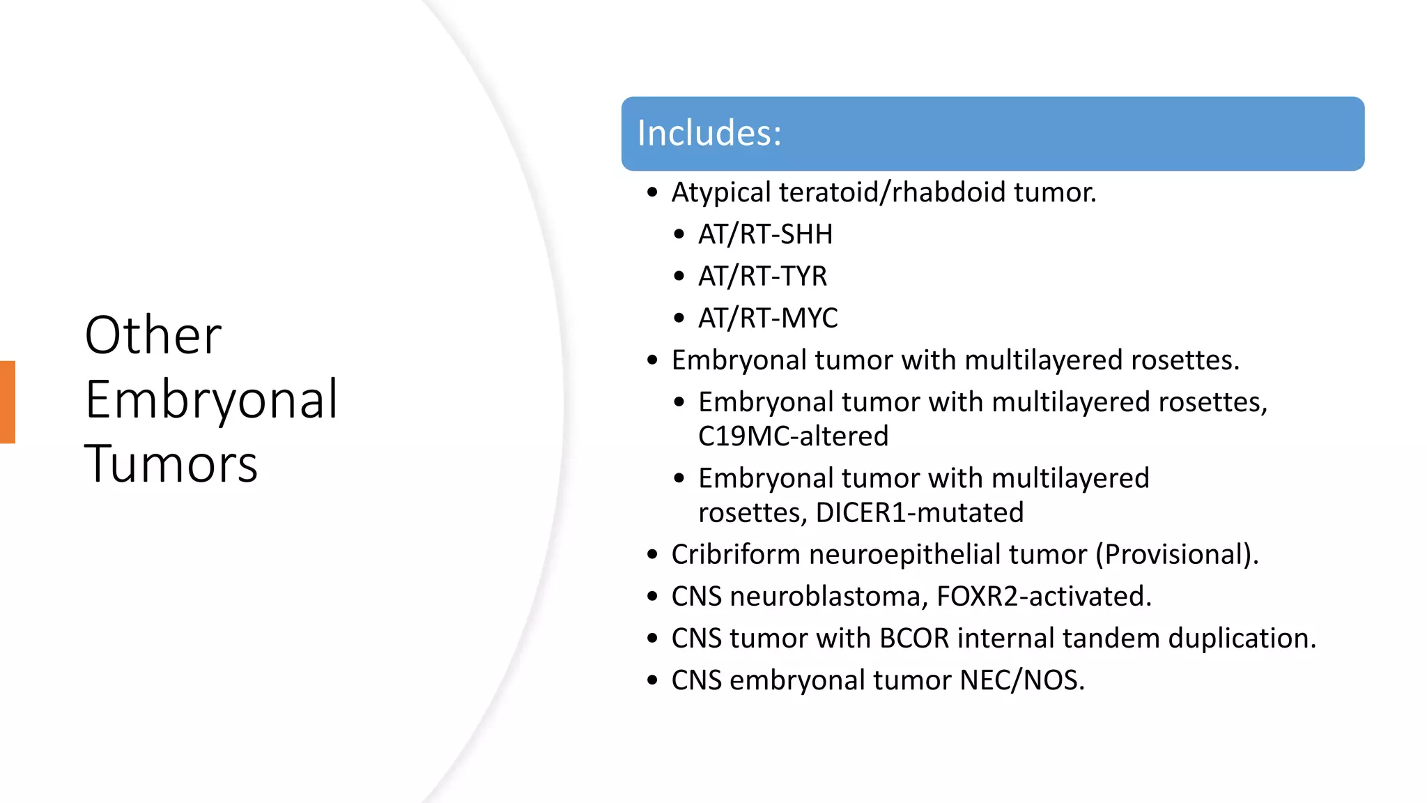
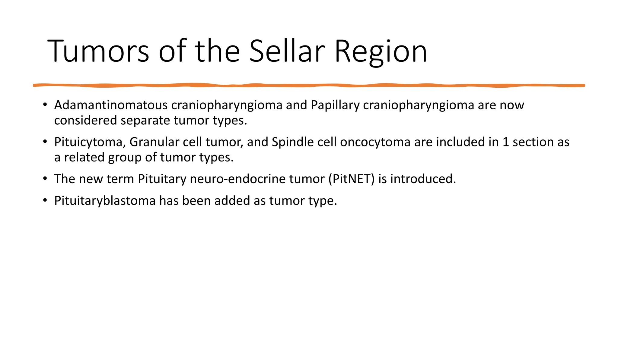
The fifth edition of the WHO Classification of Tumors of the Central Nervous System (WHO CNS5) incorporates numerous molecular changes with clinicopathologic utility that are important for the most accurate classification of CNS neoplasms.
WHO CNS5 does not recommend specific methods for molecular assessment.
WHO CNS5 has grouped tumors according to the genetic changes that enable a complete diagnosis.
IDH (Astrocytoma, Oligodendroglioma and Glioblastoma) and H3 (Diffuse midline glioma, Diffuse hemispheric glioma).
Some by looser oncogenic associations. Like MAPK pathway alteration (Multinodular and Vacuolating Neuronal Tumor).
Some are classified by histological similarities even though molecular signatures vary.
Atypical teratoid/rhabdoid tumor, Ganglioglioma, Papillary glioneuronal tumor.
Many by using molecular features to define new types and subtypes.
Medulloblastoma.
The term “type" is used instead of “entity” and “subtype” is used instead of “variant".
The fifth edition of the WHO Classification of Tumors of the Central Nervous System follows the recommendations of the 2019 cIMPACT-NOW Utrecht meeting.
Names have been simplified, and only location, age, or genetic modifiers with clinical utility have been used.
Extra-ventricular neurocytoma vs Central neurocytoma.
The characteristics of tumors that are highly characteristic are included in tumor definitions and descriptions, even if they do not appear in the tumor name itself.
chordoid gliomas occurring in the third ventricle
Sometimes tumor names reflect morphologic features that are not present in every example, and they may also reflect historical associations.
Some myxopapillary ependymomas are minimally myxoid, and some may not be overtly papillary.
Xanthomatous change may be limited to a small fraction of cells in pleomorphic xanthoastrocytomas.
Medulloblast has not been identified in developmental studies, in cases of Medulloblastoma.
As they would be disruptive to clinicians and may lead to confusion, they were not changed.
Tumors are now graded within types, modifier terms like "anaplastic" are not routinely used.
1. The 2021 WHO Classification
of Tumors of the Central
Nervous System: a summary
By: David N Louis
Neuro Oncol. 2021;23(8):1231-1251.
PRESENTED BY: DR MUHAMMAD ZAMIL
•
3. CNS Tumor Taxonomy
• The fifth edition of the WHO Classification of Tumors of the Central Nervous System (WHO CNS5)
incorporates numerous molecular changes with clinicopathologic utility that are important for the most
accurate classification of CNS neoplasms.
• WHO CNS5 does not recommend specific methods for molecular assessment.
• WHO CNS5 has grouped tumors according to the genetic changes that enable a complete diagnosis.
• IDH (Astrocytoma, Oligodendroglioma and Glioblastoma) and H3 (Diffuse midline glioma, Diffuse
hemispheric glioma).
• Some by looser oncogenic associations. Like MAPK pathway alteration (Multinodular and Vacuolating
Neuronal Tumor).
• Some are classified by histological similarities even though molecular signatures vary.
• Atypical teratoid/rhabdoid tumor, Ganglioglioma, Papillary glioneuronal tumor.
• Many by using molecular features to define new types and subtypes.
• Medulloblastoma.
• The term “type" is used instead of “entity” and “subtype” is used instead of “variant".
4. CNS Tumor Nomenclature
• The fifth edition of the WHO Classification of Tumors of the Central Nervous System follows the recommendations of the
2019 cIMPACT-NOW Utrecht meeting.
• Names have been simplified, and only location, age, or genetic modifiers with clinical utility have been used.
• Extra-ventricular neurocytoma vs Central neurocytoma.
• The characteristics of tumors that are highly characteristic are included in tumor definitions and descriptions, even if they do
not appear in the tumor name itself.
• chordoid gliomas occurring in the third ventricle
• Sometimes tumor names reflect morphologic features that are not present in every example, and they may also reflect
historical associations.
• Some myxopapillary ependymomas are minimally myxoid, and some may not be overtly papillary.
• Xanthomatous change may be limited to a small fraction of cells in pleomorphic xanthoastrocytomas.
• Medulloblast has not been identified in developmental studies, in cases of Medulloblastoma.
• As they would be disruptive to clinicians and may lead to confusion, they were not changed.
• Tumors are now graded within types, modifier terms like "anaplastic" are not routinely used.
5. Gene and Protein Nomenclature for CNS
Tumor Classification
• WHO CNS5 uses the HUGO Gene Nomenclature Committee (HGNC) system for naming gene symbols and
gene name.
• Gene symbols are presented in italics, but proteins and gene groups are not italicized
• A sequence alteration relative to a transcript reference sequence is reported using a “c. ” prefix for the
coding DNA sequence, followed by the nucleotide number and nucleotide change. The predicted
protein sequence change then follows a “p.” prefix with the reference amino acid, the amino acid
number, and the variant amino acid resulting from the mutation.
• BRAF:c.1799T>A p.Val600Glu
• For some genes like H3 Histone group are typically described in the context of the protein sequence lacks
the initiating methionine, resulting in a single amino acid difference in numbering compared with the
predicted sequence derived from the corresponding gene transcript.
• To address this issue, the fifth edition uses the legacy protein numbering system in parentheses
after the protein-level variant description.
• H3-3A:c.103G>A p.Gly35Arg (G34R), or H3-3A:c.83A>T p.Lys28Met (K27M)
6. CNS Tumor Grading
• 2 Aspect of Grading have been changed.
• Arabic Numerical
• Use of Arabic numerical is now favored as Roman Numerical can be mixed with each other and an uncaught
typographical error can result in unintended clinical consequences.
• Grading within tumor types, has been extended to many categories. This is done due to following reasons:
• To provide more flexibility in using grade relative to the tumor type.
• To emphasize biological similarities within tumor types rather than approximate clinical behavior.
• To conform with WHO grading in non-CNS tumor types.
• WHO CNS5 has generally retained the ranges of grades used for tumor types in prior editions, for example IDH mutant
Astrocytomas extend from CNS WHO grade 2-4 and Meningiomas from CNS WHO grade 1-3
• Molecular parameters have now been added as biomarkers of grading and for further estimating prognosis within multiple
tumor types, for example, CDKN2A/B homozygous deletion in IDH mutant astrocytoma, TERT promoter mutation, EGFR
amplification, and +7/−10 copy number changes in IDH-wildtype diffuse astrocytomas.
• This allows a glioblastoma, IDH-wildtype CNS WHO grade 4 designation even in cases that otherwise appear histologically
lower grade.
7. NOS (Not
Otherwise
Specified)
and NEC (Not
Elsewhere
Classified)
Diagnoses.
• NOS: the diagnostic information (histological or
molecular) necessary to assign a specific WHO
diagnosis is not available.
• NEC: That the necessary diagnostic testing has been
successfully performed but that the results do not
readily allow for a WHO diagnosis.
8. Novel
Diagnostic
Technologies
• Over the past decade, methylome profiling has
emerged as a powerful approach to CNS tumor
classification, as detailed in a variety of publications
over the past few years.
• Methylome profiling is the use of arrays to
determine DNA methylation patterns across the
genome.
9. Integrated and
Layered Diagnoses
• The fifth edition of the WHO Classification of Tumors of
the Central Nervous System follows, The International
Society of Neuropathology—Haarlem consensus guidelines.
11. Gliomas,
Glioneuronal
Tumors, and
Neuronal Tumors
• Divided into 6 different categories.
• Adult-type diffuse gliomas
• Pediatric-type diffuse low-grade gliomas
• Pediatric-type diffuse high-grade gliomas
• Circumscribed astrocytic gliomas
• Glioneuronal and neuronal tumors
• Ependymomas
12. Gliomas, Glioneuronal
Tumors, and Neuronal Tu
mors
• 14 Newly recognized types have been added.
• For some these, integrating histological appearances and
molecular features is required to arrive at a diagnosis.
• There have also been some nomenclature changes to
existing entities.
• Diffuse Midline Glioma H3 K27- altered from H3 K27M-
mutant
• Division of diffuse gliomas into adult-type and pediatric
type.
13. Gliomas, Glioneuronal Tumors, and Neuronal Tumors
• Simplification of the classification of common, adult type, diffuse gliomas.
• It Includes 3 types.
• Astrocytoma, IDH-mutant
• Oligodendroglioma, IDH-mutant and 1p/19q-codeleted
• Glioblastoma, IDH-wildtype.
• Gliosarcoma and Giant Cell Glioblastoma are not listed in the classification, they're discussed in their respective chapters.
• Nomenclature and grading of common, adult-type, diffuse astrocytic gliomas.
• All IDH mutant diffuse astrocytic tumors are considered a single type, Astrocytoma, IDH-mutant. It is then graded as 2, 3 or 4.
• Grading is no longer entirely histologic, e.g. CDKN2A/B homozygous deletion results in a CNS WHO grade of 4, even in the
absence of microvascular proliferation or necrosis.
• 3 genetic parameters are used as criteria for a diagnosis of Glioblastoma, IDH-wildtype.
• TERT promoter mutation
• EGFR gene amplification
• Combined gain of entire chromosome 7 and loss of entire chromosome 10
14. Gliomas,
Glioneuronal Tumors,
and Neuronal Tumors
Pediatric-type low-grade and high-grade diffuse gliomas.
• The low grade group includes.
• Diffuse astrocytoma, MYB- or MYBL1-altered
• Angiocentric glioma
• Polymorphous low-grade neuroepithelial tumor of the young
• Diffuse low-grade glioma, MAPK pathway-altered
• The high grade group includes.
• Diffuse midline glioma, H3 K27-altered
• Diffuse hemispheric glioma, H3 G34-mutant.
• Diffuse pediatric-type high-grade glioma, H3-wildtype and IDH-
wildtype
• Infant-type hemispheric glioma.
Neuronal and Glioneuronal Tumor
• 3 new types have been added.
• Diffuse glioneuronal tumor with oligodendroglioma-like features and
nuclear clusters (Provisional)
• Myxoid glioneuronal tumor
• Multinodular and vacuolating neuronal tumor
15. Gliomas, Glioneuronal Tumors, and Neuronal Tumors
• Ependymomas
• Classified by combination of histopathological, molecular features and anatomic site.
• Divided into molecular groups.
• Supratentorial
• Supratentorial ependymoma
• Supratentorial ependymoma, ZFTA fusion-Positive
• Supratentorial ependymoma, YAP1 fusion-Positive
• Posterior fossa (PF)
• Posterior fossa ependymoma
• Posterior fossa group A (PFA) ependymoma
• Posterior fossa group B (PFB) ependymoma
• Spinal compartments
• Spinal ependymoma, MYCN-amplified
• Myxopapillary Ependymoma is now considered WHO Grade 2 rather than 1
• Papillary, clear cell, and tanycytic morphological variants are no longer listed as subtypes of ependymoma.
• Graded as WHO grade 2 or 3 , anaplastic ependymoma is no longer listed.
16. Choroid Plexus
Tumors
This family of tumors has
been separated from the
category of primary
neuroepithelial tumors.
Classification is largely
unchanged.
17. Medulloblastomas
4 principal molecular groups.
• Medulloblastoma, WNT-activated
• Medulloblastoma, SHH-activated and TP53-wildtype
• SHH-1
• SHH-2
• SHH-3
• SHH-4
• Medulloblastoma, SHH-activated and TP53-mutant
• Medulloblastoma, non-WNT/non-SHH
• 8 Molecular sub-groups (Group ¾ Subgroup 1-8).
Histopathological Classification listed in 2016 has
now been described as morphological patterns
of Medulloblastoma, histologically defined.
18. Other
Embryonal
Tumors
Includes:
• Atypical teratoid/rhabdoid tumor.
• AT/RT-SHH
• AT/RT-TYR
• AT/RT-MYC
• Embryonal tumor with multilayered rosettes.
• Embryonal tumor with multilayered rosettes,
C19MC-altered
• Embryonal tumor with multilayered
rosettes, DICER1-mutated
• Cribriform neuroepithelial tumor (Provisional).
• CNS neuroblastoma, FOXR2-activated.
• CNS tumor with BCOR internal tandem duplication.
• CNS embryonal tumor NEC/NOS.
19. Pineal Tumors
Pineocytoma
Pineal parenchymal tumor
of intermediate
differentiation
Pineoblastoma
Pineoblastoma, miRNA
processing-altered_1
Pineoblastoma, miRNA
processing-altered_2
Pineoblastoma, RB1-
altered (pineal
retinoblastoma)
Pineoblastoma,
MYC/FOXR2-activated
Papillary tumor of the
pineal region
Desmoplastic myxoid
tumor of the pineal region,
SMARCB1-mutant
20. Meningiomas
• Considered a single type.
• Broad morphological sub-types, 15 subtypes.
• Choroid and Clear cell Meningioma have higher rate of recurrence, hence assigned WHO Grade 2.
• Grading should not be done on the basis of a rhabdoid cytology or papillary architecture alone.
• Several molecular biomarkers are also associated with classification and grading of meningiomas
• SMARCE1 --- clear cell subtype.
• BAP1 --- rhabdoid and papillary subtypes
• KLF4/TRAF7 --- secretory subtype
• TERT promoter mutation and/or homozygous deletion of CDKN2A/B --- CNS WHO grade 3
• H3K27me3 loss of nuclear expression --- potentially worse prognosis
• Methylome profiling --- prognostic subtyping
21. Mesenchymal, Non-Meningothelial Tumors
• Only those entities that occur uniquely in the CNS.
• Common soft tissue tumors, found in the CNS are no longer included given that their
diagnostic features are identical to their soft tissue counterparts, e.g. Leiomyoma.
• New Types include
• Intracranial mesenchymal tumor, FET-CREB fusion-positive (Provisional).
• CIC-rearranged sarcoma
• Primary intracranial sarcoma, DICER1-mutant
22. Nerve Tumors
• Paraganglioma are now included in nervous tumor.
• The paraganglioma of the cauda equina/filum Terminale region is now recognized as a
distinct
tumor type as, Cauda equina neuroendocrine tumor (previously paraganglioma).
• Melanotic schwannoma name has been changed to Malignant melanotic nerve sheath
tumor.
• A new subtype has been added to the neurofibroma section: Atypical neurofibromatous
neoplasm of unknown biological potential (ANNUBP)
23. Lymphomas and Histiocytic Tumors
• Includes those lymphoid and histiocytic tumor entities that occur
relatively often in the CNS or that have special histological or molecular
features when they occur in the CNS.
24. Tumors of the Sellar Region
• Adamantinomatous craniopharyngioma and Papillary craniopharyngioma are now
considered separate tumor types.
• Pituicytoma, Granular cell tumor, and Spindle cell oncocytoma are included in 1 section as
a related group of tumor types.
• The new term Pituitary neuro-endocrine tumor (PitNET) is introduced.
• Pituitaryblastoma has been added as tumor type.
25. Metastatic
Tumors
• Divided in 2 categories
• Metastases to the brain and spinal cord
parenchyma
• Metastases to the meninges
26. Genetic Tumor Syndromes
• This section has been expanded, now covering 8 disorders not covered in the prior Blue
Book.
• Carney complex
• DICER1 syndrome
• Familial paraganglioma syndrome
• Melanoma-astrocytoma syndrome
• Familial retinoblastoma
• BAP1 tumor predisposition syndrome
• Fanconi anemia
• ELP1-medulloblastoma syndrome