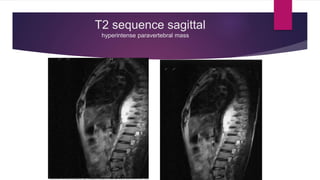
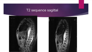
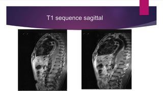
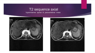
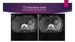
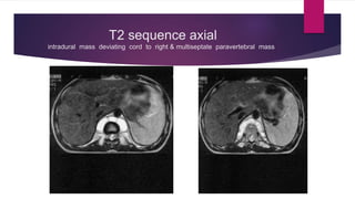
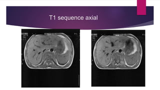
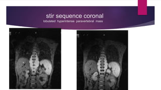
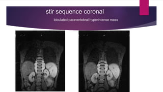
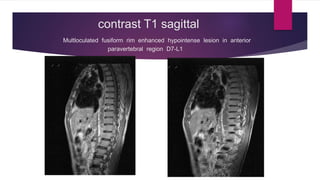
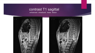
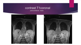
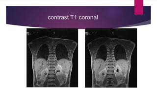
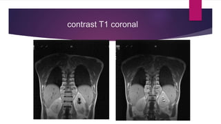
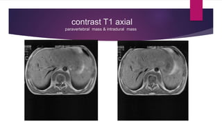
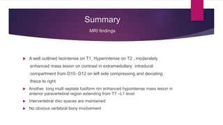
Intradural extramedullary mass:
a case on MRI
DR REKHA KHARE MD RADIOLOGY
DR VIMLESH CONSULTANT NEUROLOGY
Clinical picture
 A young boy about 18year came to radiology department for MRI thoracic spine
 His problem was for about 6 months.
 Symptoms were persistent progressive lower back pain and lower extremity weakness
on left more than right. Pain was radiating to his left thigh region .
 There was no remarkable medical history
 There was no significant family history
 Routine blood and urine examination was with in normal limit
 Neurological findings : recorded in case file by neurologist were
Radicular pain and Babinski sign present on left side
T2 sequence sagittal
hyperintense paravertebral mass
T2 sagittal
hyperintense spinal & paravertebral mass
T2 sequence sagittal
T1 sequence sagittal
hypointense spinal & paravertebral mass
T1 sequence sagittal
T2 sequence axial
hyperintense spinal & paravertebral mass
T2 sequence axial
intradural mass on left deviating theca
T2 sequence axial
intradural mass deviating cord to right & multiseptate paravertebral mass
T1 sequence axial
hypointense paravertebral mass
T1 sequence axial
stir sequence coronal
lobulated hyperintense paravertebral mass
stir sequence coronal
lobulated paravertebral hyperintense mass
contrast T1 sagittal
Multloculated fusiform rim enhanced hypointense lesion in anterior
paravertebral region D7-L1
contrast T1 sagittal
enhanced intradural mass lesion
contrast T1coronal
paravertebral mass
contrast T1 coronal
contrast T1 coronal
contrast T1 axial
paravertebral mass & intradural mass
contrast T1axial
rim enhanced hypointense paravertebral mass lesion & enhanced intradural
mass
Summary
MRI findings
 A well outlined Isointense on T1, Hyperintense on T2 , moderately
enhanced mass lesion on contrast in extramedullary intradural
compartment from D10- D12 on left side compressing and deviating
theca to right
 Another long multi septate fusiform rim enhanced hypointense mass lesion in
anterior paravertebral region extending from T7 –L1 level
 Intervertebral disc spaces are maintained
 No obvious vertebral bony involvement
Radiological impression
 In correlation with clinical picture of radicular pain and Babinski sign contrast MRI
findings …….. lesion is considered to be
extramedullary intradural lesion with paraspinal mass lesion
 Provisional preoperative diagnosis was considered Neurofibroma
 Case has been referred to higher centre for further surgical
management and histopathological confirmation
 Patient didn’t turn back for follow up
Spinal tumours
 Spinal tumours account for 15% of all CNS tumour
 Incidence is 0.5-2.5/ 100,000
 History is for months or years of Radiculopathy or symptoms of Myelopathy
 It can be Primary in origin or Metastatic
 Diagnosis is based on clinical symptoms, duration of presentation ,site in cord and
MRI findings
 Confirmation requires Biopsy
 Treatment for many spinal tumour is surgical if possible
Spinal Tumour:
Extradural
• Tuberculosis/ Potts spine
• Metastasis
• Osteoblastoma
• Osteochondroma
• Chondrosarcoma
• Giant cell tumour
• Myeloma
Intradural-
Extramedullary
• Nerve sheath tumour:
Schwannoma
neurofibroma
• Meningioma
• Metastasis
• Haemangioblastoma
• Paraglioma
Intradural-
Intramedullary
• Ependymoma
• Astrocytoma
• Haemangioblastoma
• Metastasis
Spinal tumour
Intramedullary mass
Intradural mass
Extradural mass
Imaging modality
 MRI is the modality of choice for assessment of intra spinal lesion as it has
excellent anatomical contrast and structural resolution
 MRI is able to image all compartments
 MRI can assess enhancement, cystic change and blood products etc.
Other Imaging modality
 CT remains next best modality to assess the bony structure although it is usually
not required for intradural tumours as the vertebral bodies are essentially
unaffected
 Myelography is of historical importance but sometimes now is done only in
patients for whom MRI is contraindicated
 Ultrasound except in the infants plays no role in diagnosis
 Spinal angiography is useful selectively in those patients who have vascular
lesion
MRI Protocol
Routinely following sequences are done
 T1 sagittal and axial
 T2 sagittal and axial
 T1 contrast + sagittal ,axial and coronal
MRI: General signal characteristics
 T1---Most intradural masses are Isointense or hypointense
Hyperintense if Haemorrhagic or fat content in mass
Ganglioglioma usually mixed signal on T1( unique finding )
 T2----Spinal cord tumours are usually Hyperintense
Tumour with abundant collagen are Hypointense on T2
Calcification in tumour reflects Hypointensity T2
 Peritumoural oedema is as perilesional T2 hyperintensity
 Massive oedema disproportionate is typical of Metastasis
 Prominent vascular flow voids indicates vascular lesion like Haemangioblastoma
or spinal dural AV fistula
Nerve sheath tumour: NSTs
 MRI can not distinguish Neurofibroma from Schwannoma ,may manifest as round
paravertebral masses that span one or two vertebral bodies. They are soft tissue
attenuation masses
 MRI Imaging findings: T1- isointense and hyperintense on contrast
T2 Hyperintense
Target lesion : Decreased central signal with increased
peripheral signal common in schwannoma
Sometimes split fat sign( thin peripheral rim of fat) or
fasicular sign( multiple small ring like structure) could be in schwannoma
*Paraspinal extension common in Neurofibroma can cause bone erosion
Nerve sheath tumour
 Neurofibroma: Most common NST in young patient & have no
predilection for either sex
Arises from dorsal sensory nerve roots of thoracic spine usually
Not always associated with Neurofibromatosis 1.
Usually painless, slow growing and aymptomatic
Dumbell shaped thoracic neurofibroma represents distinct type of tumour it can involve spinal
canal & thoracic cavity
*Autosomal dominant inherited condition arises from nonmyelinating type schwaan cell that exhibit biallelic
inactivation of NF1 that codes for the protein Neurofibromin which is responsible for regulating RAS mediated cell
growth signalling pathway
Schwannoma
Benign slow growing tumours of schwaan cell origin
Peak presentation in 5-6th decade with no sex predilection
When scwannoma present with NF 2 it can present in 3rd decade
Most common tumour of peripheral nerves and common posterior fossa masses
Arises from ventral motor nerve root of thoracic spine
Clinical symptoms depends on location of the tumour
Neurofibroma
NSTs contd…..
Meningioma:
 2nd most common intradural extramedullary tumour
 It is benign slow growing tumour that arises from arachnoid cells
 Average age 40-60years
 Females are affected more than males
 Usually in thoracic vertebra
MRI imaging finding: T1- hypo/iso intense
T2 –hyper / iso intense
Contrast/Gadolinium- homogenous enhancement
CT/ plain film- calcification +
Intramedullary tumour
Ependymoma
 Most common intramedullary intradural mass
 Usually seen in cervical spine or conus
 Arises from ependymal cells of spinal cord
 Presents with more neurological deficit than radiculopathy
 Peak incidence at 30-50 year
Imaging finding: Besides Hypointense T1,Hyperintense T2 and contrast
enhanced tumour spinal cord is enlarged and scalloping
or sometimes vertebral body erosion
Intramedullary tumour contd…….
Astrocytoma
 Arises from astrocytes
 Peak incidence 20-40 years of age
Imaging findings:
Difficult to differentiate Astrocytoma from Ependymoma on imaging
It needs Biopsy
Hypointense , enhanced T1 Hyperintense T2 with enlarge spinal
cord
Spinal tumour/ mass
Intramedullary
Intradural
extramedullary Extradural
References
 Text book of Radiology and Imaging By; David Sutton
 Neurogenic tumours Dr Jeremy Jones etal Radiopaedia. Org.com
 Intradural spinal tumour- Lieberman’s eRadiology eradiology.bidmc.harvard.edu/Learning
Lab/central/Bhatt.pdf
 Intradural spinal mass lesion ( an approach) Prof Frank Gaillard Radiopaedia.org.www
 Intradural nerve sheath tumour-UpToDate www.uptodate.com/contents/intradural-nerve-sheath-tumors
 Intradural extramedullary tumour Definition
www. spine-health.com/glossary/intradural-extramedullary-tumor
Thank you
HAVE A NICE DAY

Intradural extramedullary mass - a case on MRI

  • 1.
    Intradural extramedullary mass: acase on MRI DR REKHA KHARE MD RADIOLOGY DR VIMLESH CONSULTANT NEUROLOGY
  • 2.
    Clinical picture  Ayoung boy about 18year came to radiology department for MRI thoracic spine  His problem was for about 6 months.  Symptoms were persistent progressive lower back pain and lower extremity weakness on left more than right. Pain was radiating to his left thigh region .  There was no remarkable medical history  There was no significant family history  Routine blood and urine examination was with in normal limit  Neurological findings : recorded in case file by neurologist were Radicular pain and Babinski sign present on left side
  • 3.
  • 4.
    T2 sagittal hyperintense spinal& paravertebral mass
  • 5.
  • 6.
    T1 sequence sagittal hypointensespinal & paravertebral mass
  • 7.
  • 8.
    T2 sequence axial hyperintensespinal & paravertebral mass
  • 9.
    T2 sequence axial intraduralmass on left deviating theca
  • 10.
    T2 sequence axial intraduralmass deviating cord to right & multiseptate paravertebral mass
  • 11.
  • 12.
  • 13.
    stir sequence coronal lobulatedhyperintense paravertebral mass
  • 14.
    stir sequence coronal lobulatedparavertebral hyperintense mass
  • 15.
    contrast T1 sagittal Multloculatedfusiform rim enhanced hypointense lesion in anterior paravertebral region D7-L1
  • 16.
    contrast T1 sagittal enhancedintradural mass lesion
  • 17.
  • 18.
  • 19.
  • 20.
    contrast T1 axial paravertebralmass & intradural mass
  • 21.
    contrast T1axial rim enhancedhypointense paravertebral mass lesion & enhanced intradural mass
  • 22.
    Summary MRI findings  Awell outlined Isointense on T1, Hyperintense on T2 , moderately enhanced mass lesion on contrast in extramedullary intradural compartment from D10- D12 on left side compressing and deviating theca to right  Another long multi septate fusiform rim enhanced hypointense mass lesion in anterior paravertebral region extending from T7 –L1 level  Intervertebral disc spaces are maintained  No obvious vertebral bony involvement
  • 23.
    Radiological impression  Incorrelation with clinical picture of radicular pain and Babinski sign contrast MRI findings …….. lesion is considered to be extramedullary intradural lesion with paraspinal mass lesion  Provisional preoperative diagnosis was considered Neurofibroma  Case has been referred to higher centre for further surgical management and histopathological confirmation  Patient didn’t turn back for follow up
  • 24.
    Spinal tumours  Spinaltumours account for 15% of all CNS tumour  Incidence is 0.5-2.5/ 100,000  History is for months or years of Radiculopathy or symptoms of Myelopathy  It can be Primary in origin or Metastatic  Diagnosis is based on clinical symptoms, duration of presentation ,site in cord and MRI findings  Confirmation requires Biopsy  Treatment for many spinal tumour is surgical if possible
  • 25.
    Spinal Tumour: Extradural • Tuberculosis/Potts spine • Metastasis • Osteoblastoma • Osteochondroma • Chondrosarcoma • Giant cell tumour • Myeloma Intradural- Extramedullary • Nerve sheath tumour: Schwannoma neurofibroma • Meningioma • Metastasis • Haemangioblastoma • Paraglioma Intradural- Intramedullary • Ependymoma • Astrocytoma • Haemangioblastoma • Metastasis
  • 26.
  • 27.
    Imaging modality  MRIis the modality of choice for assessment of intra spinal lesion as it has excellent anatomical contrast and structural resolution  MRI is able to image all compartments  MRI can assess enhancement, cystic change and blood products etc.
  • 28.
    Other Imaging modality CT remains next best modality to assess the bony structure although it is usually not required for intradural tumours as the vertebral bodies are essentially unaffected  Myelography is of historical importance but sometimes now is done only in patients for whom MRI is contraindicated  Ultrasound except in the infants plays no role in diagnosis  Spinal angiography is useful selectively in those patients who have vascular lesion
  • 29.
    MRI Protocol Routinely followingsequences are done  T1 sagittal and axial  T2 sagittal and axial  T1 contrast + sagittal ,axial and coronal
  • 30.
    MRI: General signalcharacteristics  T1---Most intradural masses are Isointense or hypointense Hyperintense if Haemorrhagic or fat content in mass Ganglioglioma usually mixed signal on T1( unique finding )  T2----Spinal cord tumours are usually Hyperintense Tumour with abundant collagen are Hypointense on T2 Calcification in tumour reflects Hypointensity T2  Peritumoural oedema is as perilesional T2 hyperintensity  Massive oedema disproportionate is typical of Metastasis  Prominent vascular flow voids indicates vascular lesion like Haemangioblastoma or spinal dural AV fistula
  • 31.
    Nerve sheath tumour:NSTs  MRI can not distinguish Neurofibroma from Schwannoma ,may manifest as round paravertebral masses that span one or two vertebral bodies. They are soft tissue attenuation masses  MRI Imaging findings: T1- isointense and hyperintense on contrast T2 Hyperintense Target lesion : Decreased central signal with increased peripheral signal common in schwannoma Sometimes split fat sign( thin peripheral rim of fat) or fasicular sign( multiple small ring like structure) could be in schwannoma *Paraspinal extension common in Neurofibroma can cause bone erosion
  • 32.
    Nerve sheath tumour Neurofibroma: Most common NST in young patient & have no predilection for either sex Arises from dorsal sensory nerve roots of thoracic spine usually Not always associated with Neurofibromatosis 1. Usually painless, slow growing and aymptomatic Dumbell shaped thoracic neurofibroma represents distinct type of tumour it can involve spinal canal & thoracic cavity *Autosomal dominant inherited condition arises from nonmyelinating type schwaan cell that exhibit biallelic inactivation of NF1 that codes for the protein Neurofibromin which is responsible for regulating RAS mediated cell growth signalling pathway
  • 33.
    Schwannoma Benign slow growingtumours of schwaan cell origin Peak presentation in 5-6th decade with no sex predilection When scwannoma present with NF 2 it can present in 3rd decade Most common tumour of peripheral nerves and common posterior fossa masses Arises from ventral motor nerve root of thoracic spine Clinical symptoms depends on location of the tumour
  • 34.
  • 35.
    NSTs contd….. Meningioma:  2ndmost common intradural extramedullary tumour  It is benign slow growing tumour that arises from arachnoid cells  Average age 40-60years  Females are affected more than males  Usually in thoracic vertebra MRI imaging finding: T1- hypo/iso intense T2 –hyper / iso intense Contrast/Gadolinium- homogenous enhancement CT/ plain film- calcification +
  • 36.
    Intramedullary tumour Ependymoma  Mostcommon intramedullary intradural mass  Usually seen in cervical spine or conus  Arises from ependymal cells of spinal cord  Presents with more neurological deficit than radiculopathy  Peak incidence at 30-50 year Imaging finding: Besides Hypointense T1,Hyperintense T2 and contrast enhanced tumour spinal cord is enlarged and scalloping or sometimes vertebral body erosion
  • 37.
    Intramedullary tumour contd……. Astrocytoma Arises from astrocytes  Peak incidence 20-40 years of age Imaging findings: Difficult to differentiate Astrocytoma from Ependymoma on imaging It needs Biopsy Hypointense , enhanced T1 Hyperintense T2 with enlarge spinal cord
  • 38.
  • 39.
    References  Text bookof Radiology and Imaging By; David Sutton  Neurogenic tumours Dr Jeremy Jones etal Radiopaedia. Org.com  Intradural spinal tumour- Lieberman’s eRadiology eradiology.bidmc.harvard.edu/Learning Lab/central/Bhatt.pdf  Intradural spinal mass lesion ( an approach) Prof Frank Gaillard Radiopaedia.org.www  Intradural nerve sheath tumour-UpToDate www.uptodate.com/contents/intradural-nerve-sheath-tumors  Intradural extramedullary tumour Definition www. spine-health.com/glossary/intradural-extramedullary-tumor
  • 40.