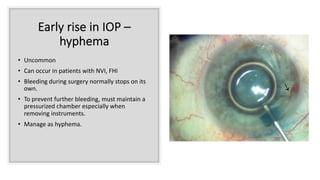
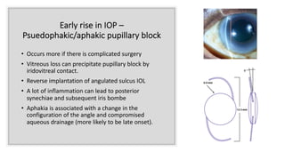
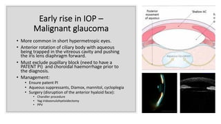
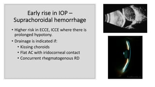
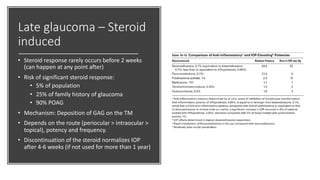
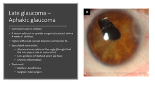
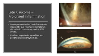
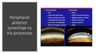
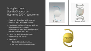
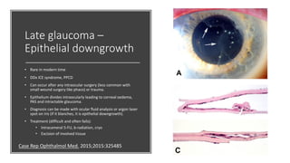
Glaucoma post cataract surgery:
Mechanisms, prevention and
management
Dr Haitham Al-Mahrouqi
Cornea and Anterior Segment
Al-Nahdha Hospital
Cataract and
glaucoma
• The crystalline Lens can cause
glaucoma and lens extraction (does
not need to be cataractous) can be a
mode of managing glaucoma.
• Glaucoma accelerates cataract and
controlled IOP can prevent cataract
development.
• Glaucoma can be a consequence of
cataract surgery.
Glaucoma post cataract surgery
• Early
• Retained viscoelastic (peak IOP rise at 4-6 hours)
• Inflammation (uveitis or trabeculitis)
• Hyphema
• Pseudophakic/Aphakic Pupillary block
• Malignant glaucoma
• Suprachoroidal hemorrhage
• Late
• Steroid induced
• Trabecular meshwork anatomical changes e.g. Aphkia
• Peripheral anterior synechiae
• Epithelial ingrowth
• Retained lens matter
• Uvieits – glaucoma – hyphema (UGH) syndrome.
Early rise in IOP –
Retained viscoelastic device
• Viscoelastic is very important in cataract surgery.
• It is imperative not to leave a lot of residual
viscoelastic material especially in glaucoma
patients.
• Should not inflate the bag with dispersive
viscoelastic.
• Takes about 3-5 days to be absorbed
• Manage medically or surgically by removal of
viscelastic if uncontrolled IOP.
Early rise in IOP –
Inflammation
• Mild inflammation is normal after cataract surgery.
• Inflammation is higher with prolonged and
complicated surgery.
• Although the inflammation can be transient, glaucoma
patients are more likely to have high IOP (due to
compromised drainage) and more likely to be affected
due to compromised optic nerve.
• Toxic anterior segment syndrome: Severe
inflammation within 24 hrs of surgery due to toxicity
from materials used. DDx Endophthalmitis
• Re-activation of HSV keratitis/uveitis: Prophylaxis pre-
op is important.
Early rise in IOP –
hyphema
• Uncommon
• Can occur in patients with NVI, FHI
• Bleeding during surgery normally stops on its
own.
• To prevent further bleeding, must maintain a
pressurized chamber especially when
removing instruments.
• Manage as hyphema.
Early rise in IOP –
Psuedophakic/aphakic pupillary block
• Occurs more if there is complicated surgery
• Vitreous loss can precipitate pupillary block by
iridovitreal contact.
• Reverse implantation of angulated sulcus IOL
• A lot of inflammation can lead to posterior
synechiae and subsequent iris bombe
• Aphakia is associated with a change in the
configuration of the angle and compromised
aqueous drainage (more likely to be late onset).
Early rise in IOP –
Malignant glaucoma
• More common in short hypermetropic eyes.
• Anterior rotation of ciliary body with aqueous
being trapped in the vitreous cavity and pushing
the iris lens diaphragm forward.
• Must exclude pupillary block (need to have a
PATENT PI) and choroidal haemorrhage prior to
the diagnosis.
• Management:
• Ensure patent PI
• Aqueous suppressants, Diamox, mannitol, cycloplegia
• Surgery (disruption of the anterior hyaloid face):
• Chandler procedure
• Yag iridozonulohyeloidectomy
• PPV
Early rise in IOP –
Suprachoroidal hemorrhage
• Higher risk in ECCE, ICCE where there is
prolonged hypotony.
• Drainage is indicated if:
• Kissing choroids
• Flat AC with iridocorneal contact
• Concurrent rhegmatogenous RD
Late glaucoma – Steroid
induced
• Steroid response rarely occurs before 2 weeks
(can happen at any point after)
• Risk of significant steroid response:
• 5% of population
• 25% of family history of glaucoma
• 90% POAG
• Mechanism: Deposition of GAG on the TM
• Depends on the route (periocular > intraocular >
topical), potency and frequency.
• Discontinuation of the steroid normalizes IOP
after 4-6 weeks (if not used for more than 1 year)
Late glaucoma –
Aphakic glaucoma
• Commonly seen in children
• A reason why not to operate congenital cataract before
4 weeks in children.
• Higher with small corneal diameter and shorter AL.
• Speculated mechanism:
• Abnormal maturation of the angle (thought that
the lens plays a role in maturation)
• Lens proteins left behind which are toxic
• Chronic inflammation
• Treatment:
• Medical: Avoid brimo
• Surgical: Tube surgery
Late glaucoma –
Prolonged inflammation
• Inadequate control of the inflammation
induced by e.g. retained lens material,
viscoelastic, pre-existing uveitis, HSV
uveitis.
• Can lead to posterior synechiae and
peripheral anterior synechiae.
PAS
Retained lens material
Peripheral
anterior
synechiae vs
iris processes
Late glaucoma:
Uveitis-Glaucoma-
Hyphema (UGH) syndrome
• Classically described with anterior
chamber IOLs with poor fixation.
• Continuous staffing of the IOL with iris
produces pigment release,
inflammation, NVI, recurrent hyphema,
corneal oedema and CME.
• Can occur with single piece IOLs
implanted in the sulcus.
• Treatment:
• Control the inflammation
• IOL may need to be explanted
Single piece
3-piece
Kelman AC IOL3- piece
Late glaucoma –
Epithelial downgrowth
• Rare in modern time
• DDx ICE syndrome, PPCD
• Can occur after any intraocular surgery (less common with
small wound surgery like phaco) or trauma.
• Epithelium divides intraocularly leading to corneal oedema,
PAS and intractable glaucoma.
• Diagnosis can be made with ocular fluid analysis or argon laser
spot on iris (if it blanches, it is epithelial downgrowth).
• Treatment (difficult and often fails):
• Intracameral 5-FU, b-radiation, cryo
• Excision of involved tissue
Case Rep Ophthalmol Med. 2015;2015:325485
Glaucoma
post cataract
surgery -
conclusion
There are many causes
Can be early or late
Management includes full
assessment and managing the
underlying cause.
Thank you

Glaucoma post cataract surgery

  • 1.
    Glaucoma post cataractsurgery: Mechanisms, prevention and management Dr Haitham Al-Mahrouqi Cornea and Anterior Segment Al-Nahdha Hospital
  • 2.
    Cataract and glaucoma • Thecrystalline Lens can cause glaucoma and lens extraction (does not need to be cataractous) can be a mode of managing glaucoma. • Glaucoma accelerates cataract and controlled IOP can prevent cataract development. • Glaucoma can be a consequence of cataract surgery.
  • 3.
    Glaucoma post cataractsurgery • Early • Retained viscoelastic (peak IOP rise at 4-6 hours) • Inflammation (uveitis or trabeculitis) • Hyphema • Pseudophakic/Aphakic Pupillary block • Malignant glaucoma • Suprachoroidal hemorrhage • Late • Steroid induced • Trabecular meshwork anatomical changes e.g. Aphkia • Peripheral anterior synechiae • Epithelial ingrowth • Retained lens matter • Uvieits – glaucoma – hyphema (UGH) syndrome.
  • 4.
    Early rise inIOP – Retained viscoelastic device • Viscoelastic is very important in cataract surgery. • It is imperative not to leave a lot of residual viscoelastic material especially in glaucoma patients. • Should not inflate the bag with dispersive viscoelastic. • Takes about 3-5 days to be absorbed • Manage medically or surgically by removal of viscelastic if uncontrolled IOP.
  • 5.
    Early rise inIOP – Inflammation • Mild inflammation is normal after cataract surgery. • Inflammation is higher with prolonged and complicated surgery. • Although the inflammation can be transient, glaucoma patients are more likely to have high IOP (due to compromised drainage) and more likely to be affected due to compromised optic nerve. • Toxic anterior segment syndrome: Severe inflammation within 24 hrs of surgery due to toxicity from materials used. DDx Endophthalmitis • Re-activation of HSV keratitis/uveitis: Prophylaxis pre- op is important.
  • 6.
    Early rise inIOP – hyphema • Uncommon • Can occur in patients with NVI, FHI • Bleeding during surgery normally stops on its own. • To prevent further bleeding, must maintain a pressurized chamber especially when removing instruments. • Manage as hyphema.
  • 7.
    Early rise inIOP – Psuedophakic/aphakic pupillary block • Occurs more if there is complicated surgery • Vitreous loss can precipitate pupillary block by iridovitreal contact. • Reverse implantation of angulated sulcus IOL • A lot of inflammation can lead to posterior synechiae and subsequent iris bombe • Aphakia is associated with a change in the configuration of the angle and compromised aqueous drainage (more likely to be late onset).
  • 8.
    Early rise inIOP – Malignant glaucoma • More common in short hypermetropic eyes. • Anterior rotation of ciliary body with aqueous being trapped in the vitreous cavity and pushing the iris lens diaphragm forward. • Must exclude pupillary block (need to have a PATENT PI) and choroidal haemorrhage prior to the diagnosis. • Management: • Ensure patent PI • Aqueous suppressants, Diamox, mannitol, cycloplegia • Surgery (disruption of the anterior hyaloid face): • Chandler procedure • Yag iridozonulohyeloidectomy • PPV
  • 9.
    Early rise inIOP – Suprachoroidal hemorrhage • Higher risk in ECCE, ICCE where there is prolonged hypotony. • Drainage is indicated if: • Kissing choroids • Flat AC with iridocorneal contact • Concurrent rhegmatogenous RD
  • 10.
    Late glaucoma –Steroid induced • Steroid response rarely occurs before 2 weeks (can happen at any point after) • Risk of significant steroid response: • 5% of population • 25% of family history of glaucoma • 90% POAG • Mechanism: Deposition of GAG on the TM • Depends on the route (periocular > intraocular > topical), potency and frequency. • Discontinuation of the steroid normalizes IOP after 4-6 weeks (if not used for more than 1 year)
  • 11.
    Late glaucoma – Aphakicglaucoma • Commonly seen in children • A reason why not to operate congenital cataract before 4 weeks in children. • Higher with small corneal diameter and shorter AL. • Speculated mechanism: • Abnormal maturation of the angle (thought that the lens plays a role in maturation) • Lens proteins left behind which are toxic • Chronic inflammation • Treatment: • Medical: Avoid brimo • Surgical: Tube surgery
  • 12.
    Late glaucoma – Prolongedinflammation • Inadequate control of the inflammation induced by e.g. retained lens material, viscoelastic, pre-existing uveitis, HSV uveitis. • Can lead to posterior synechiae and peripheral anterior synechiae. PAS Retained lens material
  • 13.
  • 14.
    Late glaucoma: Uveitis-Glaucoma- Hyphema (UGH)syndrome • Classically described with anterior chamber IOLs with poor fixation. • Continuous staffing of the IOL with iris produces pigment release, inflammation, NVI, recurrent hyphema, corneal oedema and CME. • Can occur with single piece IOLs implanted in the sulcus. • Treatment: • Control the inflammation • IOL may need to be explanted Single piece 3-piece Kelman AC IOL3- piece
  • 15.
    Late glaucoma – Epithelialdowngrowth • Rare in modern time • DDx ICE syndrome, PPCD • Can occur after any intraocular surgery (less common with small wound surgery like phaco) or trauma. • Epithelium divides intraocularly leading to corneal oedema, PAS and intractable glaucoma. • Diagnosis can be made with ocular fluid analysis or argon laser spot on iris (if it blanches, it is epithelial downgrowth). • Treatment (difficult and often fails): • Intracameral 5-FU, b-radiation, cryo • Excision of involved tissue Case Rep Ophthalmol Med. 2015;2015:325485
  • 16.
    Glaucoma post cataract surgery - conclusion Thereare many causes Can be early or late Management includes full assessment and managing the underlying cause.
  • 17.