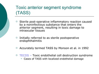
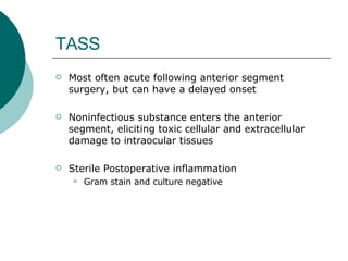
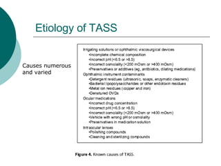
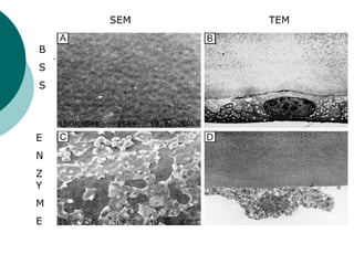
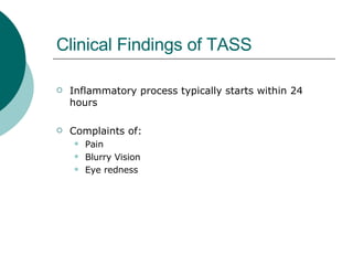
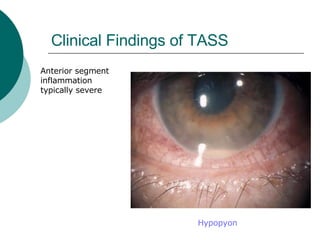
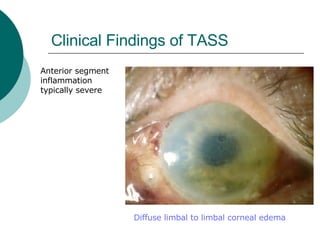
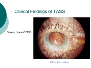
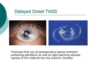
Journal Club June 14, 2006 Terry J. Alexandrou, MD Department of Ophthalmology and Visual Science University of Chicago
Toxic Anterior Segment Syndrome Review/Update Mamalis N, Edelhauser HF, Dawson DG, Chew J, LeBoyer RM, Werner L.  J Cataract Refract Surg 2006; 32:324-333. Journal Club Terry J. Alexandrou, MD Department of Ophthalmology and Visual Science University of Chicago
Toxic anterior segment syndrome (TASS) Sterile post-operative inflammatory reaction caused by a noninfectious substance that enters the anterior segment, resulting in toxic damage to intraocular tissues Initially referred to as sterile postoperative endophthalmitis Accurately termed TASS by Monson et al. in 1992 TECDS –  Toxic endothelial cell destruction syndrome Cases of TASS with localized endothelial damage
TASS Most often acute following anterior segment surgery, but can have a delayed onset Noninfectious substance enters the anterior segment, eliciting toxic cellular and extracellular damage to intraocular tissues Sterile Postoperative inflammation Gram stain and culture negative
Histopathology Hallmark is toxic anterior segment damage Cellular necrosis Apoptosis Extracellular damage Results in a severe acute inflammatory immune response
Etiology of TASS Causes numerous and varied
Preservatives Corneal endothelium extremely sensitive to preservatives Any medication injected into the eye must be preservative free
Preservatives  Inadvertently injected into the eye Liu et al. describe cases of TASS after intraocular use of Eye Stream (preserved with benzalkonium chloride (BAK)) All patients demonstrated generalized corneal edema Most eyes ended up with a visual acuity of CF secondary to persistent corneal edema No treatment other than corneal transplant beneficial in these patients
Preservatives  Inadvertently injected into the eye Elfeftheriadis et al. reported similar cases with intraocular use of benzalkonium (BAK) in cataract surgery patients Significant corneal edema and endothelial damage
Intraocular Anesthetics Intracameral use of preservatve-free bupivacaine hydrochloride 0.5% and preservative-free lidocaine hydrochloride 2.0% have caused signficant corneal thickening and opacification post-op Intracameral use of preservative-free lidocaine 1.0% appears safe for cataract surgery assuming it is immediately followed by phaco, in which the most of the lidocaine is washed out
Sterilization/Detergents Any substance used in cleaning or sterilizing ophthalmic instruments may cause TASS Detergents may accumulate as residues on reusable instruments Enzymes and other active ingredients only deactivated at temp >140°C  (most autoclaves reach 120 to 130°C) Must flush instruments with sterile deionized water
Sterilization/Detergents Parikh et al. report a dose related increase in corneal thickness from endothelial cell damage due to enzymatic detergents (in vitro data) Detergent residues may cause more localized toxicity (TECDS)
B S S E N Z Y M E SEM TEM
Sterilization/Detergents TASS may also be secondary to endotoxin contamination during sterilization Autoclaves may harbor gram negative bacteria Although bacteria may be destroyed during autoclaving, heat stable lipopolysaccharide endotoxin from gram negative cell walls may remain attached to instruments Can only be removed with rinsing and wiping with alcohol or acetone
Sterilization/Detergents Oxidized metal deposits may form on reusable tubing and metal hubs of cannulas when gas sterilization is used
Antibiotic Agents Gentamycin and Vancomycin -possible toxicity Cephalosporins -Kraman et al. and Montan et al. both demonstrated no signs of local toxicity Prophylactic intracameral antibiotic use not currently routinely recommended for endophthalmitis prophylaxis after cataract surgery in most countries
Water 2002 TASS outbreak caused by sulfate water impurities in the autoclave
Clinical Findings of TASS Inflammatory process typically starts within 24 hours Complaints of: Pain Blurry Vision Eye redness
Clinical Findings of TASS Anterior segment inflammation typically severe Hypopyon
Clinical Findings of TASS Anterior segment inflammation typically severe Diffuse limbal to limbal corneal edema
Clinical Findings of TASS Severe cases of TASS Fibrin Formation
Clinical Findings of TASS May result in TM damage - Secondary glaucoma  Fibrin Formation
Differentiating TASS from Infectious Bacterial Endophthalmitis Difficult to differentiate, however few helpful diffferences: TASS typically within 24 hours (compared to 4-7 days) TASS almost always limited to anterior segment TASS commonly presents with diffuse corneal edema TASS improves with topical and/or oral steroids None of these signs are specific enough to diagnose TASS or rule out infectious endophthalmitis
Differentiating TASS from Infectious Bacterial Endophthalmitis Severe anterior segment inflammation with hypopyon or fibrin formation seen in both Other signs of infection are often present in infectious endophthalmitis Lid swelling, chemosis, injection, discharge
Differentiating TASS from Infectious Bacterial Endophthalmitis TASS gram stain and culture negative, however a certain percentage of patients with infectious bacterial endophthalmitis are gram stain and culture negative
Delayed Onset TASS Jehan et al. reported 10 cases of delayed onset inflamattion following cataract surgery Onset 1 to 21 days post-op All AC taps gram stain and culture negative Patients improved with topical antiinflammatory meds Theorized that a residual polishing compound on the IOL was the cause
Delayed Onset TASS Another potential source is ophthalmic ointment Werner et al described an outbreak of delayed onset TASS
Delayed Onset TASS Small globules formed in the anterior chamber
Delayed Onset TASS Oily material coating the anterior surface of the IOL
Delayed Onset TASS Theorized that use of postoperative topical ointment containing petroleum as well as tight patching allowed ingress of the material into the anterior chamber
Treatment
Treatment PREVENTION
Treatment Once the toxic agent enters the eye and causes damage, treatment will do little other than suppress the secondary immune inflammatory response
Treatment Once infectious etiology has been ruled out: Intense topical steroid drops – q1 to q2 hours Must follow closely during the first few days to ensure the inflammatory reaction is stabilizing, and not worsening
Treatment Monitor IOP -initially low - after few days IOP can rise dramatically as aqueous humor production resumes, and TM has been damaged secondary to the toxic insult
Clinical Course Depends on: Type and amount of substance introduced into the eye Duration of exposure How soon treatment was initiated Ranges from clearing within days to weeks for mild cases, to persistent damage (nonclearing corneal edema, significant TM damage and uncontrolled IOP) in severe cases
Final thoughts Prevention is the key Disposable instruments If reusable, then should be rinsed with sterile, deionized water at the end of the sterilization process Vigilance when using any agent in the eye

Toxic Anterior Segment Syndrome

  • 1.
    Journal Club June14, 2006 Terry J. Alexandrou, MD Department of Ophthalmology and Visual Science University of Chicago
  • 2.
    Toxic Anterior SegmentSyndrome Review/Update Mamalis N, Edelhauser HF, Dawson DG, Chew J, LeBoyer RM, Werner L. J Cataract Refract Surg 2006; 32:324-333. Journal Club Terry J. Alexandrou, MD Department of Ophthalmology and Visual Science University of Chicago
  • 3.
    Toxic anterior segmentsyndrome (TASS) Sterile post-operative inflammatory reaction caused by a noninfectious substance that enters the anterior segment, resulting in toxic damage to intraocular tissues Initially referred to as sterile postoperative endophthalmitis Accurately termed TASS by Monson et al. in 1992 TECDS – Toxic endothelial cell destruction syndrome Cases of TASS with localized endothelial damage
  • 4.
    TASS Most oftenacute following anterior segment surgery, but can have a delayed onset Noninfectious substance enters the anterior segment, eliciting toxic cellular and extracellular damage to intraocular tissues Sterile Postoperative inflammation Gram stain and culture negative
  • 5.
    Histopathology Hallmark istoxic anterior segment damage Cellular necrosis Apoptosis Extracellular damage Results in a severe acute inflammatory immune response
  • 6.
    Etiology of TASSCauses numerous and varied
  • 7.
    Preservatives Corneal endotheliumextremely sensitive to preservatives Any medication injected into the eye must be preservative free
  • 8.
    Preservatives Inadvertentlyinjected into the eye Liu et al. describe cases of TASS after intraocular use of Eye Stream (preserved with benzalkonium chloride (BAK)) All patients demonstrated generalized corneal edema Most eyes ended up with a visual acuity of CF secondary to persistent corneal edema No treatment other than corneal transplant beneficial in these patients
  • 9.
    Preservatives Inadvertentlyinjected into the eye Elfeftheriadis et al. reported similar cases with intraocular use of benzalkonium (BAK) in cataract surgery patients Significant corneal edema and endothelial damage
  • 10.
    Intraocular Anesthetics Intracameraluse of preservatve-free bupivacaine hydrochloride 0.5% and preservative-free lidocaine hydrochloride 2.0% have caused signficant corneal thickening and opacification post-op Intracameral use of preservative-free lidocaine 1.0% appears safe for cataract surgery assuming it is immediately followed by phaco, in which the most of the lidocaine is washed out
  • 11.
    Sterilization/Detergents Any substanceused in cleaning or sterilizing ophthalmic instruments may cause TASS Detergents may accumulate as residues on reusable instruments Enzymes and other active ingredients only deactivated at temp >140°C (most autoclaves reach 120 to 130°C) Must flush instruments with sterile deionized water
  • 12.
    Sterilization/Detergents Parikh etal. report a dose related increase in corneal thickness from endothelial cell damage due to enzymatic detergents (in vitro data) Detergent residues may cause more localized toxicity (TECDS)
  • 13.
    B S SE N Z Y M E SEM TEM
  • 14.
    Sterilization/Detergents TASS mayalso be secondary to endotoxin contamination during sterilization Autoclaves may harbor gram negative bacteria Although bacteria may be destroyed during autoclaving, heat stable lipopolysaccharide endotoxin from gram negative cell walls may remain attached to instruments Can only be removed with rinsing and wiping with alcohol or acetone
  • 15.
    Sterilization/Detergents Oxidized metaldeposits may form on reusable tubing and metal hubs of cannulas when gas sterilization is used
  • 16.
    Antibiotic Agents Gentamycinand Vancomycin -possible toxicity Cephalosporins -Kraman et al. and Montan et al. both demonstrated no signs of local toxicity Prophylactic intracameral antibiotic use not currently routinely recommended for endophthalmitis prophylaxis after cataract surgery in most countries
  • 17.
    Water 2002 TASSoutbreak caused by sulfate water impurities in the autoclave
  • 18.
    Clinical Findings ofTASS Inflammatory process typically starts within 24 hours Complaints of: Pain Blurry Vision Eye redness
  • 19.
    Clinical Findings ofTASS Anterior segment inflammation typically severe Hypopyon
  • 20.
    Clinical Findings ofTASS Anterior segment inflammation typically severe Diffuse limbal to limbal corneal edema
  • 21.
    Clinical Findings ofTASS Severe cases of TASS Fibrin Formation
  • 22.
    Clinical Findings ofTASS May result in TM damage - Secondary glaucoma Fibrin Formation
  • 23.
    Differentiating TASS fromInfectious Bacterial Endophthalmitis Difficult to differentiate, however few helpful diffferences: TASS typically within 24 hours (compared to 4-7 days) TASS almost always limited to anterior segment TASS commonly presents with diffuse corneal edema TASS improves with topical and/or oral steroids None of these signs are specific enough to diagnose TASS or rule out infectious endophthalmitis
  • 24.
    Differentiating TASS fromInfectious Bacterial Endophthalmitis Severe anterior segment inflammation with hypopyon or fibrin formation seen in both Other signs of infection are often present in infectious endophthalmitis Lid swelling, chemosis, injection, discharge
  • 25.
    Differentiating TASS fromInfectious Bacterial Endophthalmitis TASS gram stain and culture negative, however a certain percentage of patients with infectious bacterial endophthalmitis are gram stain and culture negative
  • 26.
    Delayed Onset TASSJehan et al. reported 10 cases of delayed onset inflamattion following cataract surgery Onset 1 to 21 days post-op All AC taps gram stain and culture negative Patients improved with topical antiinflammatory meds Theorized that a residual polishing compound on the IOL was the cause
  • 27.
    Delayed Onset TASSAnother potential source is ophthalmic ointment Werner et al described an outbreak of delayed onset TASS
  • 28.
    Delayed Onset TASSSmall globules formed in the anterior chamber
  • 29.
    Delayed Onset TASSOily material coating the anterior surface of the IOL
  • 30.
    Delayed Onset TASSTheorized that use of postoperative topical ointment containing petroleum as well as tight patching allowed ingress of the material into the anterior chamber
  • 31.
  • 32.
  • 33.
    Treatment Once thetoxic agent enters the eye and causes damage, treatment will do little other than suppress the secondary immune inflammatory response
  • 34.
    Treatment Once infectiousetiology has been ruled out: Intense topical steroid drops – q1 to q2 hours Must follow closely during the first few days to ensure the inflammatory reaction is stabilizing, and not worsening
  • 35.
    Treatment Monitor IOP-initially low - after few days IOP can rise dramatically as aqueous humor production resumes, and TM has been damaged secondary to the toxic insult
  • 36.
    Clinical Course Dependson: Type and amount of substance introduced into the eye Duration of exposure How soon treatment was initiated Ranges from clearing within days to weeks for mild cases, to persistent damage (nonclearing corneal edema, significant TM damage and uncontrolled IOP) in severe cases
  • 37.
    Final thoughts Preventionis the key Disposable instruments If reusable, then should be rinsed with sterile, deionized water at the end of the sterilization process Vigilance when using any agent in the eye