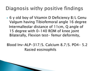
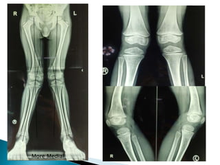
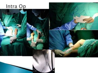
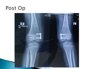
Guide: Dr. Jaswinder Sir
Presenter: Shubhanshu Ranjan Singh
 6 y old boy of Vitamin D Deficiency B/L Genu
Valgum having Tibiofemoral angle 16 degree
Intermalleolar distance of 11cm, Q angle of
15 degree with 0-140 ROM of knee joint
Bilaterally, Flexion test- femur deformity,
Blood Inv-ALP-317/S. Calcium 8.7/S. PO4- 5.2
Rasied eosinophils
More Medial
 Varun Kumar 6y/ M from Patna
 Gait- Adduction, patella centralised knee touching
knee
 C/O abonormal walking/ future concern
 Deformity first notice by her mother in 2019 and
progessive in nature for which patient got treatment
that time locally for 2 years and deformity get fixed.
 History of vitamin D deficiency (no previous record)
for which he took one weekly sachet and calcium
syrup
 No history of trauma/ infection/swelling/ burning
and pain in micturation/depression in between
thorax and abdomen
 Birth history- mother didn’t took iron and
calcium tablet during pregnancy, rest
uneventful and normal
 Developmental milestones attend
appropiately at his age group
 Family History- Mother h/o hypothyroidism
since 9 years on medication
 2019 diagnosed with vitamin D deficiency
for which she took tab calcium and vit d
sachet
 At age of 1 year left mid upper limb burn scar
mark present
6 y/ M, FTNVD, C/o abnormal walking and
future concern of progression Mother noticed
deformity @ age 3 years age
Deformity progressive
No h/o trauma, infection or any swelling
F/H Mother hypothyroidsm since 9 years
 Height 115
 Weight 25
 General Exam- good nutrition, no skin lesion,
no polydactyly, no short stature spinal deformity
(for Rickets/ skeletal dysplasia/multiple hereditary
exostosis)
Gait- Adduction, Knee touch each other wide space ankle
(circumduction gait)
B/L Genu valgus no patellofemoral instability
ROM normal
 Valgum Alignment – Inward angulation of
extremity in coronal plane (Distal deviate
lateral wrt proximal)
 Valgum called be deformity
depends on
 Age
 Degree of Valgus – 7 degree normal upto 7
year
 Physiological- knock knee, apparent
(ligamentous laxity, rotational deformity, fat
thigh)
 Pathological- Idiopathic, trauma (malunion,
physeal arrest, metaphyseal tibial fracture, )
tumor, tumor like conditions (fibrous dysplesis,
enchondromatosis, MHE), Infection,
 Metabolic (Rickets, renal osteodystrophy)
 Inflammatory- RA
 Syndrome – Ellis Van Creveld syndrome
 Polio, Cerebral palsy
 Skeletal dysplasia- focal fibrocartilaginous
dysplasia, Osteogenic imperfecta
 Assymetric Involvement
 Short stature
 Limb length discrepancy
 Supine knee flexed touching each other both
feet medial malleoli approximated
 If defomity resolve than its femur otherwise
in tibia
 Tibio femoral angle- Clinical radiological
 Intermaleolar distance- Bet medial malleoli
<5/5-10/>10
standing erect wirth hips knee fully extended
and neutral rotation with patella both knee
touching each other
Marking ASIS, centre of patella centre of ankle,
malleoli
 Goniometer ?
 Rotational Profile Assesment
Foot Progression angle
Thigh foot angle
Medial hip rotation 70
Lateral hip rotation 30
Sole of foot (straight)
?femoral anteversion/ tibial torsion
 FPA-line of progession vs foot 10-15
external N
 Thigh foot axis- 20 IR +, ER –
 Apex @ Patella b/w ligamentum patellae and
extension of quadriceps resultant distally
 Patellar instability or not
 Measure – Standing/ Supine 30 flexed(patella
fixed into femoral condyle)(8-10M/15F)
 For ligamentous laxity
 Center of rotation of angulation- Apex of
deformity where osteotomy done in
orthoscanogram drawn.
 Imaging – Full length Xray of bilateral Lower
limbs, xray of wrist, knee
 Lab- Ca/P/PO4/S. VitD/ALP,CBC,ESR, PTYH U.
Ca/P
 Treatment-
Observation – Physiological
Acute Correction-Corrective Osteotomy
Gradual Correction-Guided growth
modulation/ six axis correction device or
ilizarov
 Mechanical Axis deviation ???? Stevans Zones
 Xray Wrist – Metapyseal splaying
/cupping/fraying/osteopenia/ Epiphyseal
thininng
 X-ray Ankle
 In our patient growth potential remaining
So,Guided growth modulation
Figure of 8 plate 16mm applied B/L
Now this is standard treatment for skeletally
immature
Removal?
 Iliotibial band contracture
Hip- abduction/ apparent lenthening/ true
shortening
ANKLE/FOOT- talipes equino varus
Leg- external tibial torsion
Knee- flexion contracture/ genu valgum
Hip- flexion/abduction/ER
Pelvic- pelvic obliquity
Spine- lumber lordosis/ scoliosis

Genu Valgum

  • 1.
    Guide: Dr. JaswinderSir Presenter: Shubhanshu Ranjan Singh
  • 2.
     6 yold boy of Vitamin D Deficiency B/L Genu Valgum having Tibiofemoral angle 16 degree Intermalleolar distance of 11cm, Q angle of 15 degree with 0-140 ROM of knee joint Bilaterally, Flexion test- femur deformity, Blood Inv-ALP-317/S. Calcium 8.7/S. PO4- 5.2 Rasied eosinophils
  • 3.
  • 6.
     Varun Kumar6y/ M from Patna  Gait- Adduction, patella centralised knee touching knee  C/O abonormal walking/ future concern  Deformity first notice by her mother in 2019 and progessive in nature for which patient got treatment that time locally for 2 years and deformity get fixed.  History of vitamin D deficiency (no previous record) for which he took one weekly sachet and calcium syrup  No history of trauma/ infection/swelling/ burning and pain in micturation/depression in between thorax and abdomen
  • 7.
     Birth history-mother didn’t took iron and calcium tablet during pregnancy, rest uneventful and normal  Developmental milestones attend appropiately at his age group  Family History- Mother h/o hypothyroidism since 9 years on medication
  • 8.
     2019 diagnosedwith vitamin D deficiency for which she took tab calcium and vit d sachet  At age of 1 year left mid upper limb burn scar mark present
  • 9.
    6 y/ M,FTNVD, C/o abnormal walking and future concern of progression Mother noticed deformity @ age 3 years age Deformity progressive No h/o trauma, infection or any swelling F/H Mother hypothyroidsm since 9 years
  • 10.
     Height 115 Weight 25  General Exam- good nutrition, no skin lesion, no polydactyly, no short stature spinal deformity (for Rickets/ skeletal dysplasia/multiple hereditary exostosis) Gait- Adduction, Knee touch each other wide space ankle (circumduction gait) B/L Genu valgus no patellofemoral instability ROM normal
  • 11.
     Valgum Alignment– Inward angulation of extremity in coronal plane (Distal deviate lateral wrt proximal)  Valgum called be deformity depends on  Age  Degree of Valgus – 7 degree normal upto 7 year
  • 12.
     Physiological- knockknee, apparent (ligamentous laxity, rotational deformity, fat thigh)  Pathological- Idiopathic, trauma (malunion, physeal arrest, metaphyseal tibial fracture, ) tumor, tumor like conditions (fibrous dysplesis, enchondromatosis, MHE), Infection,  Metabolic (Rickets, renal osteodystrophy)  Inflammatory- RA  Syndrome – Ellis Van Creveld syndrome  Polio, Cerebral palsy  Skeletal dysplasia- focal fibrocartilaginous dysplasia, Osteogenic imperfecta
  • 13.
     Assymetric Involvement Short stature  Limb length discrepancy
  • 14.
     Supine kneeflexed touching each other both feet medial malleoli approximated  If defomity resolve than its femur otherwise in tibia
  • 15.
     Tibio femoralangle- Clinical radiological  Intermaleolar distance- Bet medial malleoli <5/5-10/>10 standing erect wirth hips knee fully extended and neutral rotation with patella both knee touching each other Marking ASIS, centre of patella centre of ankle, malleoli
  • 16.
     Goniometer ? Rotational Profile Assesment Foot Progression angle Thigh foot angle Medial hip rotation 70 Lateral hip rotation 30 Sole of foot (straight) ?femoral anteversion/ tibial torsion
  • 17.
     FPA-line ofprogession vs foot 10-15 external N  Thigh foot axis- 20 IR +, ER –
  • 18.
     Apex @Patella b/w ligamentum patellae and extension of quadriceps resultant distally  Patellar instability or not  Measure – Standing/ Supine 30 flexed(patella fixed into femoral condyle)(8-10M/15F)
  • 19.
  • 20.
     Center ofrotation of angulation- Apex of deformity where osteotomy done in orthoscanogram drawn.
  • 23.
     Imaging –Full length Xray of bilateral Lower limbs, xray of wrist, knee  Lab- Ca/P/PO4/S. VitD/ALP,CBC,ESR, PTYH U. Ca/P  Treatment- Observation – Physiological Acute Correction-Corrective Osteotomy Gradual Correction-Guided growth modulation/ six axis correction device or ilizarov
  • 24.
     Mechanical Axisdeviation ???? Stevans Zones  Xray Wrist – Metapyseal splaying /cupping/fraying/osteopenia/ Epiphyseal thininng  X-ray Ankle
  • 25.
     In ourpatient growth potential remaining So,Guided growth modulation Figure of 8 plate 16mm applied B/L Now this is standard treatment for skeletally immature Removal?
  • 26.
     Iliotibial bandcontracture Hip- abduction/ apparent lenthening/ true shortening ANKLE/FOOT- talipes equino varus Leg- external tibial torsion Knee- flexion contracture/ genu valgum Hip- flexion/abduction/ER Pelvic- pelvic obliquity Spine- lumber lordosis/ scoliosis