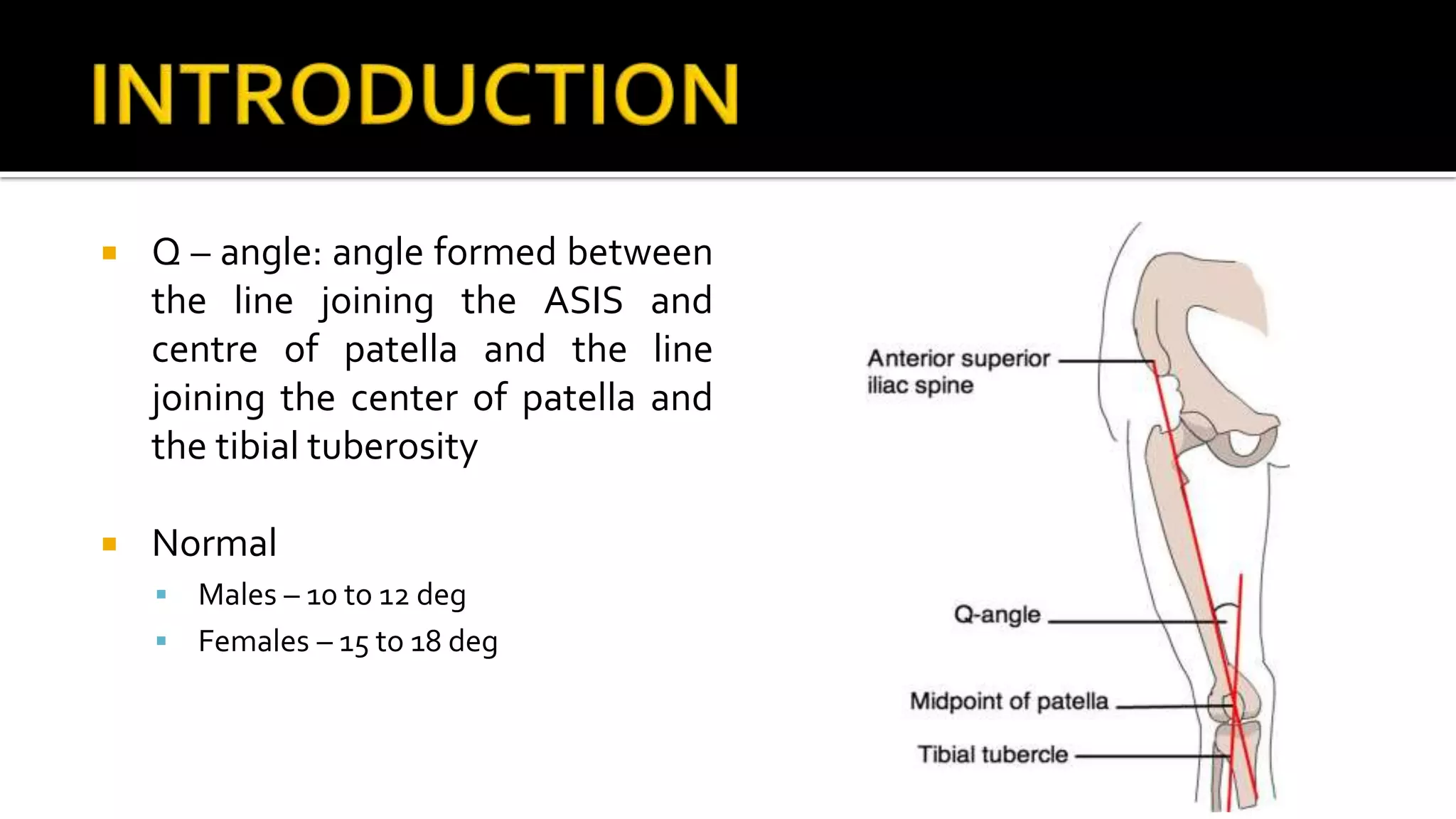
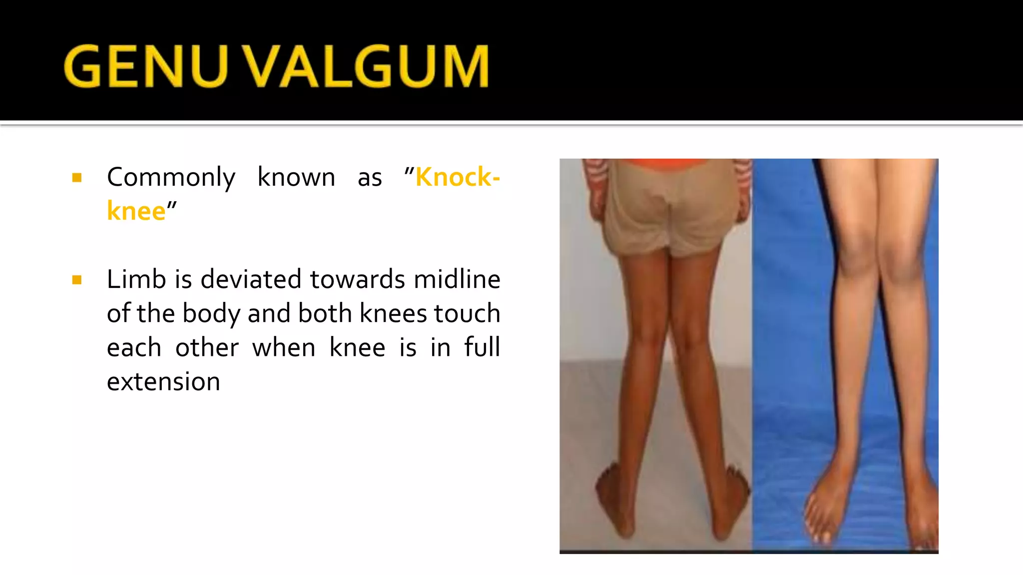
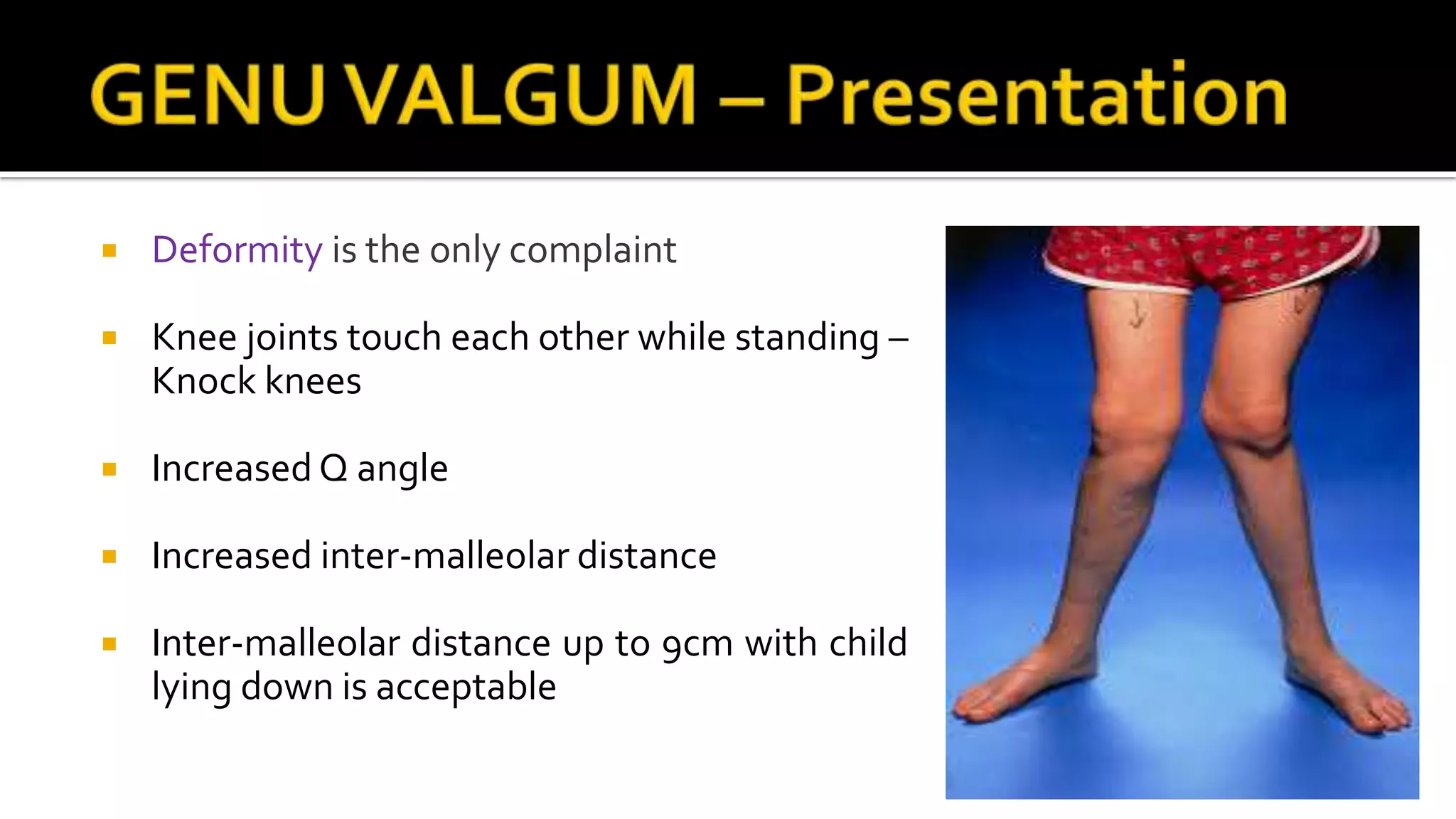
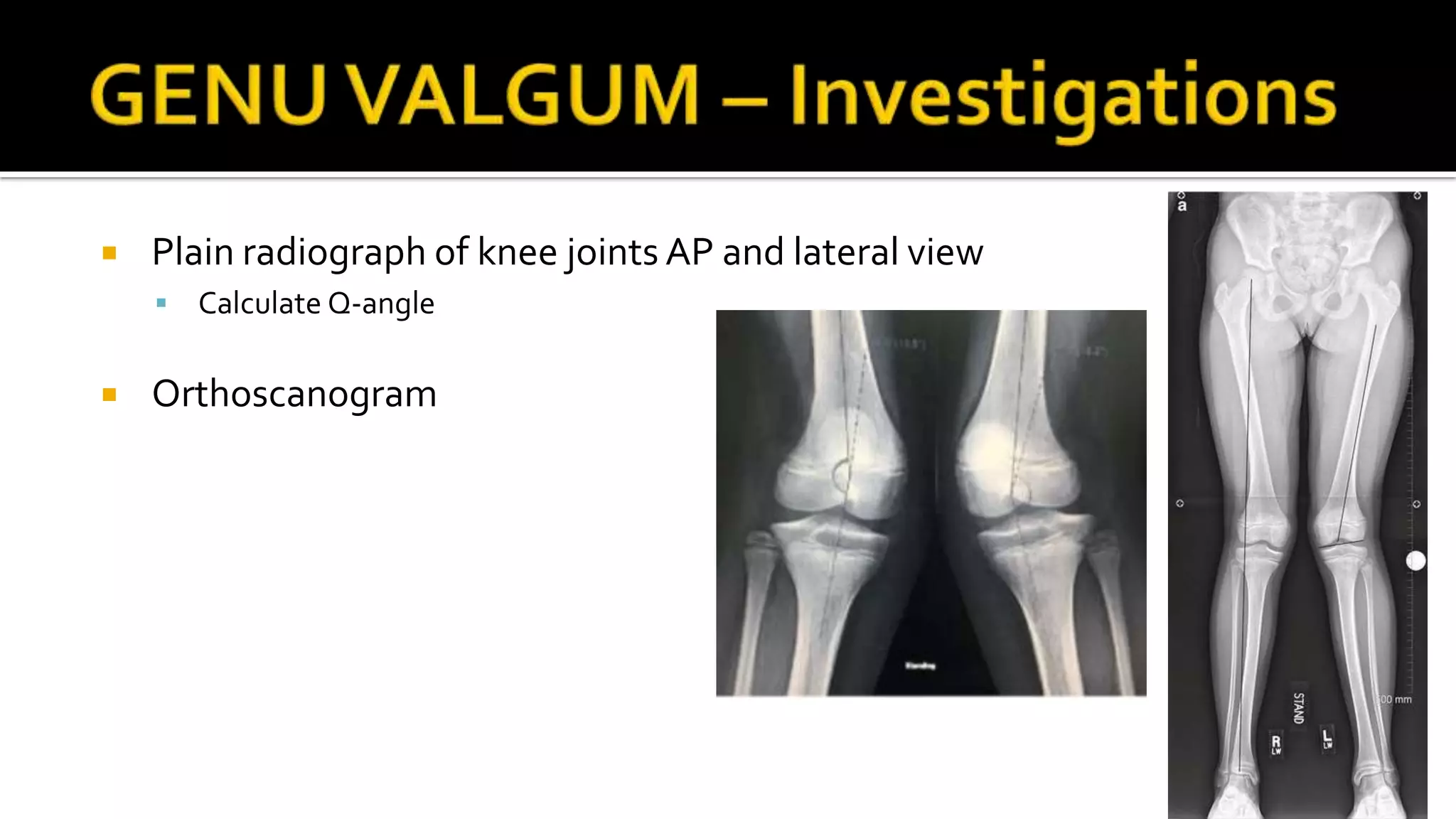
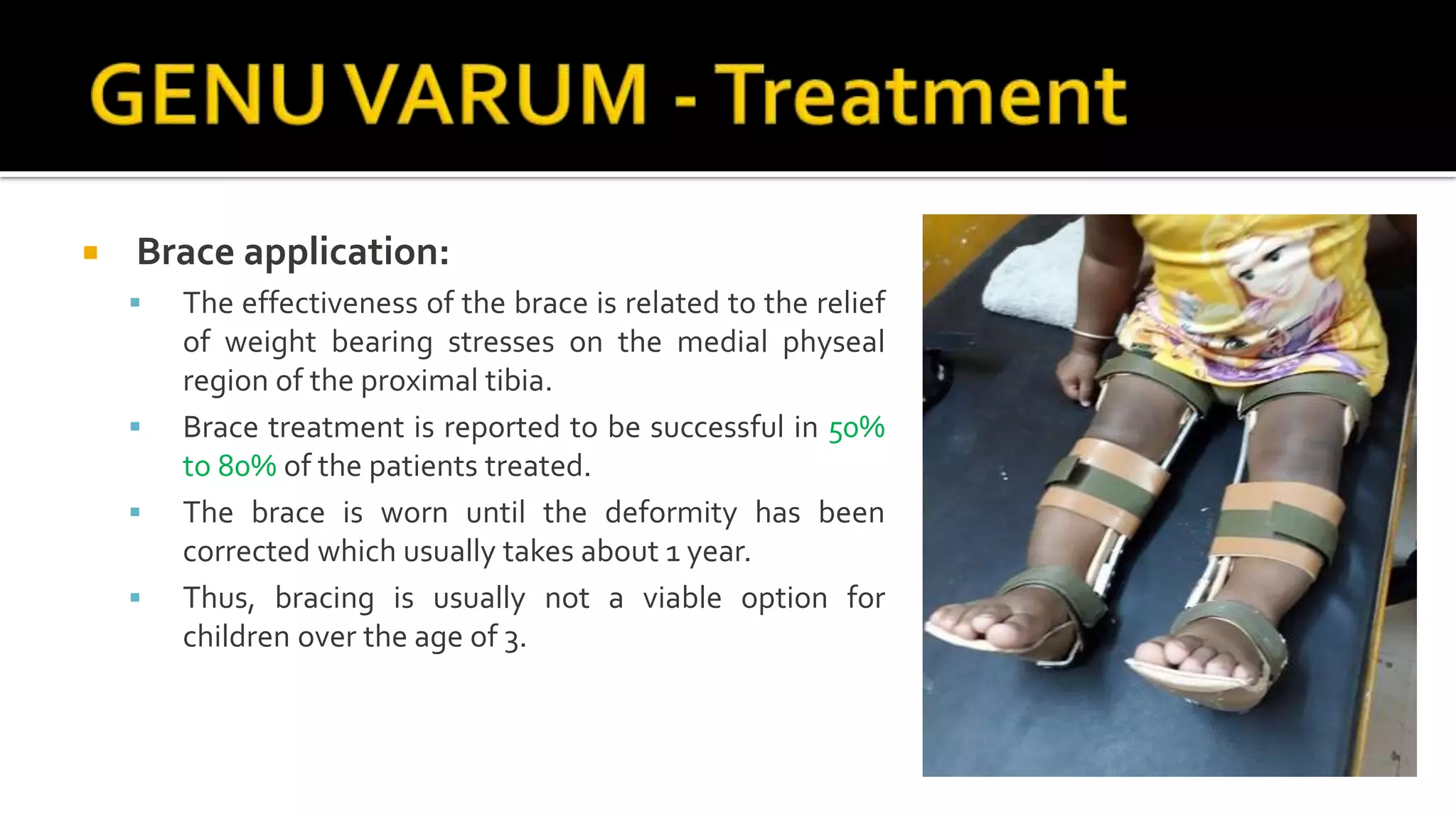
DR. BIPUL BORTHAKUR
Professor, Dept of Orthopaedics, SMCH
 Q – angle: angle formed between
the line joining the ASIS and
centre of patella and the line
joining the center of patella and
the tibial tuberosity
 Normal
 Males – 10 to 12 deg
 Females – 15 to 18 deg
 Commonly known as ”Knock-
knee”
 Limb is deviated towards midline
of the body and both knees touch
each other when knee is in full
extension
 Genu valgum is a normal physiological process in children
 Up to 4 years of age.
 Therefore it is critical to differentiate between a physiological and
pathological process
 Distal femur is the most common location of primary pathologic genu
valgum but can arise from tibia
 Bilateral GenuValgum
 Physiological
 Renal osteodystrophy (Renal rickets)
 Skeletal dysplasia
 Morquio syndrome
 Spondyloepiphyseal dysplasia
 Chondroctodermal dysplasia
 UnilateralGenuValgum
 Physeal injury –Trauma, Infection and
Vascular insult
 Proximal metaphyseal tibia fracture
 Secondary arthritis
 Benign tumour
 Fibrous dysplasia
 Osteochondromas
 Ollier’s disease
 Ligamentous injury
 Paget’s disease
 Deformity is the only complaint
 Knee joints touch each other while standing –
Knock knees
 Increased Q angle
 Increased inter-malleolar distance
 Inter-malleolar distance up to 9cm with child
lying down is acceptable
 Plain radiograph of knee joints AP and lateral view
 Calculate Q-angle
 Orthoscanogram
 Non – operative
 Observation of deformity and parent counselling
 Consider as first line of management for physiological Genu valgum in children of < 6
years
 Treatment of primary cause
 Operative treatment
 Hemiepiphysiodesis or physeal tethering
 Bone stapling
 Done before completing growth
 Put into the bone around growth plate
 Corrective Osteotomy
 Done at the apex of the deformity at femur and /or tibia
 10 years old with Inter-malleolar distance > 10cm
 Can be a lateral open wedge or medial close wedge
osteotomy
 TREATMENT FACTS OF GENUVALGUM
 < 4 yrs— No treatment. Only observation.
 4-10 yrs—Heel raise, knock-knee brace.
 10-14 yrs—Epiphyseal stapling.
 14-16 yrs—wait until skeletal maturity, as it is too late for stapling and too early for
osteotomy, as it may recur
 > 16 yrs— Osteotomy.
 Angular deformity of proximal tibia in which the child appear
“bowlegged”.
 Maximum varus is present at 6-12 months of age
 Bowlegs after 2 years of age considered abnormal
 Physiological
 Pathological
 Blount’s disease
 Hypophosphatemic or nutritional rickets
 Post-traumatic
 Post-infectious
 Congenital deformities
 Focal fibrocartilaginous dysplasia
 Metaphysealchondrodysplasia
 Fibrous dysplasia
 Osteogenesis imperfecta
 Renal osteodystrophy
 CAUSES IN ADULTS
 May be sequel of childhood deformity and if so usually cause no problems. However, if
the deformity is associated with joint instability, this can lead to osteoarthritis of the
medial compartment.
 Other causes include:
 Fracture of the lower part of the femur or the upper part of the tibia with malunion.
 Osteoarthritis
 Rarefying diseases of the bone such as osteomalacia
 Other bone-softening diseases such as Paget’s disease.
 HISTORY:
 The stature and nutritional status of the child
 Developmental milestones
 Other nutritional or medical problems
 History of trauma or infections
 EXAMINATION:
 Short stature :
 Suggests the possibility of vitamin D refractory (hypo-phosphatemic) rickets or bone
dysplasia (achondroplasia or metaphyseal dysplasia).
 The inter-condylar distance:
 Performed with the medial malleoli in contact
 Done in stance and supine.
 Greater than 6 cm is abnormal.
 Ruling out the deformity of the feet: e.g. metatarsus varus or valgus
which may represent torsional deformity of the limb
 Plain radiograph of knee joints AP and lateral views
 Orthoscanogram
 Complete bone metabolic profile
 Serum Ca, Ph, ALP
 SerumVitamin D level
 Complete blood count
 RFT, urine Ca level
 Non-operative
 In the vast majority of cases, genu varum will correct with growth.
 In physiological genu varum, education and assurance of the parents is
important and just follow its natural course by reassessing the child in 6
months.
 Treatment of underlying causes
 Brace application
 Brace application:
 The effectiveness of the brace is related to the relief
of weight bearing stresses on the medial physeal
region of the proximal tibia.
 Brace treatment is reported to be successful in 50%
to 80% of the patients treated.
 The brace is worn until the deformity has been
corrected which usually takes about 1 year.
 Thus, bracing is usually not a viable option for
children over the age of 3.
 Operative:
 Tibial osteotomy – The shinbone is cut just below
the knee and reshaped to correct the alignment
 Guided growth - This surgery of the growth plate
stops the growth on the healthy side of the
shinbone which gives the abnormal side a chance
to catch up, straightening the leg with the child’s
natural growth
Genu varus and valgus

Genu varus and valgus

  • 1.
    DR. BIPUL BORTHAKUR Professor,Dept of Orthopaedics, SMCH
  • 2.
     Q –angle: angle formed between the line joining the ASIS and centre of patella and the line joining the center of patella and the tibial tuberosity  Normal  Males – 10 to 12 deg  Females – 15 to 18 deg
  • 3.
     Commonly knownas ”Knock- knee”  Limb is deviated towards midline of the body and both knees touch each other when knee is in full extension
  • 4.
     Genu valgumis a normal physiological process in children  Up to 4 years of age.  Therefore it is critical to differentiate between a physiological and pathological process  Distal femur is the most common location of primary pathologic genu valgum but can arise from tibia
  • 5.
     Bilateral GenuValgum Physiological  Renal osteodystrophy (Renal rickets)  Skeletal dysplasia  Morquio syndrome  Spondyloepiphyseal dysplasia  Chondroctodermal dysplasia  UnilateralGenuValgum  Physeal injury –Trauma, Infection and Vascular insult  Proximal metaphyseal tibia fracture  Secondary arthritis  Benign tumour  Fibrous dysplasia  Osteochondromas  Ollier’s disease  Ligamentous injury  Paget’s disease
  • 6.
     Deformity isthe only complaint  Knee joints touch each other while standing – Knock knees  Increased Q angle  Increased inter-malleolar distance  Inter-malleolar distance up to 9cm with child lying down is acceptable
  • 7.
     Plain radiographof knee joints AP and lateral view  Calculate Q-angle  Orthoscanogram
  • 8.
     Non –operative  Observation of deformity and parent counselling  Consider as first line of management for physiological Genu valgum in children of < 6 years  Treatment of primary cause
  • 9.
     Operative treatment Hemiepiphysiodesis or physeal tethering  Bone stapling  Done before completing growth  Put into the bone around growth plate
  • 10.
     Corrective Osteotomy Done at the apex of the deformity at femur and /or tibia  10 years old with Inter-malleolar distance > 10cm  Can be a lateral open wedge or medial close wedge osteotomy
  • 11.
     TREATMENT FACTSOF GENUVALGUM  < 4 yrs— No treatment. Only observation.  4-10 yrs—Heel raise, knock-knee brace.  10-14 yrs—Epiphyseal stapling.  14-16 yrs—wait until skeletal maturity, as it is too late for stapling and too early for osteotomy, as it may recur  > 16 yrs— Osteotomy.
  • 12.
     Angular deformityof proximal tibia in which the child appear “bowlegged”.  Maximum varus is present at 6-12 months of age  Bowlegs after 2 years of age considered abnormal
  • 13.
     Physiological  Pathological Blount’s disease  Hypophosphatemic or nutritional rickets  Post-traumatic  Post-infectious  Congenital deformities  Focal fibrocartilaginous dysplasia  Metaphysealchondrodysplasia  Fibrous dysplasia  Osteogenesis imperfecta  Renal osteodystrophy
  • 14.
     CAUSES INADULTS  May be sequel of childhood deformity and if so usually cause no problems. However, if the deformity is associated with joint instability, this can lead to osteoarthritis of the medial compartment.  Other causes include:  Fracture of the lower part of the femur or the upper part of the tibia with malunion.  Osteoarthritis  Rarefying diseases of the bone such as osteomalacia  Other bone-softening diseases such as Paget’s disease.
  • 15.
     HISTORY:  Thestature and nutritional status of the child  Developmental milestones  Other nutritional or medical problems  History of trauma or infections
  • 16.
     EXAMINATION:  Shortstature :  Suggests the possibility of vitamin D refractory (hypo-phosphatemic) rickets or bone dysplasia (achondroplasia or metaphyseal dysplasia).  The inter-condylar distance:  Performed with the medial malleoli in contact  Done in stance and supine.  Greater than 6 cm is abnormal.  Ruling out the deformity of the feet: e.g. metatarsus varus or valgus which may represent torsional deformity of the limb
  • 17.
     Plain radiographof knee joints AP and lateral views  Orthoscanogram  Complete bone metabolic profile  Serum Ca, Ph, ALP  SerumVitamin D level  Complete blood count  RFT, urine Ca level
  • 18.
     Non-operative  Inthe vast majority of cases, genu varum will correct with growth.  In physiological genu varum, education and assurance of the parents is important and just follow its natural course by reassessing the child in 6 months.  Treatment of underlying causes  Brace application
  • 19.
     Brace application: The effectiveness of the brace is related to the relief of weight bearing stresses on the medial physeal region of the proximal tibia.  Brace treatment is reported to be successful in 50% to 80% of the patients treated.  The brace is worn until the deformity has been corrected which usually takes about 1 year.  Thus, bracing is usually not a viable option for children over the age of 3.
  • 20.
     Operative:  Tibialosteotomy – The shinbone is cut just below the knee and reshaped to correct the alignment  Guided growth - This surgery of the growth plate stops the growth on the healthy side of the shinbone which gives the abnormal side a chance to catch up, straightening the leg with the child’s natural growth