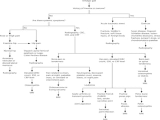
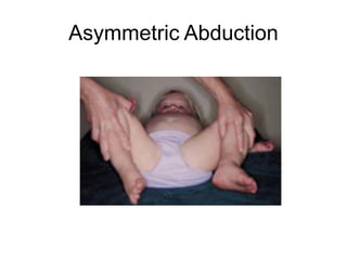
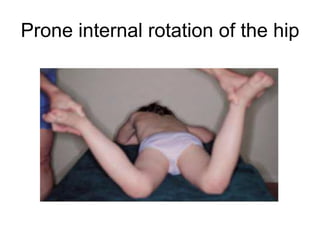
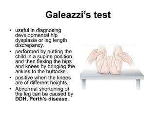
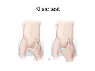
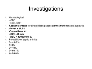
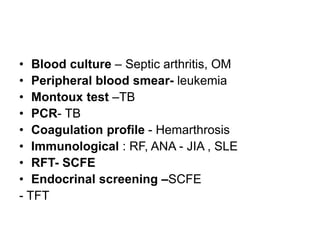
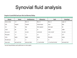
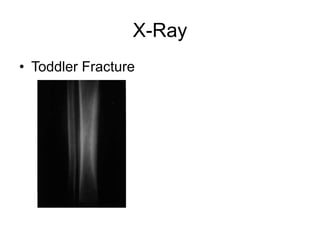
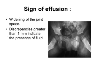
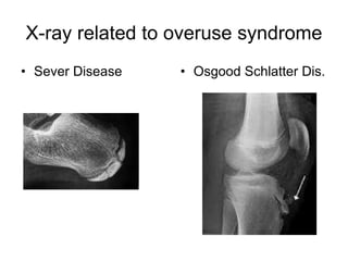
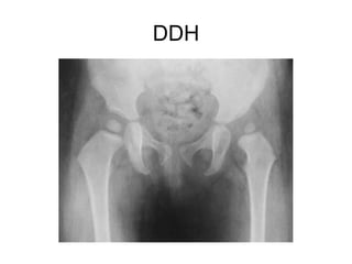
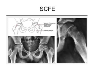
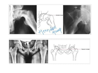
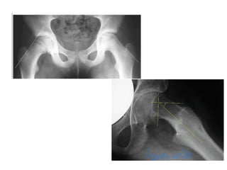
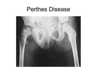
This document provides guidance on evaluating a limping child. It discusses that limp can be caused by pain, weakness or deformity and management depends on the underlying cause. A thorough history, physical exam, and investigations may be needed. The physical exam involves assessing gait, leg length, range of motion, and neurological function. Potential investigations include blood tests, imaging like x-rays, ultrasound and MRI to identify causes such as fractures, infections, tumors or developmental disorders. The goal is to determine the cause and provide appropriate treatment ranging from reassurance to surgery.
2. LIMP
• Limp is defined as, an uneven, jerky gait
caused by pain, weakness or deformity.
• Limp can be caused by both benign and
life threatening conditions
• Management varies from simple
reassurance to major surgery depending
upon the cause.
3. PATHOPHYSIOLOGY
• Three main factors cause a child to limp.
• Pain, weakness, structural or mechanical
abnormalities of the spine, pelvis and
lower extremities.
4. NORMAL GAIT
• A normal gait is composed of symmetrical,
alternating, rhythmical motions involving two
phases: stance and swing.
• The stance phase normally encompasses 60%
of the gait cycle during which the foot is in
contact with the ground.
• And the swing phase 40%, during which the foot
is in the air.
• Mature Gait – by 3 years, by 7 years the gait will
be approximately that of the adult.
14. History
• Sex – Girls – DDH
–Boys – Perthes , SCFE
• Onset
• Painless or Painful
• Acute or Chronic
• History of trauma
15. History
• Fever, night sweats, Anorexia, weight loss (Inf. , Malignancy).
• Recent history of viral illness or streptococcal infection (post
infectious arthritis)
• Recent history of dysentry ( Reiters syndome)
• Recent history of new or increased sports activity
• Recent history of intramuscular injection (can cause muscle
inflammation or sterile abscess)
• History of endocrine dysfunction (may predispose to slipped capital
femoral epiphysis)
• Family history of connective tissue disorder, inflammatory bowel
disease, hemoglobinopathy, bleeding disorder, or neuromuscular
disorder
• Prenatal and birth history
16. Physical Examination
• Standing:
• - back should be examined for scoliosis ,local
tenderness, range of motion.
• -if there is pelvic tilt is present , it can be measured by
placing blocks under the shorter leg until the pelvis in
level (horizontal)
• -trendelenburg test
• Measurement of thigh and calf circumference should
reveal atrophy (more than 1 to 2 cm of difference
between sides) in a patient with any hip or knee
condition that has limited function for more than one to
two months.
17. Physical Examination
• Supine:
• - each joint should be examined separately
• -look for swelling, feel for tenderness, assess the ROM
• -for hip flexion contracture --------Thomas test
• -neurological examination should be performed
• -check for leg length discrepancy , the short leg must be
differentiated from apparent shortening that is caused by
scoliosis or pelvic obliquity or joint contracture.
• Prone:
• -hip rotation
• *-femoral anteversion
20. Galeazzi’s test
• useful in diagnosing
developmental hip
dysplasia or leg length
discrepancy.
• performed by putting the
child in a supine position
and then flexing the hips
and knees by bringing the
ankles to the buttocks .
• positive when the knees
are of different heights.
• Abnormal shortening of
the leg can be caused by
DDH, Perth's disease.
38. Ultrasonography
• Effusion
• -Widening of space between capsule and bone
of > 2mm indicates effusion.
• -Echo-free transient synovitis
• -Positively echogenic septic arthritis
• useful in the detection of early slips by
demonstrating joint effusion and a “step”
between the femoral neck and the epiphysis
created by slipping-SCFE
• Acute osteomyelitis- periosteal thickening ,
subpriosteal thickening
39. CT scan
• deep soft tissue infections of the Para spinal and
retroperitoneal regions
• Three dimensional images of shape of femoral
head and acetabulum in perthes disease
• SCFE- confirm closure of the proximal femoral
physis.
• Provide three dimensional reconstructed CT
images used to assess the severity of residual
deformity of the upper femur,[ especially when
reconstructive osteotomy is being considered.
40. MRI
• Evaluation of the spine (for discitis or spinal
tumors), soft tissue tumors and abscesses in the
Para spinal and retroperitoneal regions,
osteomyelitis of the pelvis and long bones.
• Legg-Calvé-Perthes disease
• Highly specific for detection of AVN
• MRI with gadolinium-contrast arthrography- the
evaluation of the adolescent patient with hip
dysplasia and pain for assessing of the condition
of the labrum and the articular cartilage of the
hip joint
41. Radionuclide scans :
• sensitive means of detecting
alterations in the metabolic rate of
bone and thus a sensitive means
of localizing pathology
• lacks specificity because such
alterations in bone metabolism
can occur in Legg- Calvé-Perthes
disease, osteomyelitis,osteoid
osteoma, and malignant bone
tumors.
• Decrease uptake in AVN
• increased uptake in the capital
femoral physis of an involved hip,
decreased uptake in the presence
of AVN, and increased uptake in
the joint space in the presence of
chondrolysis