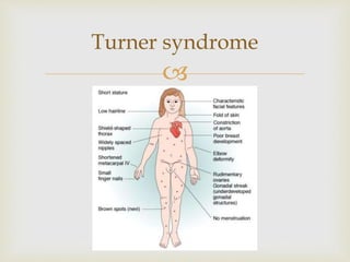
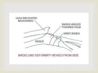
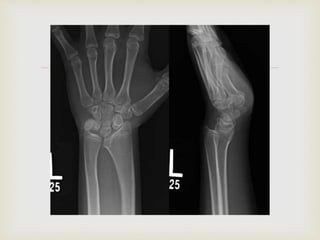
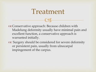
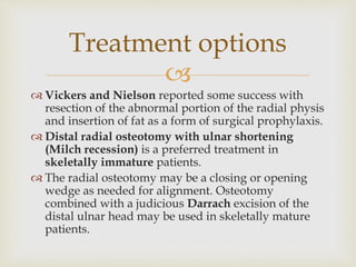
Madelung deformity is an abnormality of the palmar ulnar part of the distal radial physis in which progressive ulnar and volar tilt develops at the distal radial articular surface, with dorsal subluxation of the distal ulna.
3.
First described by Malgaigne and later by Madelung.
An abnormality of the palmar ulnar part of the distal
radial physis in which progressive ulnar and volar
tilt develops at the distal radial articular surface,
with dorsal subluxation of the distal ulna.
Introduction
4.
Congenital , transmitted as AD.
Vickers described an abnormal ligament that tethers
the lunate to the distal radius proximal to the physis.
This ligament is believed to impede the growth of the
ulnopalmar aspect of the distal radius and is
commonly known as the ligament of Vickers.
Mutations in the homeobox gene SHOX.
Madelung-like deformities have occurred after trauma
and also after infection or neoplasm.
Causes
10.
Vender and Watson classified Madelung and
Madelung-like deformities into four groups:
1. Posttraumatic,
2. Dysplastic (dyschondrosteosis diaphyseal aclasis),
3. Genetic (e.g. Turner syndrome)
4. Idiopathic.
Classification
11.
Clinical feature
More commonly bilateral and
affects girls more frequently
than boys.
Volar subluxation of the hand,
with prominence of the distal
ulna and volar and ulnar
angulation of the distal
radius.
The deformity usually
manifests in late childhood or
early adolescence, with
decreased motion and
minimal pain. As growth
occurs, the deformity worsens
in appearance.
12.
The radius is curved, with its convexity dorsal and radial,
and there is a similar angulation of the distal radial
articular surface. The forearm is relatively short.
The distal radial epiphysis is triangular because of the
failure of growth in the ulnar and volar aspects of the
physis; early closure of these aspects of the physis also is
frequent.
The ulna is subluxated dorsally, the ulnar head is
enlarged, and the overall length of the ulna is decreased.
The carpus appears to have subluxated ulnarward and
palmarward into the distal radioulnar joint, which
usually is spread apart.
The carpus appears wedge shaped, with its apex proximal
within the lunate.
Radiography
16.
Conservative approach: Because children with
Madelung deformity usually have minimal pain and
excellent function, a conservative approach is
warranted initially.
Surgery should be considered for severe deformity
or persistent pain, usually from ulnocarpal
impingement of the carpus.
Treatment
17.
Vickers and Nielson reported some success with
resection of the abnormal portion of the radial physis
and insertion of fat as a form of surgical prophylaxis.
Distal radial osteotomy with ulnar shortening
(Milch recession) is a preferred treatment in
skeletally immature patients.
The radial osteotomy may be a closing or opening
wedge as needed for alignment. Osteotomy
combined with a judicious Darrach excision of the
distal ulnar head may be used in skeletally mature
patients.
Treatment options
19.
Watson balanced radial osteotomy
Watson et al
performed balanced
radial osteotomies
combined with a
matched ulnar
resection.
They reported that
radial length was
preserved better using
this technique.
20.
Carter and Ezaki recommended excision of the
ligament of Vickers alone in very young patients or
in combination with a dome distal radial osteotomy
if considerable deformity already exists.
The dome osteotomy tends to provide better volar
coverage to the lunate and corrects some of the ulnar
positive variance. Ulnar shortening may be required
at a later date if ulnar wrist pain persists in
association with positive ulnar variance.
Carter and Ezaki