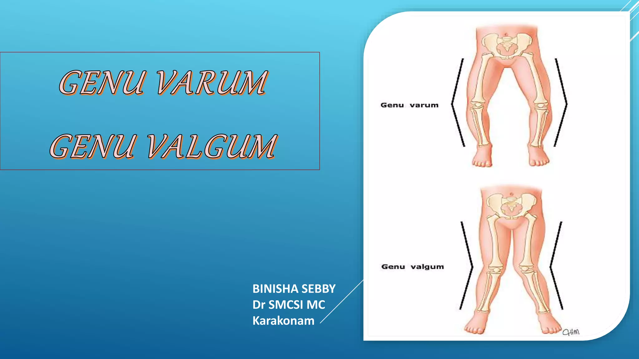
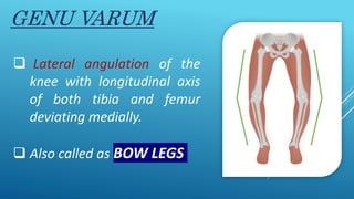
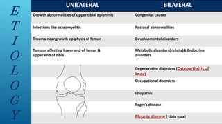
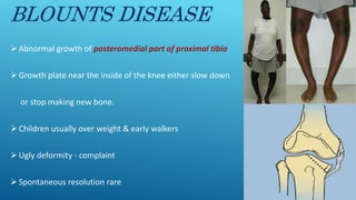
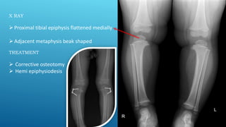
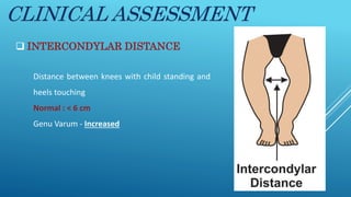
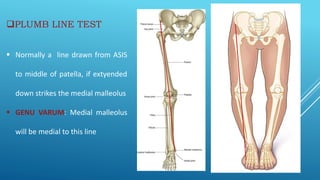
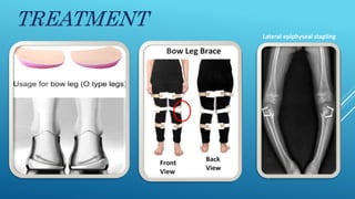
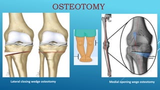
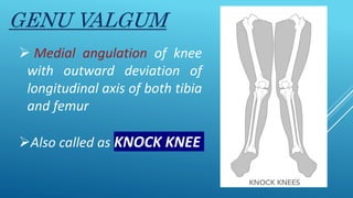
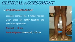
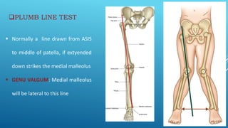
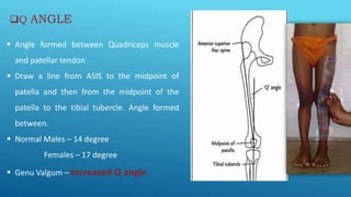
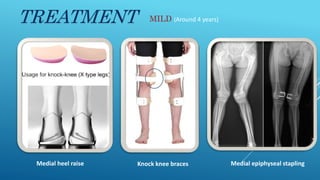
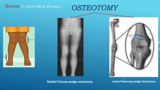
BINISHA SEBBY
Dr SMCSI MC
Karakonam
ANATOMIC AXIS
OF FEMUR
ANATOMIC AXIS
OF TIBIA
(5-7degree)
Normal knee is
approximately 6° valgus
PHYSIOLOGICAL
Bow legs &
Knock knees
 Considered as normal stages of development
 Usually disappears when child grows ,around 7-8 yrs
 By age of 10 , still marked – Operative correction
 Stapling of physes
 Corrective osteotomy
 Hemi epiphysiodesis
GENU VARUM
 Lateral angulation of the
knee with longitudinal axis
of both tibia and femur
deviating medially.
 Also called as BOW LEGS
E
T
I
O
L
O
G
Y
UNILATERAL BILATERAL
Growth abnormalities of upper tibial epiphysis Congenital causes
Infections like osteomyelitis Postural abnormalities
Trauma near growth epiphysis of femur Developmental disorders
Tumour affecting lower end of femur &
upper end of tibia
Metabolic disorders(rickets)& Endocrine
disorders
Degenerative disorders (Osteoarthritis of
knee)
Occupational disorders
Idiopathic
Paget’s disease
Blounts disease ( tibia vara)
BLOUNTS DISEASE
Abnormal growth of posteromedial part of proximal tibia
Growth plate near the inside of the knee either slow down
or stop making new bone.
Children usually over weight & early walkers
Ugly deformity - complaint
Spontaneous resolution rare
X RAY
Proximal tibial epiphysis flattened medially
Adjacent metaphysis beak shaped
TREATMENT
 Corrective osteotomy
 Hemi epiphysiodesis
CLINICAL FEATURES
PRIMARY DEFORMITY SECONDARY DEFORMITY
Lateral angulation of knee Internal torsion of distal tibia
In toeing of both feet
Patella face outward while walking
Tight medial & lax lateral structures
CLINICAL ASSESSMENT
 INTERCONDYLAR DISTANCE
Distance between knees with child standing and
heels touching
Normal : < 6 cm
Genu Varum - Increased
PLUMB LINE TEST
 Normally a line drawn from ASIS
to middle of patella, if extyended
down strikes the medial malleolus
 GENU VARUM: Medial malleolus
will be medial to this line
TREATMENT
Lateral epiphyseal stapling
Lateral closing wedge osteotomy Medial opening wege osteotomy
OSTEOTOMY
GENU VALGUM
 Medial angulation of knee
with outward deviation of
longitudinal axis of both tibia
and femur
Also called as KNOCK KNEE
UNILATERAL BILATERAL
Trauma Congenital disorders
Osteomyelitis Idiopathic
Tumors Developmental disorders(eg. Epiphyseal dysplasia)
Endocrine disorders(eg. Thyroid disorders)
Metabolic disorders(eg. Rickets)
Paralytic disorders
Traumatic disorders
Infective disorders
Inflammatory disorders(Rheumatoid arthritis)
Degenerative disorders
E
T
I
O
L
O
G
Y
CLINICAL FEATURES
PRIMARY DEFORMITY SECONDARY DEFORMITY
Medial angulation of knee Distal end of femur & proximal tibia
rotated externally
Compensatory internal torsion of distal
tibia
Lateral dislocation of patella
Tight lateral & lax medial structures
Flat foot
CLINICAL ASSESSMENT
 INTERMALLEOLAR GAP
Distance between the 2 medial malleoli
when knees are lightly touching and
patella facing forwards.
Normal : <8 cm
Genu valgum – Increased, >10 cm
PLUMB LINE TEST
 Normally a line drawn from ASIS
to middle of patella, if extyended
down strikes the medial malleolus
 GENU VALGUM: Medial malleolus
will be lateral to this line
Q ANGLE
 Angle formed between Quadriceps muscle
and patellar tendon
 Draw a line from ASIS to the midpoint of
patella and then from the midpoint of the
patella to the tibial tubercle. Angle formed
between.
 Normal Males – 14 degree
Females – 17 degree
 Genu Valgum – Increased Q angle
TREATMENT MILD (Around 4 years)
Medial heel raise Knock knee braces Medial epiphyseal stapling
Severe ( > 10 cm IM at 10 years )
OSTEOTOMY
Medial Closing wedge osteotomy Lateral Opening wedge Osteotomy
GENU VALGUM &  VARUM.pptx

GENU VALGUM & VARUM.pptx

  • 1.
  • 2.
    ANATOMIC AXIS OF FEMUR ANATOMICAXIS OF TIBIA (5-7degree) Normal knee is approximately 6° valgus
  • 3.
    PHYSIOLOGICAL Bow legs & Knockknees  Considered as normal stages of development  Usually disappears when child grows ,around 7-8 yrs  By age of 10 , still marked – Operative correction  Stapling of physes  Corrective osteotomy  Hemi epiphysiodesis
  • 4.
    GENU VARUM  Lateralangulation of the knee with longitudinal axis of both tibia and femur deviating medially.  Also called as BOW LEGS
  • 5.
    E T I O L O G Y UNILATERAL BILATERAL Growth abnormalitiesof upper tibial epiphysis Congenital causes Infections like osteomyelitis Postural abnormalities Trauma near growth epiphysis of femur Developmental disorders Tumour affecting lower end of femur & upper end of tibia Metabolic disorders(rickets)& Endocrine disorders Degenerative disorders (Osteoarthritis of knee) Occupational disorders Idiopathic Paget’s disease Blounts disease ( tibia vara)
  • 6.
    BLOUNTS DISEASE Abnormal growthof posteromedial part of proximal tibia Growth plate near the inside of the knee either slow down or stop making new bone. Children usually over weight & early walkers Ugly deformity - complaint Spontaneous resolution rare
  • 7.
    X RAY Proximal tibialepiphysis flattened medially Adjacent metaphysis beak shaped TREATMENT  Corrective osteotomy  Hemi epiphysiodesis
  • 8.
    CLINICAL FEATURES PRIMARY DEFORMITYSECONDARY DEFORMITY Lateral angulation of knee Internal torsion of distal tibia In toeing of both feet Patella face outward while walking Tight medial & lax lateral structures
  • 9.
    CLINICAL ASSESSMENT  INTERCONDYLARDISTANCE Distance between knees with child standing and heels touching Normal : < 6 cm Genu Varum - Increased
  • 10.
    PLUMB LINE TEST Normally a line drawn from ASIS to middle of patella, if extyended down strikes the medial malleolus  GENU VARUM: Medial malleolus will be medial to this line
  • 11.
  • 12.
    Lateral closing wedgeosteotomy Medial opening wege osteotomy OSTEOTOMY
  • 13.
    GENU VALGUM  Medialangulation of knee with outward deviation of longitudinal axis of both tibia and femur Also called as KNOCK KNEE
  • 14.
    UNILATERAL BILATERAL Trauma Congenitaldisorders Osteomyelitis Idiopathic Tumors Developmental disorders(eg. Epiphyseal dysplasia) Endocrine disorders(eg. Thyroid disorders) Metabolic disorders(eg. Rickets) Paralytic disorders Traumatic disorders Infective disorders Inflammatory disorders(Rheumatoid arthritis) Degenerative disorders E T I O L O G Y
  • 15.
    CLINICAL FEATURES PRIMARY DEFORMITYSECONDARY DEFORMITY Medial angulation of knee Distal end of femur & proximal tibia rotated externally Compensatory internal torsion of distal tibia Lateral dislocation of patella Tight lateral & lax medial structures Flat foot
  • 16.
    CLINICAL ASSESSMENT  INTERMALLEOLARGAP Distance between the 2 medial malleoli when knees are lightly touching and patella facing forwards. Normal : <8 cm Genu valgum – Increased, >10 cm
  • 17.
    PLUMB LINE TEST Normally a line drawn from ASIS to middle of patella, if extyended down strikes the medial malleolus  GENU VALGUM: Medial malleolus will be lateral to this line
  • 18.
    Q ANGLE  Angleformed between Quadriceps muscle and patellar tendon  Draw a line from ASIS to the midpoint of patella and then from the midpoint of the patella to the tibial tubercle. Angle formed between.  Normal Males – 14 degree Females – 17 degree  Genu Valgum – Increased Q angle
  • 19.
    TREATMENT MILD (Around4 years) Medial heel raise Knock knee braces Medial epiphyseal stapling
  • 20.
    Severe ( >10 cm IM at 10 years ) OSTEOTOMY Medial Closing wedge osteotomy Lateral Opening wedge Osteotomy