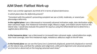
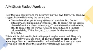
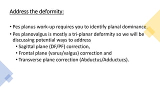
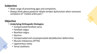
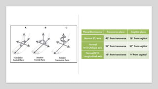
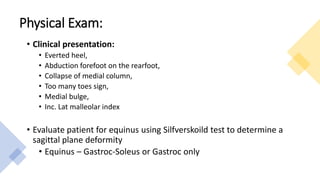
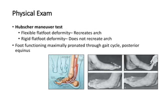
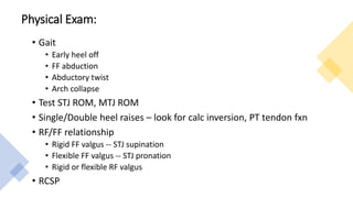
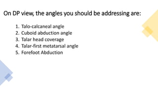
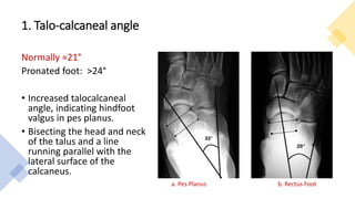
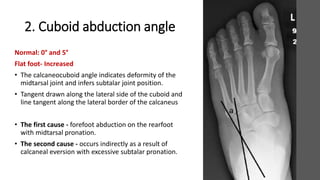
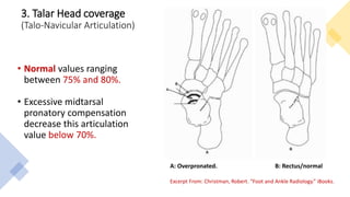
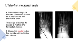
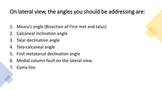
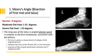
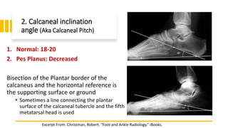
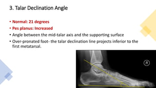
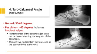
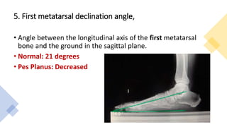
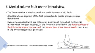
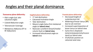
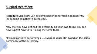
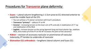
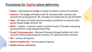
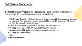
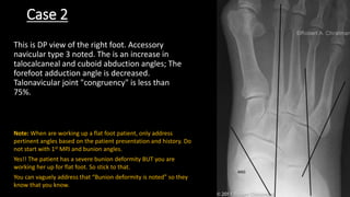
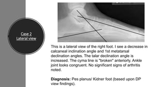
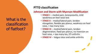
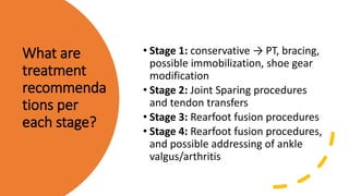
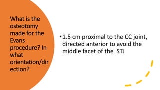
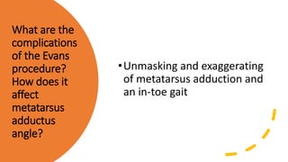
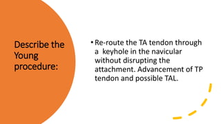
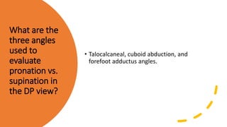
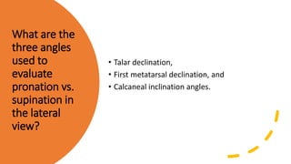
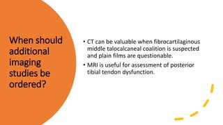
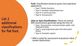
AJM Sheet: Flat Foot
Work-up
Review Questions Included
AJM Sheet: How to “Work-Up” a Surgical Patient
• With regard to specific surgery and the interviews, it’s always
important to “know your program”. In other words, programs
tend to have favorite procedures that they routinely do. For a
given bunion deformity, one program may primarily do Austin-
Akins, whereas other programs may never do an Akin, and still
others may always do a Lapidus in the exact same situation.
Some people may feel very strongly in favor of the lateral
release, while others may never do it for any situation. This
could even happen between two attendings at the same
program in the same room during your interview!
• If you give a hard, definitive answer for a procedure choice, one
attending may completely agree with you while another may think
it’s completely the wrong choice. So if you are asked what type of
procedure you would do for a given situation, be as general as
possible, but always give the reason/specific indications why you
are choosing that procedure or group of procedures. Name a
couple different similar procedures instead of sticking by your guns
with one procedure. Additionally, your interviewers may not expect
you to know for sure what procedure to choose, but they will
definitely expect you to be able to completely work-up the patient
and know which procedures are acceptable for which indications.
AJM Sheet: How to “Work-Up” a Surgical Patient
• The two work-ups that you should have down cold are the HAV and
flatfoot work-ups. Practice, practice, practice working through these
situations out loud, and practice, practice, practice going through the
radiographic analyses of these deformities out loud. Again, RC and I
found it helpful while studying for interviews to pick up random podiatry
textbooks and just flip through the pages, alternating our description of
the radiographs out loud.
• There are of course many, many radiographic angles that you can use to
describe during either of these work-ups, so focus the majority of your
energy on those that will have the most impact on your treatment
choice. Here’s the way that I think about these deformities. This
certainly isn’t the “right” way; it’s just the way that helped me as I first
started doing this out loud:
AJM Sheet: How to “Work-Up” a Surgical Patient
AJM Sheet: Flatfoot Work-up
Here I use a similar approach, but think of it in terms of planal dominance:
In which plane does the deformity present?
“Consistent with the patient’s presenting complaint we see a (mild, moderate, or severe) pes
planovalgus deformity.
In the sagittal plane I see a (decreased or increased) calcaneal inclination angle, talar declination angle,
talar-calcaneal angle, first metatarsal inclination angle, Meary’s angle, and medial column fault on the
lateral view. I would also evaluate the patient for equinus using the Silfverskiold test to determine a
sagittal plane deformity.
In the transverse plane I see a (decreased or increased) talar-calcaneal angle, cuboid abduction angle,
talar head coverage, talar-first metatarsal angle, metatarsus adductus angle on the AP view.
In the frontal plane we can see the Cyma Line is (anteriorly displaced, posteriorly displaced or normal)
on the lateral view, and that the subtalar joint alignment, ankle joint alignment and calcaneal position
are (normal or abnormal) on the long leg calcaneal axial views.”
AJM Sheet: Flatfoot Work-up
Now that you have defined the deformity on your own terms, you can now
suggest how to fix it using the same tools.
• “I would consider performing a (Gastroc recession, TAL, Cotton
osteotomy, medial column arthrodesis, etc.) to correct for the sagittal
plane deformity, a (Evans osteotomy, CC joint distraction arthrodesis,
etc.) to correct for the transverse plane deformity, and a (medial
calcaneal slide, STJ implant, etc.) to correct for the frontal plane
deformity.
This is a little philosophic, but radiographic angles aren’t real. They only
come into reality if you use them, so only use them as tools to your
advantage. You can use them to first define the deformity on your own
terms, and then to show that your intervention was successful.
Address the deformity:
• Pes planus work-up requires you to identify planal dominance.
• Pes planovalgus is mostly a tri-planar deformity so we will be
discussing potential ways to address
• Sagittal plane (DF/PF) correction,
• Frontal plane (varus/valgus) correction and
• Transverse plane correction (Abductus/Adductucs).
Subjective
• Wide range of presenting ages and complaints.
• Always think about posterior tibialis tendon dysfunction when someone
complains of “medial ankle pain.”
Objective
• Underlying Orthopedic Etiologies:
• Compensated forefoot varus
• Forefoot valgus
• Rearfoot valgus
• Equinus
• Compensated and uncompensated ab/adduction deformities
• Muscle imbalances (PTTD)
• Ligamentous laxity
• Tarsal coalitions
Physical Exam:
• Clinical presentation:
• Everted heel,
• Abduction forefoot on the rearfoot,
• Collapse of medial column,
• Too many toes sign,
• Medial bulge,
• Inc. Lat malleolar index
• Evaluate patient for equinus using Silfverskoild test to determine a
sagittal plane deformity
• Equinus – Gastroc-Soleus or Gastroc only
Physical Exam
• Hubscher maneuver test
• Flexible flatfoot deformity– Recreates arch
• Rigid flatfoot deformity– Does not recreate arch
• Foot functioning maximally pronated through gait cycle, posterior
equinus
Physical Exam:
• Gait
• Early heel off
• FF abduction
• Abductory twist
• Arch collapse
• Test STJ ROM, MTJ ROM
• Single/Double heel raises – look for calc inversion, PT tendon fxn
• RF/FF relationship
• Rigid FF valgus -- STJ supination
• Flexible FF valgus -- STJ pronation
• Rigid or flexible RF valgus
• RCSP
Radiographs
Practice reading x-rays in a systematic way which works for you whether it
is from proximal to distal or vice-versa. It will get you in a rhythm and also
help you to not miss any significant angles while reading under pressure
on externships.
There are basically 3 components that are involved in producing the
alignment abnormalities of symptomatic adult flatfoot:
1. Collapse of the longitudinal arch
2. Hindfoot valgus
3. Forefoot abduction
On DP view, the angles you should be addressing are:
1. Talo-calcaneal angle
2. Cuboid abduction angle
3. Talar head coverage
4. Talar-first metatarsal angle
5. Forefoot Abduction
1. Talo-calcaneal angle
Normally ≈21°
Pronated foot: >24°
• Increased talocalcaneal
angle, indicating hindfoot
valgus in pes planus.
• Bisecting the head and neck
of the talus and a line
running parallel with the
lateral surface of the
calcaneus.
a. Pes Planus b. Rectus Foot
2. Cuboid abduction angle
Normal: 0° and 5°
Flat foot- Increased
• The calcaneocuboid angle indicates deformity of the
midtarsal joint and infers subtalar joint position.
• Tangent drawn along the lateral side of the cuboid and
line tangent along the lateral border of the calcaneus
• The first cause - forefoot abduction on the rearfoot
with midtarsal pronation.
• The second cause - occurs indirectly as a result of
calcaneal eversion with excessive subtalar pronation.
3. Talar Head coverage
(Talo-Navicular Articulation)
• Normal values ranging
between 75% and 80%.
• Excessive midtarsal
pronatory compensation
decrease this articulation
value below 70%.
A: Overpronated. B: Rectus/normal
Excerpt From: Christman, Robert. “Foot and Ankle Radiology.” iBooks.
4. Talar-first metatarsal angle
• A line drawn through the
mid-axis of the talus should
be in line with the first
metatarsal shaft
• This angle evaluates the
degree of midfoot and
forefoot abduction.
• If it is angled medial to the
first metatarsal it indicates
pes planus.
On lateral view, the angles you should be addressing are:
1. Meary’s angle (Bisection of First met and talus)
2. Calcaneal inclination angle
3. Talar declination angle
4. Talo-calcaneal angle
5. First metatarsal declination angle
6. Medial column fault on the lateral view.
7. Cyma line
1. Meary’s Angle (Bisection
of First met and talus)
Normal - 0 degrees
Moderate Flat Foot: 1-15 degrees
Severe Flat Foot: >15 degrees
• The long axis of the talus is angled plantar-ward
in relation to the first metatarsal, consistent with
pes planus.
• This line is used as a measurement of collapse of the
longitudinal arch.
• Collapse may occur at the TN joint, NC, or cun-met joints.
• Long axis of talus should nearly bisect navicular and 1st
metatarsal shaft
2. Calcaneal inclination
angle (Aka Calcaneal Pitch)
1. Normal: 18-20
2. Pes Planus: Decreased
Bisection of the Plantar border of the
calcaneus and the horizontal reference is
the supporting surface or ground
• Sometimes a line connecting the plantar
surface of the calcaneal tubercle and the fifth
metatarsal head is used
Excerpt From: Christman, Robert. “Foot and Ankle Radiology.” iBooks.
3. Talar Declination Angle
• Normal: 21 degrees
• Pes planus: Increased
• Angle between the mid-talar axis and the supporting surface
• Over-pronated foot- the talar declination line projects inferior to the
first metatarsal.
4. Talo-Calcaneal Angle
(Kite’s Angle)
• Normal: 30-40 degrees.
• Pes planus: >40 degrees indicates
hindfoot valgus.
• Plantar border of the calcaneus (or a line
can be drawn bisecting the long axis of the
calcaneus).
• Through two midpoints in the talus, one at
the body and one at the neck.
5. First metatarsal declination angle,
• Angle between the longitudinal axis of the first metatarsal
bone and the ground in the sagittal plane.
• Normal: 21 degrees
• Pes Planus: Decreased
6. Medial column fault on the lateral view.
• The Talo-navicular, Naviculo-cuneiform, and Calcaneo-cuboid Faults.
• A fault is when a segment of the foot hyperextends, that is, shows excessive
dorsiflexion.
• Hyperextension is viewed as a collapse of a portion of the arch of the foot. No
matter which joint(s) is involved, as the forefoot is dorsiflexed, the dorsal surfaces of
the joints may appear compressed, the plantar joint space appears wider, and a sag
in the involved segment is perceived.
Excerpt From: Christman, Robert. “Foot and Ankle Radiology.” iBooks.
7. Cyma Line
Anterior break of the cyma
line in Pes Planus due to
over pronation.
Summary
AP View
1. Increased Talo-calcaneal angle
2. Increased Cuboid abduction angle
3. Decreased Talar head coverage
4. Talar-first metatarsal
angle abnormal with medial
deviation
Lateral View
1. Increase in Meary’s angle with long axis of
talus pointing plantar-ward.
2. Decreased Calcaneal inclination angle
3. Increased Talar declination angle
4. Increased Talo-calcaneal angle
5. Decreased First metatarsal declination
angle
6. Medial column fault on the lateral view.
7. Anterior Break in Cyma line
Angles and their planal dominance
Transverse plane deformity:
• Kite’s angle (Lat. talo-
calcaneal angle)
• Cuboid Abduction angle
• Talonavicular Coverage
• Metatarsus Adductus (FF to
RF Adduction)
Sagittal plane deformity:
• 1st met declination
• Calcaneal Inclination Angle
• Talar declination
• Meary’s angle (talus-first metatarsal
angle on lateral view)
• Navicular cuneiform breach/medial
column fault on lateral view
• Increased talocalcaneal angle on
lateral view
Frontal plane deformity:
• Decreased height of
sustentaculum tali
• Widening of lesser tarsus (AP)
due to calc eversion
• Increased superimposition of
lesser tarsus on lateral view
• Cyma line is displaced
(anteriorly/posteriorly/inconsi
stent) on lateral view
• STJ/AJ/Calc position on
calcaneal axial view
Conservative Treatment:
• Orthotics
• NSAIDs
• Activity modification
• PT/stretching for equinus
Surgical treatment:
Procedure Selection: Can be combined or performed independently
(depending on patient’s pathology).
Now that you have defined the deformity on your own terms, you can
now suggest how to fix it using the same tools.
“I would consider performing a …. Evans or kouts etc” based on the planal
dominance of the deformity.
Procedures for Transverse plane deformity:
• Evans – Lateral column lengthening 1-1.5cm prox to CCJ directed anterior to
avoid the middle facet of the STJ.
• You can achieve a Tri-planar correction with Evan’s procedure.
• Transverse *primary* – adducts FF on RF
• Coronal – increasing tension on the lever arm of PL and aids in stabilization of 1st ray
and stabilization of the MLA
• Sagittal – increase tension on the spring ligament long and short plantar ligs, stabilizes
MLA, and creates pf of the FF on the RF increases the pitch of the calcaneus
• Kidner – excision of accessory navicular or prominence of navicular
tuberosity, PT tendon to underside of navicular
• Distraction CCJ arthrodesis – lengthens lateral column and fuses CCJ.
Procedures for Sagittal plane deformity:
• Cotton – opening dorsal wedge in medial cuneiform, corrects ff supinatus
• Lowman – TN wedge arthrodesis with TA rerouted under navicular and
sutured into spring ligament, TAL, tenodesis of medial arch w/ slip of Achilles
• Hoke – NC fusion of medial and intermediate cuneiforms to navicular with
plantar based wedge removal, TAL
• Miller – Lapidus + navicular medial cuneiform fusion, PT tendon and spring
lig advancement using osteoperiosteal flap
• Young’s Tenosuspension – Reroute TA tendon through keyhole slot in the
navicular without detaching from insertion, PT advanced under navicular
• TAL – corrects GS equinus
• Gastroc lengthening – corrects gastroc equinus
• Medial column arthrodesis
Procedures for Coronal plane deformity:
• STJ arthrodesis or triple arthrodesis (depending on the level of
deformity)
• Kouts – medial calcaneal slide, shifts Achilles to the supinatory side of
STJ
• Arthroeresis – sinus tarsi implant, prevents excess pronation
• Silver – lateral opening base wedge w/graft in calc
• Selakovich – opening wedge osteotomy with bone graft under sus tali
• Baker – osteotomy inferior to STJ post facet with bone graft
• Chambers – bone graft under sinus tarsi to raise the posterior STJ facet
Soft Tissue Procedures:
General Surgical Procedures: Indications - Keep in mind that it is very
common to do combinations of these procedures.
• FDL Tendon Transfer: FDL is sectioned as distal as possible (consider anastomosis
of stump to FHL) and either attach proximal FDL to the PT, within the PT sheath
or into the navicular under tension.
• Cobb: Split TA tendon, transfer to the PT or into the navicular
• Young’s Tenosuspension: TA rerouted through navicular
• Anastomosis of PB and PL: Removes PB as deforming force
• STJ implant (arthroeresis)
• TAL
• Gastroc recession
Read a flatfoot x-ray
Case 1
Case 1
This is a DP view of the right foot, skeletally
mature patient. There is an increase in
talocalcaneal and cuboid abduction angles; The
talar first-met angle is also increased and angled
medially. Talonavicular joint "congruency" is
approximately 50%. The forefoot adduction
angle is decreased.
Case 1
Lateral view
This is a lateral view of the right foot. I see a decrease in
calcaneal inclination angle and 1st metatarsal declination
angles. The talar declination angle and Kite’s angle is
increased. Increase in Meary’s angle with long axis of
talus pointing plantar-ward. The cyma line is "broken"
anteriorly.
Diagnosis: Pes Planus.
Case 2
Case 2
This is DP view of the right foot. Accessory
navicular type 3 noted. The is an increase in
talocalcaneal and cuboid abduction angles; The
forefoot adduction angle is decreased.
Talonavicular joint "congruency" is less than
75%.
Note: When are working up a flat foot patient, only address
pertinent angles based on the patient presentation and history. Do
not start with 1st MPJ and bunion angles.
Yes!! The patient has a severe bunion deformity BUT you are
working her up for flat foot. So stick to that.
You can vaguely address that “Bunion deformity is noted” so they
know that you know.
Case 2
Lateral view
This is a lateral view of the right foot. I see a decrease in
calcaneal inclination angle and 1st metatarsal
declination angles. The talar declination angle is
increased. The cyma line is "broken" anteriorly. Ankle
joint looks congruent. No significant signs of arthritis
noted.
Diagnosis: Pes planus/ Kidner foot (based upon DP
view findings).
Review Questions
How does the
calcaneal
inclination
angle change
with
pronation/supi
nation?
•It does not because it is a
structural angle
What is the
classification
of flatfoot?
PTTD classification
Johnson and Storm with Myerson Modification
• STAGE I -- medial pain, tenosynovitis, mild
weakness on heel raise test
• STAGE II -- medial/lateral pain, tendon
elongation, flexible pes planus, weakness on heel
raise, + too many toes
• STAGE III -- medial/lateral pain, tendon
degeneration, fixed pes planus, no inversion on
heel raise, + too many tos, STJ arthritis
• STAGE IV -- Valgus talus and ankle arthritis
What are
treatment
recommenda
tions per
each stage?
• Stage 1: conservative → PT, bracing,
possible immobilization, shoe gear
modification
• Stage 2: Joint Sparing procedures
and tendon transfers
• Stage 3: Rearfoot fusion procedures
• Stage 4: Rearfoot fusion procedures,
and possible addressing of ankle
valgus/arthritis
What is the
osteotomy
made for the
Evans
procedure? In
what
orientation/dir
ection?
•1.5 cm proximal to the CC joint,
directed anterior to avoid the
middle facet of the STJ
What are the
complications
of the Evans
procedure?
How does it
affect
metatarsus
adductus
angle?
•Unmasking and exaggerating
of metatarsus adduction and
an in-toe gait
Describe the
Young
procedure:
•Re-route the TA tendon through
a keyhole in the navicular
without disrupting the
attachment. Advancement of TP
tendon and possible TAL.
Describe the
Dwyer
procedure:
• Opening wedge osteotomy lateral calc
• Closing wedge-medial calc
What paper is
the
benchmark for
rearfoot
motion after
isolated joint
fusions? And
what was the
big picture?
• Astion, 1996, 2 degrees of motion
are preserved in STJ and CCJ after
fusion of TNJ.
• TNJ is the key articulation of
hindfoot motion.
What are
Glissane’s
rules for
arthrodesis?
• Complete removal of all cartilage,
fibrous tissue and other materials
preventing bone to bone contact
• Accurate and close fitting surfaces
• Optimal position of fusion
• Maintain apposition or undisturbed
until fusion complete
For
arthroereisis
to be effect,
what must be
reducible?
•Heel valgus and Forefoot varus
or supinatus
What are the
three angles
used to
evaluate
pronation vs.
supination in
the DP view?
• Talocalcaneal, cuboid abduction, and
forefoot adductus angles.
What are the
three angles
used to
evaluate
pronation vs.
supination in
the lateral
view?
• Talar declination,
• First metatarsal declination, and
• Calcaneal inclination angles.
Why is it
useful to
determine
whether the
foot is
pronated or
supinated?
• Foot position can have a profound effect on
the form of bones. One needs to be able to
predict these appearances, and not
misinterpret "odd-appearing" bone forms as
pathology.
What are the
best views to
order?
• At a minimum, order dorsoplantar and
lateral views.
• Lateral oblique view should be added when
considering accessory navicular;
• Include the Harris-Beath view for suspected
middle talocalcaneal coalition;
• Add the medial oblique view when
calcanonavicular bar is suspected.
When should
additional
imaging
studies be
ordered?
• CT can be valuable when fibrocartilaginous
middle talocalcaneal coalition is suspected
and plain films are questionable.
• MRI is useful for assessment of posterior
tibial tendon dysfunction.
List 2
additional
classifications
for flat foot.
Funk: Classification based on gross intra-operative
appearance
• Type I: Tendon Avulsions
• Type II: Complete midsubstance tear
• Type III: In-continuity tears
• Type IV: Tenosynovitis
Jahss or Janis Classifications: There are several
MRI classifications generally along the lines of:
• Type I: Tenosynovitis, increased tendon width, mild
longitudinal splits
• Type II: Long longitudinal splits with attenuated
tendon
• Type III: Complete rupture
• MRI is useful for assessment of posterior tibial
tendon dysfunction.
Additional Reading:
[Hix J, et al. Calcaneal osteotomies for the treatment of adult-acquired flatfoot. Clin Podiatr Med Surg.
2007 Oct; 24(4): 699-719.]
[Mosier-LaClair S, et al. Operative treatment of the difficult stage 2 adult acquired flatfoot deformity.
Foot Ankle Clin. 2001 Mar; 6(1): 95-119.]
[Roye DP, Raimondo RA. Surgical treatment of the child’s and adolescent’s flexible flatfoot. Clin
Podiatr Med Surg. 2000 Jul; 17(3): 515-30.]
[Toolan BC, Sangeorzan, Hansen. Complex reconstruction for the treatment of dorsolateral peritalar
subluxation of the foot. JBJS-Am. 1999 Nov; 81(11): 1545-60.]
[Weinraub GM, Heilala MA. Adult flatfoot/posterior tibial tendon dysfunction: outcomes analysis of
surgical treatment utilizing an algorithmic approach. J Foot Ankle Surg. 2001 Jan-Feb; 40(1): 54-7.]
References
Images:
http://uwmsk.org/footalignment/doku.php?id=pes_pl
anus
Images Excerpt From: Christman, Robert. “Foot and
Ankle Radiology.” iBooks.

Flat foot work up

  • 1.
    AJM Sheet: FlatFoot Work-up Review Questions Included
  • 2.
    AJM Sheet: Howto “Work-Up” a Surgical Patient • With regard to specific surgery and the interviews, it’s always important to “know your program”. In other words, programs tend to have favorite procedures that they routinely do. For a given bunion deformity, one program may primarily do Austin- Akins, whereas other programs may never do an Akin, and still others may always do a Lapidus in the exact same situation. Some people may feel very strongly in favor of the lateral release, while others may never do it for any situation. This could even happen between two attendings at the same program in the same room during your interview!
  • 3.
    • If yougive a hard, definitive answer for a procedure choice, one attending may completely agree with you while another may think it’s completely the wrong choice. So if you are asked what type of procedure you would do for a given situation, be as general as possible, but always give the reason/specific indications why you are choosing that procedure or group of procedures. Name a couple different similar procedures instead of sticking by your guns with one procedure. Additionally, your interviewers may not expect you to know for sure what procedure to choose, but they will definitely expect you to be able to completely work-up the patient and know which procedures are acceptable for which indications. AJM Sheet: How to “Work-Up” a Surgical Patient
  • 4.
    • The twowork-ups that you should have down cold are the HAV and flatfoot work-ups. Practice, practice, practice working through these situations out loud, and practice, practice, practice going through the radiographic analyses of these deformities out loud. Again, RC and I found it helpful while studying for interviews to pick up random podiatry textbooks and just flip through the pages, alternating our description of the radiographs out loud. • There are of course many, many radiographic angles that you can use to describe during either of these work-ups, so focus the majority of your energy on those that will have the most impact on your treatment choice. Here’s the way that I think about these deformities. This certainly isn’t the “right” way; it’s just the way that helped me as I first started doing this out loud: AJM Sheet: How to “Work-Up” a Surgical Patient
  • 5.
    AJM Sheet: FlatfootWork-up Here I use a similar approach, but think of it in terms of planal dominance: In which plane does the deformity present? “Consistent with the patient’s presenting complaint we see a (mild, moderate, or severe) pes planovalgus deformity. In the sagittal plane I see a (decreased or increased) calcaneal inclination angle, talar declination angle, talar-calcaneal angle, first metatarsal inclination angle, Meary’s angle, and medial column fault on the lateral view. I would also evaluate the patient for equinus using the Silfverskiold test to determine a sagittal plane deformity. In the transverse plane I see a (decreased or increased) talar-calcaneal angle, cuboid abduction angle, talar head coverage, talar-first metatarsal angle, metatarsus adductus angle on the AP view. In the frontal plane we can see the Cyma Line is (anteriorly displaced, posteriorly displaced or normal) on the lateral view, and that the subtalar joint alignment, ankle joint alignment and calcaneal position are (normal or abnormal) on the long leg calcaneal axial views.”
  • 6.
    AJM Sheet: FlatfootWork-up Now that you have defined the deformity on your own terms, you can now suggest how to fix it using the same tools. • “I would consider performing a (Gastroc recession, TAL, Cotton osteotomy, medial column arthrodesis, etc.) to correct for the sagittal plane deformity, a (Evans osteotomy, CC joint distraction arthrodesis, etc.) to correct for the transverse plane deformity, and a (medial calcaneal slide, STJ implant, etc.) to correct for the frontal plane deformity. This is a little philosophic, but radiographic angles aren’t real. They only come into reality if you use them, so only use them as tools to your advantage. You can use them to first define the deformity on your own terms, and then to show that your intervention was successful.
  • 7.
    Address the deformity: •Pes planus work-up requires you to identify planal dominance. • Pes planovalgus is mostly a tri-planar deformity so we will be discussing potential ways to address • Sagittal plane (DF/PF) correction, • Frontal plane (varus/valgus) correction and • Transverse plane correction (Abductus/Adductucs).
  • 8.
    Subjective • Wide rangeof presenting ages and complaints. • Always think about posterior tibialis tendon dysfunction when someone complains of “medial ankle pain.” Objective • Underlying Orthopedic Etiologies: • Compensated forefoot varus • Forefoot valgus • Rearfoot valgus • Equinus • Compensated and uncompensated ab/adduction deformities • Muscle imbalances (PTTD) • Ligamentous laxity • Tarsal coalitions
  • 10.
    Physical Exam: • Clinicalpresentation: • Everted heel, • Abduction forefoot on the rearfoot, • Collapse of medial column, • Too many toes sign, • Medial bulge, • Inc. Lat malleolar index • Evaluate patient for equinus using Silfverskoild test to determine a sagittal plane deformity • Equinus – Gastroc-Soleus or Gastroc only
  • 11.
    Physical Exam • Hubschermaneuver test • Flexible flatfoot deformity– Recreates arch • Rigid flatfoot deformity– Does not recreate arch • Foot functioning maximally pronated through gait cycle, posterior equinus
  • 12.
    Physical Exam: • Gait •Early heel off • FF abduction • Abductory twist • Arch collapse • Test STJ ROM, MTJ ROM • Single/Double heel raises – look for calc inversion, PT tendon fxn • RF/FF relationship • Rigid FF valgus -- STJ supination • Flexible FF valgus -- STJ pronation • Rigid or flexible RF valgus • RCSP
  • 13.
    Radiographs Practice reading x-raysin a systematic way which works for you whether it is from proximal to distal or vice-versa. It will get you in a rhythm and also help you to not miss any significant angles while reading under pressure on externships. There are basically 3 components that are involved in producing the alignment abnormalities of symptomatic adult flatfoot: 1. Collapse of the longitudinal arch 2. Hindfoot valgus 3. Forefoot abduction
  • 14.
    On DP view,the angles you should be addressing are: 1. Talo-calcaneal angle 2. Cuboid abduction angle 3. Talar head coverage 4. Talar-first metatarsal angle 5. Forefoot Abduction
  • 15.
    1. Talo-calcaneal angle Normally≈21° Pronated foot: >24° • Increased talocalcaneal angle, indicating hindfoot valgus in pes planus. • Bisecting the head and neck of the talus and a line running parallel with the lateral surface of the calcaneus. a. Pes Planus b. Rectus Foot
  • 16.
    2. Cuboid abductionangle Normal: 0° and 5° Flat foot- Increased • The calcaneocuboid angle indicates deformity of the midtarsal joint and infers subtalar joint position. • Tangent drawn along the lateral side of the cuboid and line tangent along the lateral border of the calcaneus • The first cause - forefoot abduction on the rearfoot with midtarsal pronation. • The second cause - occurs indirectly as a result of calcaneal eversion with excessive subtalar pronation.
  • 17.
    3. Talar Headcoverage (Talo-Navicular Articulation) • Normal values ranging between 75% and 80%. • Excessive midtarsal pronatory compensation decrease this articulation value below 70%. A: Overpronated. B: Rectus/normal Excerpt From: Christman, Robert. “Foot and Ankle Radiology.” iBooks.
  • 18.
    4. Talar-first metatarsalangle • A line drawn through the mid-axis of the talus should be in line with the first metatarsal shaft • This angle evaluates the degree of midfoot and forefoot abduction. • If it is angled medial to the first metatarsal it indicates pes planus.
  • 19.
    On lateral view,the angles you should be addressing are: 1. Meary’s angle (Bisection of First met and talus) 2. Calcaneal inclination angle 3. Talar declination angle 4. Talo-calcaneal angle 5. First metatarsal declination angle 6. Medial column fault on the lateral view. 7. Cyma line
  • 20.
    1. Meary’s Angle(Bisection of First met and talus) Normal - 0 degrees Moderate Flat Foot: 1-15 degrees Severe Flat Foot: >15 degrees • The long axis of the talus is angled plantar-ward in relation to the first metatarsal, consistent with pes planus. • This line is used as a measurement of collapse of the longitudinal arch. • Collapse may occur at the TN joint, NC, or cun-met joints. • Long axis of talus should nearly bisect navicular and 1st metatarsal shaft
  • 21.
    2. Calcaneal inclination angle(Aka Calcaneal Pitch) 1. Normal: 18-20 2. Pes Planus: Decreased Bisection of the Plantar border of the calcaneus and the horizontal reference is the supporting surface or ground • Sometimes a line connecting the plantar surface of the calcaneal tubercle and the fifth metatarsal head is used Excerpt From: Christman, Robert. “Foot and Ankle Radiology.” iBooks.
  • 22.
    3. Talar DeclinationAngle • Normal: 21 degrees • Pes planus: Increased • Angle between the mid-talar axis and the supporting surface • Over-pronated foot- the talar declination line projects inferior to the first metatarsal.
  • 23.
    4. Talo-Calcaneal Angle (Kite’sAngle) • Normal: 30-40 degrees. • Pes planus: >40 degrees indicates hindfoot valgus. • Plantar border of the calcaneus (or a line can be drawn bisecting the long axis of the calcaneus). • Through two midpoints in the talus, one at the body and one at the neck.
  • 24.
    5. First metatarsaldeclination angle, • Angle between the longitudinal axis of the first metatarsal bone and the ground in the sagittal plane. • Normal: 21 degrees • Pes Planus: Decreased
  • 25.
    6. Medial columnfault on the lateral view. • The Talo-navicular, Naviculo-cuneiform, and Calcaneo-cuboid Faults. • A fault is when a segment of the foot hyperextends, that is, shows excessive dorsiflexion. • Hyperextension is viewed as a collapse of a portion of the arch of the foot. No matter which joint(s) is involved, as the forefoot is dorsiflexed, the dorsal surfaces of the joints may appear compressed, the plantar joint space appears wider, and a sag in the involved segment is perceived. Excerpt From: Christman, Robert. “Foot and Ankle Radiology.” iBooks.
  • 26.
    7. Cyma Line Anteriorbreak of the cyma line in Pes Planus due to over pronation.
  • 27.
    Summary AP View 1. IncreasedTalo-calcaneal angle 2. Increased Cuboid abduction angle 3. Decreased Talar head coverage 4. Talar-first metatarsal angle abnormal with medial deviation Lateral View 1. Increase in Meary’s angle with long axis of talus pointing plantar-ward. 2. Decreased Calcaneal inclination angle 3. Increased Talar declination angle 4. Increased Talo-calcaneal angle 5. Decreased First metatarsal declination angle 6. Medial column fault on the lateral view. 7. Anterior Break in Cyma line
  • 28.
    Angles and theirplanal dominance Transverse plane deformity: • Kite’s angle (Lat. talo- calcaneal angle) • Cuboid Abduction angle • Talonavicular Coverage • Metatarsus Adductus (FF to RF Adduction) Sagittal plane deformity: • 1st met declination • Calcaneal Inclination Angle • Talar declination • Meary’s angle (talus-first metatarsal angle on lateral view) • Navicular cuneiform breach/medial column fault on lateral view • Increased talocalcaneal angle on lateral view Frontal plane deformity: • Decreased height of sustentaculum tali • Widening of lesser tarsus (AP) due to calc eversion • Increased superimposition of lesser tarsus on lateral view • Cyma line is displaced (anteriorly/posteriorly/inconsi stent) on lateral view • STJ/AJ/Calc position on calcaneal axial view
  • 29.
    Conservative Treatment: • Orthotics •NSAIDs • Activity modification • PT/stretching for equinus
  • 30.
    Surgical treatment: Procedure Selection:Can be combined or performed independently (depending on patient’s pathology). Now that you have defined the deformity on your own terms, you can now suggest how to fix it using the same tools. “I would consider performing a …. Evans or kouts etc” based on the planal dominance of the deformity.
  • 31.
    Procedures for Transverseplane deformity: • Evans – Lateral column lengthening 1-1.5cm prox to CCJ directed anterior to avoid the middle facet of the STJ. • You can achieve a Tri-planar correction with Evan’s procedure. • Transverse *primary* – adducts FF on RF • Coronal – increasing tension on the lever arm of PL and aids in stabilization of 1st ray and stabilization of the MLA • Sagittal – increase tension on the spring ligament long and short plantar ligs, stabilizes MLA, and creates pf of the FF on the RF increases the pitch of the calcaneus • Kidner – excision of accessory navicular or prominence of navicular tuberosity, PT tendon to underside of navicular • Distraction CCJ arthrodesis – lengthens lateral column and fuses CCJ.
  • 32.
    Procedures for Sagittalplane deformity: • Cotton – opening dorsal wedge in medial cuneiform, corrects ff supinatus • Lowman – TN wedge arthrodesis with TA rerouted under navicular and sutured into spring ligament, TAL, tenodesis of medial arch w/ slip of Achilles • Hoke – NC fusion of medial and intermediate cuneiforms to navicular with plantar based wedge removal, TAL • Miller – Lapidus + navicular medial cuneiform fusion, PT tendon and spring lig advancement using osteoperiosteal flap • Young’s Tenosuspension – Reroute TA tendon through keyhole slot in the navicular without detaching from insertion, PT advanced under navicular • TAL – corrects GS equinus • Gastroc lengthening – corrects gastroc equinus • Medial column arthrodesis
  • 33.
    Procedures for Coronalplane deformity: • STJ arthrodesis or triple arthrodesis (depending on the level of deformity) • Kouts – medial calcaneal slide, shifts Achilles to the supinatory side of STJ • Arthroeresis – sinus tarsi implant, prevents excess pronation • Silver – lateral opening base wedge w/graft in calc • Selakovich – opening wedge osteotomy with bone graft under sus tali • Baker – osteotomy inferior to STJ post facet with bone graft • Chambers – bone graft under sinus tarsi to raise the posterior STJ facet
  • 34.
    Soft Tissue Procedures: GeneralSurgical Procedures: Indications - Keep in mind that it is very common to do combinations of these procedures. • FDL Tendon Transfer: FDL is sectioned as distal as possible (consider anastomosis of stump to FHL) and either attach proximal FDL to the PT, within the PT sheath or into the navicular under tension. • Cobb: Split TA tendon, transfer to the PT or into the navicular • Young’s Tenosuspension: TA rerouted through navicular • Anastomosis of PB and PL: Removes PB as deforming force • STJ implant (arthroeresis) • TAL • Gastroc recession
  • 35.
  • 36.
  • 37.
    Case 1 This isa DP view of the right foot, skeletally mature patient. There is an increase in talocalcaneal and cuboid abduction angles; The talar first-met angle is also increased and angled medially. Talonavicular joint "congruency" is approximately 50%. The forefoot adduction angle is decreased.
  • 38.
    Case 1 Lateral view Thisis a lateral view of the right foot. I see a decrease in calcaneal inclination angle and 1st metatarsal declination angles. The talar declination angle and Kite’s angle is increased. Increase in Meary’s angle with long axis of talus pointing plantar-ward. The cyma line is "broken" anteriorly. Diagnosis: Pes Planus.
  • 39.
  • 40.
    Case 2 This isDP view of the right foot. Accessory navicular type 3 noted. The is an increase in talocalcaneal and cuboid abduction angles; The forefoot adduction angle is decreased. Talonavicular joint "congruency" is less than 75%. Note: When are working up a flat foot patient, only address pertinent angles based on the patient presentation and history. Do not start with 1st MPJ and bunion angles. Yes!! The patient has a severe bunion deformity BUT you are working her up for flat foot. So stick to that. You can vaguely address that “Bunion deformity is noted” so they know that you know.
  • 41.
    Case 2 Lateral view Thisis a lateral view of the right foot. I see a decrease in calcaneal inclination angle and 1st metatarsal declination angles. The talar declination angle is increased. The cyma line is "broken" anteriorly. Ankle joint looks congruent. No significant signs of arthritis noted. Diagnosis: Pes planus/ Kidner foot (based upon DP view findings).
  • 42.
  • 43.
    How does the calcaneal inclination anglechange with pronation/supi nation? •It does not because it is a structural angle
  • 44.
    What is the classification offlatfoot? PTTD classification Johnson and Storm with Myerson Modification • STAGE I -- medial pain, tenosynovitis, mild weakness on heel raise test • STAGE II -- medial/lateral pain, tendon elongation, flexible pes planus, weakness on heel raise, + too many toes • STAGE III -- medial/lateral pain, tendon degeneration, fixed pes planus, no inversion on heel raise, + too many tos, STJ arthritis • STAGE IV -- Valgus talus and ankle arthritis
  • 45.
    What are treatment recommenda tions per eachstage? • Stage 1: conservative → PT, bracing, possible immobilization, shoe gear modification • Stage 2: Joint Sparing procedures and tendon transfers • Stage 3: Rearfoot fusion procedures • Stage 4: Rearfoot fusion procedures, and possible addressing of ankle valgus/arthritis
  • 46.
    What is the osteotomy madefor the Evans procedure? In what orientation/dir ection? •1.5 cm proximal to the CC joint, directed anterior to avoid the middle facet of the STJ
  • 47.
    What are the complications ofthe Evans procedure? How does it affect metatarsus adductus angle? •Unmasking and exaggerating of metatarsus adduction and an in-toe gait
  • 48.
    Describe the Young procedure: •Re-route theTA tendon through a keyhole in the navicular without disrupting the attachment. Advancement of TP tendon and possible TAL.
  • 49.
    Describe the Dwyer procedure: • Openingwedge osteotomy lateral calc • Closing wedge-medial calc
  • 50.
    What paper is the benchmarkfor rearfoot motion after isolated joint fusions? And what was the big picture? • Astion, 1996, 2 degrees of motion are preserved in STJ and CCJ after fusion of TNJ. • TNJ is the key articulation of hindfoot motion.
  • 51.
    What are Glissane’s rules for arthrodesis? •Complete removal of all cartilage, fibrous tissue and other materials preventing bone to bone contact • Accurate and close fitting surfaces • Optimal position of fusion • Maintain apposition or undisturbed until fusion complete
  • 52.
    For arthroereisis to be effect, whatmust be reducible? •Heel valgus and Forefoot varus or supinatus
  • 53.
    What are the threeangles used to evaluate pronation vs. supination in the DP view? • Talocalcaneal, cuboid abduction, and forefoot adductus angles.
  • 54.
    What are the threeangles used to evaluate pronation vs. supination in the lateral view? • Talar declination, • First metatarsal declination, and • Calcaneal inclination angles.
  • 55.
    Why is it usefulto determine whether the foot is pronated or supinated? • Foot position can have a profound effect on the form of bones. One needs to be able to predict these appearances, and not misinterpret "odd-appearing" bone forms as pathology.
  • 56.
    What are the bestviews to order? • At a minimum, order dorsoplantar and lateral views. • Lateral oblique view should be added when considering accessory navicular; • Include the Harris-Beath view for suspected middle talocalcaneal coalition; • Add the medial oblique view when calcanonavicular bar is suspected.
  • 57.
    When should additional imaging studies be ordered? •CT can be valuable when fibrocartilaginous middle talocalcaneal coalition is suspected and plain films are questionable. • MRI is useful for assessment of posterior tibial tendon dysfunction.
  • 58.
    List 2 additional classifications for flatfoot. Funk: Classification based on gross intra-operative appearance • Type I: Tendon Avulsions • Type II: Complete midsubstance tear • Type III: In-continuity tears • Type IV: Tenosynovitis Jahss or Janis Classifications: There are several MRI classifications generally along the lines of: • Type I: Tenosynovitis, increased tendon width, mild longitudinal splits • Type II: Long longitudinal splits with attenuated tendon • Type III: Complete rupture • MRI is useful for assessment of posterior tibial tendon dysfunction.
  • 59.
    Additional Reading: [Hix J,et al. Calcaneal osteotomies for the treatment of adult-acquired flatfoot. Clin Podiatr Med Surg. 2007 Oct; 24(4): 699-719.] [Mosier-LaClair S, et al. Operative treatment of the difficult stage 2 adult acquired flatfoot deformity. Foot Ankle Clin. 2001 Mar; 6(1): 95-119.] [Roye DP, Raimondo RA. Surgical treatment of the child’s and adolescent’s flexible flatfoot. Clin Podiatr Med Surg. 2000 Jul; 17(3): 515-30.] [Toolan BC, Sangeorzan, Hansen. Complex reconstruction for the treatment of dorsolateral peritalar subluxation of the foot. JBJS-Am. 1999 Nov; 81(11): 1545-60.] [Weinraub GM, Heilala MA. Adult flatfoot/posterior tibial tendon dysfunction: outcomes analysis of surgical treatment utilizing an algorithmic approach. J Foot Ankle Surg. 2001 Jan-Feb; 40(1): 54-7.]
  • 60.

Editor's Notes

  • #37 References: Christman RA: Foot and Ankle Radiology, 2nd edition, Lippincott Williams & Wilkins, 2015. Nalaboff KM, Schweitzer ME: MRI of tarsal coalition: frequency, distribution, and innovative signs. Bull NYU Hosp Jt Dis 66(1):14-21, 2008. Lateur, LM et al: Subtalar coalition: diagnosis with the C sign on lateral radiographs of the ankle. Radiology 193:847, 1994 Crim JR, Kjeldsberg KM: Radiographic Diagnosis of Tarsal Coalition. AJR 182(2):323-328, 2004(Feb)
  • #38 References: Christman RA: Foot and Ankle Radiology, 2nd edition, Lippincott Williams & Wilkins, 2015. Nalaboff KM, Schweitzer ME: MRI of tarsal coalition: frequency, distribution, and innovative signs. Bull NYU Hosp Jt Dis 66(1):14-21, 2008. Lateur, LM et al: Subtalar coalition: diagnosis with the C sign on lateral radiographs of the ankle. Radiology 193:847, 1994 Crim JR, Kjeldsberg KM: Radiographic Diagnosis of Tarsal Coalition. AJR 182(2):323-328, 2004(Feb)
  • #39 References: Christman RA: Foot and Ankle Radiology, 2nd edition, Lippincott Williams & Wilkins, 2015. Nalaboff KM, Schweitzer ME: MRI of tarsal coalition: frequency, distribution, and innovative signs. Bull NYU Hosp Jt Dis 66(1):14-21, 2008. Lateur, LM et al: Subtalar coalition: diagnosis with the C sign on lateral radiographs of the ankle. Radiology 193:847, 1994 Crim JR, Kjeldsberg KM: Radiographic Diagnosis of Tarsal Coalition. AJR 182(2):323-328, 2004(Feb)
  • #40 References: Christman RA: Foot and Ankle Radiology, 2nd edition, Lippincott Williams & Wilkins, 2015. Nalaboff KM, Schweitzer ME: MRI of tarsal coalition: frequency, distribution, and innovative signs. Bull NYU Hosp Jt Dis 66(1):14-21, 2008. Lateur, LM et al: Subtalar coalition: diagnosis with the C sign on lateral radiographs of the ankle. Radiology 193:847, 1994 Crim JR, Kjeldsberg KM: Radiographic Diagnosis of Tarsal Coalition. AJR 182(2):323-328, 2004(Feb)
  • #41 References: Christman RA: Foot and Ankle Radiology, 2nd edition, Lippincott Williams & Wilkins, 2015. Nalaboff KM, Schweitzer ME: MRI of tarsal coalition: frequency, distribution, and innovative signs. Bull NYU Hosp Jt Dis 66(1):14-21, 2008. Lateur, LM et al: Subtalar coalition: diagnosis with the C sign on lateral radiographs of the ankle. Radiology 193:847, 1994 Crim JR, Kjeldsberg KM: Radiographic Diagnosis of Tarsal Coalition. AJR 182(2):323-328, 2004(Feb)
  • #42 References: Christman RA: Foot and Ankle Radiology, 2nd edition, Lippincott Williams & Wilkins, 2015. Nalaboff KM, Schweitzer ME: MRI of tarsal coalition: frequency, distribution, and innovative signs. Bull NYU Hosp Jt Dis 66(1):14-21, 2008. Lateur, LM et al: Subtalar coalition: diagnosis with the C sign on lateral radiographs of the ankle. Radiology 193:847, 1994 Crim JR, Kjeldsberg KM: Radiographic Diagnosis of Tarsal Coalition. AJR 182(2):323-328, 2004(Feb)