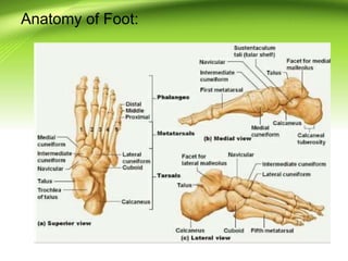
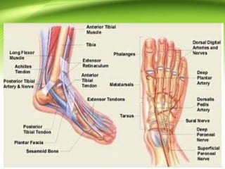
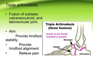
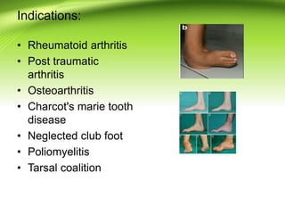
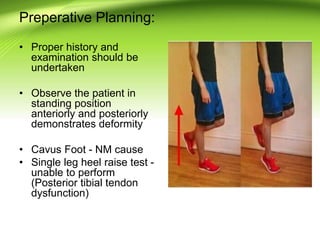
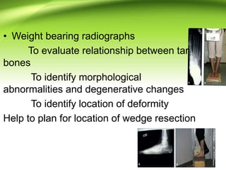
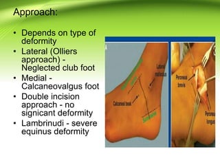
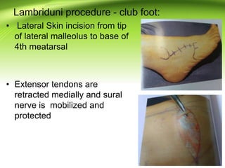
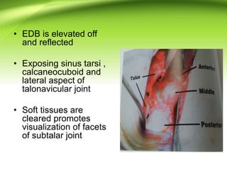
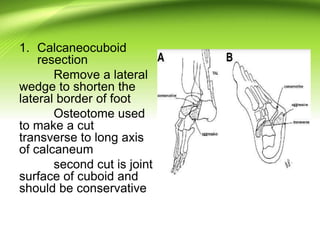
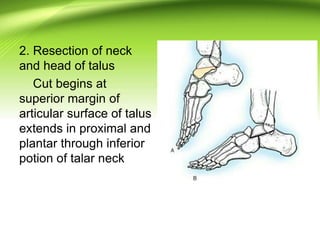
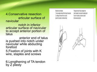
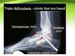
TRIPLE ARTHRODESIS
Ponnilavan
Anatomy of Foot:
Triple arthrodesis:
• Fusion of subtalar,
calcaneocuboid, and
talonavicular joint.
• Aim:
Provide hindfoot
stability
• Provide
hindfoot alignment
• Relieve pain
Indications:
• Rheumatoid arthritis
• Post traumatic
arthritis
• Osteoarthritis
• Charcot's marie tooth
disease
• Neglected club foot
• Poliomyelitis
• Tarsal coalition
Contraindications:
• Young child less than 12 years
• Inadequate blood supply
Surgery is offered when nonoperative
measures have failed to alleviate the
symptoms
Preperative Planning:
• Proper history and
examination should be
undertaken
• Observe the patient in
standing position
anteriorly and posteriorly
demonstrates deformity
• Cavus Foot - NM cause
• Single leg heel raise test -
unable to perform
(Posterior tibial tendon
dysfunction)
• Weight bearing radiographs
To evaluate relationship between tarsal
bones
To identify morphological
abnormalities and degenerative changes
To identify location of deformity
Help to plan for location of wedge resection
Approach:
• Depends on type of
deformity
• Lateral (Olliers
approach) -
Neglected club foot
• Medial -
Calcaneovalgus foot
• Double incision
approach - no
signicant deformity
• Lambrinudi - severe
equinus deformity
Lambriduni procedure - club foot:
• Lateral Skin incision from tip
of lateral malleolus to base of
4th meatarsal
• Extensor tendons are
retracted medially and sural
nerve is mobilized and
protected
• EDB is elevated off
and reflected
• Exposing sinus tarsi ,
calcaneocuboid and
lateral aspect of
talonavicular joint
• Soft tissues are
cleared promotes
visualization of facets
of subtalar joint
1. Calcaneocuboid
resection
Remove a lateral
wedge to shorten the
lateral border of foot
Osteotome used
to make a cut
transverse to long axis
of calcaneum
second cut is joint
surface of cuboid and
should be conservative
2. Resection of neck
and head of talus
Cut begins at
superior margin of
articular surface of talus
extends in proximal and
plantar through inferior
potion of talar neck
3. Completes the wedge resection and lies
along anterior surface of calcaneus parallel
to long axis of forefoot in sagital plane
Facilitates dorsiflexion of forefoot to
achieve plantigrade position
4.Conservative resection
articular surface of
navicular
notch in inferior
articular surface of navicular
to accept anterior portion of
talus
anterior end of talus
is pushed into notch under
navicular while abducting
forefoot
5.Fixation of joints with K
wire, staples and screws
6.Lengthening of TA tendon
by Z plasty
Other procedures:
• Beak triple arthrodesis
• Dunn arthrodesis
• Hoke kite arthrodesis
• Inlay grafting method
•
Postoperative protocol:
• Limb is typically immobilised in a short or long cast for
atleast 6 weeks
• Weight bearing is permitted after 6 weeks
• Ankle foot orthosis required in patient with NM disease
Week 2: wound check and remove the suture if incision is
healed
Radiographs:
1)week 6 - Good posture and evidence of healing
2)3 months - Progression of healing
3) 6 months - To confirm full union
Complications:
• Injury to neurovascular or tendinous
structures
• Wound infection
• Wound breakdown or skin necrosis
• Pseudoarthrosis
• Recurrent deformity in NM disease
• Pain from persistent malalignment,
degenerative changes and avascular
necrosis of talus
References:
1. Comprehensive orthopaedic surgery
Chapman's Volume 3
2.Operative techniques in orthopaedic
surgery Weisel 2 nd edition

Triple arthrodesis

  • 1.
  • 2.
  • 4.
    Triple arthrodesis: • Fusionof subtalar, calcaneocuboid, and talonavicular joint. • Aim: Provide hindfoot stability • Provide hindfoot alignment • Relieve pain
  • 5.
    Indications: • Rheumatoid arthritis •Post traumatic arthritis • Osteoarthritis • Charcot's marie tooth disease • Neglected club foot • Poliomyelitis • Tarsal coalition
  • 6.
    Contraindications: • Young childless than 12 years • Inadequate blood supply Surgery is offered when nonoperative measures have failed to alleviate the symptoms
  • 7.
    Preperative Planning: • Properhistory and examination should be undertaken • Observe the patient in standing position anteriorly and posteriorly demonstrates deformity • Cavus Foot - NM cause • Single leg heel raise test - unable to perform (Posterior tibial tendon dysfunction)
  • 8.
    • Weight bearingradiographs To evaluate relationship between tarsal bones To identify morphological abnormalities and degenerative changes To identify location of deformity Help to plan for location of wedge resection
  • 9.
    Approach: • Depends ontype of deformity • Lateral (Olliers approach) - Neglected club foot • Medial - Calcaneovalgus foot • Double incision approach - no signicant deformity • Lambrinudi - severe equinus deformity
  • 10.
    Lambriduni procedure -club foot: • Lateral Skin incision from tip of lateral malleolus to base of 4th meatarsal • Extensor tendons are retracted medially and sural nerve is mobilized and protected
  • 11.
    • EDB iselevated off and reflected • Exposing sinus tarsi , calcaneocuboid and lateral aspect of talonavicular joint • Soft tissues are cleared promotes visualization of facets of subtalar joint
  • 12.
    1. Calcaneocuboid resection Remove alateral wedge to shorten the lateral border of foot Osteotome used to make a cut transverse to long axis of calcaneum second cut is joint surface of cuboid and should be conservative
  • 13.
    2. Resection ofneck and head of talus Cut begins at superior margin of articular surface of talus extends in proximal and plantar through inferior potion of talar neck
  • 14.
    3. Completes thewedge resection and lies along anterior surface of calcaneus parallel to long axis of forefoot in sagital plane Facilitates dorsiflexion of forefoot to achieve plantigrade position
  • 15.
    4.Conservative resection articular surfaceof navicular notch in inferior articular surface of navicular to accept anterior portion of talus anterior end of talus is pushed into notch under navicular while abducting forefoot 5.Fixation of joints with K wire, staples and screws 6.Lengthening of TA tendon by Z plasty
  • 16.
    Other procedures: • Beaktriple arthrodesis • Dunn arthrodesis • Hoke kite arthrodesis • Inlay grafting method
  • 17.
  • 18.
    Postoperative protocol: • Limbis typically immobilised in a short or long cast for atleast 6 weeks • Weight bearing is permitted after 6 weeks • Ankle foot orthosis required in patient with NM disease Week 2: wound check and remove the suture if incision is healed Radiographs: 1)week 6 - Good posture and evidence of healing 2)3 months - Progression of healing 3) 6 months - To confirm full union
  • 19.
    Complications: • Injury toneurovascular or tendinous structures • Wound infection • Wound breakdown or skin necrosis • Pseudoarthrosis • Recurrent deformity in NM disease • Pain from persistent malalignment, degenerative changes and avascular necrosis of talus
  • 20.
    References: 1. Comprehensive orthopaedicsurgery Chapman's Volume 3 2.Operative techniques in orthopaedic surgery Weisel 2 nd edition