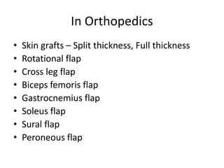
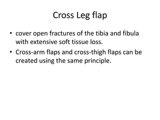
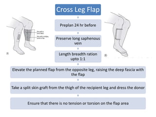
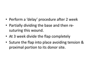
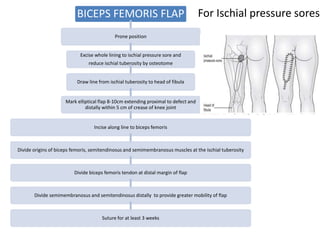
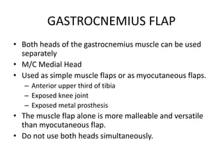
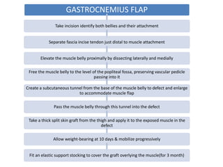
Plastic and reconstructive
surgery
Dr. Shubhanshu
Outline
• Anatomy and physiology
• Various type
• Principle of flap
• How to manage difficult and complex tissue
loss
Trauma
• Soft-tissue loss (skin, tendons, nerves, muscle)
• Upper and lower limb injury
Anatomy
Plastic surgery principles
• Adequate debridement or resection
• Wound or flap must have a good blood supply to
heal
• Gentle handling of tissue
• Minimal skin tension
• Replace defect with similar tissue – ‘like with like’
• Optimum Surgical technique
• Remember donor site ‘cost’
How does a skin graft survive?
• Imbibition plasma from the wound bed
• Inosculation of blood by after 48 hours fine
anastomotic connections
• Capillary ingrowths
• Granulation will support a graft
• Contraindicated to cover exposed
tendon/cartilage/cortical bone
Grafts
• Without their blood supply
– Split-thickness skin grafts (varying thickness)
• Thiersch grafts
• Cover all size of wound
• Contract
– Full-thickness skin grafts
– Wolfe grafts
– For Smaller area, not contract, Used in fingers
Grafts
– Composite skin grafts
• usually skin and fat, or skin and cartilage
• Rebuilding missing element like finger tip
– Nerve grafts
– Tendon grafts
Flap
• Transferred with a blood supply
– Random flaps-length to breadth ratio within 1.5:1
– Axial flaps
– Pedicled flaps
– Free flaps
• Composite flaps - osseocutaneous or myocutaneous
flaps
– Perforator flaps
Initial assessment
• Adequate removal of devitalized tissue
• vital structures reconstruction immediately or
better reconstructed later
• Degree of contamination require debridement
further
• Definitive soft-tissue cover of the wound
In Orthopedics
• Skin grafts – Split thickness, Full thickness
• Rotational flap
• Cross leg flap
• Biceps femoris flap
• Gastrocnemius flap
• Soleus flap
• Sural flap
• Peroneous flap
Concern
• Contracture
• Venous congestions
• Flap ischemia
• Loss of sensation
Skin graft
Grab skin so become tense
cut graft with humpley knife
Dress donar with calcium
alginate redress after 10 days
Apply graft directly over
wound
Apply dressing redress after
5 days
Rotational Flap
Draw Isosceles triangle around
defect apex towards centre of arc
rotation of flap
Draw arc
Raise skin subcutaneous tissue
Place flap over wound
• For Sacral pressure sore
Cross Leg flap
• cover open fractures of the tibia and fibula
with extensive soft tissue loss.
• Cross-arm flaps and cross-thigh flaps can be
created using the same principle.
Cross Leg Flap
Preplan 24 hr before
Preserve long saphenous
vein
Length breadth ration
upto 1:1
Elevate the planned flap from the opposite leg, raising the deep fascia with
the flap
Take a split skin graft from the thigh of the recipient leg and dress the donor
Ensure that there is no tension or torsion on the flap area
• Perform a ‘delay’ procedure after 2 week
• Partially dividing the base and then re-
suturing this wound.
• At 3 week divide the flap completely
• Suture the flap into place avoiding tension &
proximal portion to its donor site.
For Ischial pressure sores
BICEPS FEMORIS FLAP
Prone position
Excise whole lining to ischial pressure sore and
reduce ischial tuberosity by osteotome
Draw line from ischial tuberosity to head of fibula
Mark elliptical flap 8-10cm extending proximal to defect and
distally within 5 cm of crease of knee joint
Incise along line to biceps femoris
Divide origins of biceps femoris, semitendinosus and semimembranosus muscles at the ischial tuberosity
Divide biceps femoris tendon at distal margin of flap
Divide semimembranosus and semitendinosus distally to provide greater mobility of flap
Suture for at least 3 weeks
GASTROCNEMIUS FLAP
• Both heads of the gastrocnemius muscle can be used
separately
• M/C Medial Head
• Used as simple muscle flaps or as myocutaneous flaps.
– Anterior upper third of tibia
– Exposed knee joint
– Exposed metal prosthesis
• The muscle flap alone is more malleable and versatile
than myocutaneous flap.
• Do not use both heads simultaneously.
GASTROCNEMIUS FLAP
Take incision identify both bellies and their attachment
Separate fascia incise tendon just distal to muscle attachment
Elevate the muscle belly proximally by dissecting laterally and medially
Free the muscle belly to the level of the popliteal fossa, preserving vascular pedicle
passing into it
Create a subcutaneous tunnel from the base of the muscle belly to defect and enlarge
to accommodate muscle flap
Pass the muscle belly through this tunnel into the defect
Take a thick split skin graft from the thigh and apply it to the exposed muscle in the
defect
Allow weight-bearing at 10 days & mobilize progressively
Fit an elastic support stocking to cover the graft overlying the muscle(for 3 month)
Soleus Flap
An incision is made on the medial aspect of the leg commencing approx 10
cm below the popliteal fossa extending to the achilles tendon
Soleus and gastrocnemius muscles are identified and separated.
Hemisoleus is then divided from its insertion at the achilles tendon and
dissected cephalad to allow for a sufficient arc of rotation to cover the defect
Two sets of perforating vessels are divided to achieve this
Hemisoleus is then inset into the defect and sutured in place
split-thickness skin grafts (STSG) to cover the muscle
• Most common usage of this flap is for the distal-third defects of the
leg.
• The reverse sural flap permits the soft tissue reconstruction without
the need for microsurgery.
Sural Flap
flap is marked on the skin in the form of an ellipse centered on the raphe between the two
gastrocnemius muscle bodies, whose projection is visible on the posterior aspect of the leg
The incision starts on the lateral and superior borders of the flap and continues in the
subfascial plane until the sural nerve is identified in the median raphe
Then the incision goes on the other boundaries of the flap and the subfascial dissection
continues with the ligation of all the perforators from the gastrocnemius belly
The sural nerve is attached to the fascia at the superior border of the flap.
Peroneous Brevis flap
• Lower 1/3 leg wound
• Distal leg ankle
• Lateral Malleolous,
• Proximal calcaneus
• Achilles
• Medial malleolous
• Ankle joint
Intact peroneal artery perforators
Peroneous Brevis Flap
Identify peroneous brevis separate in distal to proximal
Preserve peroneous lie posterior surface close to posterior septum
Proximal connection of muscle to fibula was detached and muscle was dissected off
fibula in a proximal to distal direction leaving a thin layer of muscle attached to fibula
Preserve the superficial peroneal nerve, anterior tibial vessels to augment the
vascularity during harvest of flap
Covered with a split thickness skin graft
THANKYOU

Plastic and reconstructive surgery presentation.pptx

  • 1.
  • 2.
    Outline • Anatomy andphysiology • Various type • Principle of flap • How to manage difficult and complex tissue loss
  • 3.
    Trauma • Soft-tissue loss(skin, tendons, nerves, muscle) • Upper and lower limb injury
  • 4.
  • 5.
    Plastic surgery principles •Adequate debridement or resection • Wound or flap must have a good blood supply to heal • Gentle handling of tissue • Minimal skin tension • Replace defect with similar tissue – ‘like with like’ • Optimum Surgical technique • Remember donor site ‘cost’
  • 6.
    How does askin graft survive? • Imbibition plasma from the wound bed • Inosculation of blood by after 48 hours fine anastomotic connections • Capillary ingrowths • Granulation will support a graft • Contraindicated to cover exposed tendon/cartilage/cortical bone
  • 7.
    Grafts • Without theirblood supply – Split-thickness skin grafts (varying thickness) • Thiersch grafts • Cover all size of wound • Contract – Full-thickness skin grafts – Wolfe grafts – For Smaller area, not contract, Used in fingers
  • 8.
    Grafts – Composite skingrafts • usually skin and fat, or skin and cartilage • Rebuilding missing element like finger tip – Nerve grafts – Tendon grafts
  • 9.
    Flap • Transferred witha blood supply – Random flaps-length to breadth ratio within 1.5:1 – Axial flaps – Pedicled flaps – Free flaps • Composite flaps - osseocutaneous or myocutaneous flaps – Perforator flaps
  • 11.
    Initial assessment • Adequateremoval of devitalized tissue • vital structures reconstruction immediately or better reconstructed later • Degree of contamination require debridement further • Definitive soft-tissue cover of the wound
  • 12.
    In Orthopedics • Skingrafts – Split thickness, Full thickness • Rotational flap • Cross leg flap • Biceps femoris flap • Gastrocnemius flap • Soleus flap • Sural flap • Peroneous flap
  • 14.
    Concern • Contracture • Venouscongestions • Flap ischemia • Loss of sensation
  • 15.
    Skin graft Grab skinso become tense cut graft with humpley knife Dress donar with calcium alginate redress after 10 days Apply graft directly over wound Apply dressing redress after 5 days
  • 16.
    Rotational Flap Draw Isoscelestriangle around defect apex towards centre of arc rotation of flap Draw arc Raise skin subcutaneous tissue Place flap over wound • For Sacral pressure sore
  • 17.
    Cross Leg flap •cover open fractures of the tibia and fibula with extensive soft tissue loss. • Cross-arm flaps and cross-thigh flaps can be created using the same principle.
  • 18.
    Cross Leg Flap Preplan24 hr before Preserve long saphenous vein Length breadth ration upto 1:1 Elevate the planned flap from the opposite leg, raising the deep fascia with the flap Take a split skin graft from the thigh of the recipient leg and dress the donor Ensure that there is no tension or torsion on the flap area
  • 19.
    • Perform a‘delay’ procedure after 2 week • Partially dividing the base and then re- suturing this wound. • At 3 week divide the flap completely • Suture the flap into place avoiding tension & proximal portion to its donor site.
  • 20.
    For Ischial pressuresores BICEPS FEMORIS FLAP Prone position Excise whole lining to ischial pressure sore and reduce ischial tuberosity by osteotome Draw line from ischial tuberosity to head of fibula Mark elliptical flap 8-10cm extending proximal to defect and distally within 5 cm of crease of knee joint Incise along line to biceps femoris Divide origins of biceps femoris, semitendinosus and semimembranosus muscles at the ischial tuberosity Divide biceps femoris tendon at distal margin of flap Divide semimembranosus and semitendinosus distally to provide greater mobility of flap Suture for at least 3 weeks
  • 21.
    GASTROCNEMIUS FLAP • Bothheads of the gastrocnemius muscle can be used separately • M/C Medial Head • Used as simple muscle flaps or as myocutaneous flaps. – Anterior upper third of tibia – Exposed knee joint – Exposed metal prosthesis • The muscle flap alone is more malleable and versatile than myocutaneous flap. • Do not use both heads simultaneously.
  • 22.
    GASTROCNEMIUS FLAP Take incisionidentify both bellies and their attachment Separate fascia incise tendon just distal to muscle attachment Elevate the muscle belly proximally by dissecting laterally and medially Free the muscle belly to the level of the popliteal fossa, preserving vascular pedicle passing into it Create a subcutaneous tunnel from the base of the muscle belly to defect and enlarge to accommodate muscle flap Pass the muscle belly through this tunnel into the defect Take a thick split skin graft from the thigh and apply it to the exposed muscle in the defect Allow weight-bearing at 10 days & mobilize progressively Fit an elastic support stocking to cover the graft overlying the muscle(for 3 month)
  • 25.
    Soleus Flap An incisionis made on the medial aspect of the leg commencing approx 10 cm below the popliteal fossa extending to the achilles tendon Soleus and gastrocnemius muscles are identified and separated. Hemisoleus is then divided from its insertion at the achilles tendon and dissected cephalad to allow for a sufficient arc of rotation to cover the defect Two sets of perforating vessels are divided to achieve this Hemisoleus is then inset into the defect and sutured in place split-thickness skin grafts (STSG) to cover the muscle
  • 28.
    • Most commonusage of this flap is for the distal-third defects of the leg. • The reverse sural flap permits the soft tissue reconstruction without the need for microsurgery. Sural Flap flap is marked on the skin in the form of an ellipse centered on the raphe between the two gastrocnemius muscle bodies, whose projection is visible on the posterior aspect of the leg The incision starts on the lateral and superior borders of the flap and continues in the subfascial plane until the sural nerve is identified in the median raphe Then the incision goes on the other boundaries of the flap and the subfascial dissection continues with the ligation of all the perforators from the gastrocnemius belly The sural nerve is attached to the fascia at the superior border of the flap.
  • 33.
    Peroneous Brevis flap •Lower 1/3 leg wound • Distal leg ankle • Lateral Malleolous, • Proximal calcaneus • Achilles • Medial malleolous • Ankle joint Intact peroneal artery perforators
  • 34.
    Peroneous Brevis Flap Identifyperoneous brevis separate in distal to proximal Preserve peroneous lie posterior surface close to posterior septum Proximal connection of muscle to fibula was detached and muscle was dissected off fibula in a proximal to distal direction leaving a thin layer of muscle attached to fibula Preserve the superficial peroneal nerve, anterior tibial vessels to augment the vascularity during harvest of flap Covered with a split thickness skin graft
  • 36.

Editor's Notes

  • #2 Sir Harold Gillies operating during the First World War ‘the birth of plastic surgery’.
  • #5 Epidermis regenerates from deeper follicular elements, with the most superficial layer losing vascularity and acting as a barrier to fluid loss and providing important protection against invasion by microorganisms. depth of the dermis and the amounts of elastin and skin adnexal elements, such as sweat glands and hair follicles Without skin, wounds heal by secondary intention with fibrosis and contracture and underlying structures are vulnerable to necrosis, chronic infection and dysfunction.
  • #16 Do not apply grafts in the presence of Group A beta haemolytic streptococci infection Staphylococcus MRSA