
Recommended
More Related Content
What's hot
What's hot (20)
Viewers also liked
Viewers also liked (20)
Similar to Angular & torsional deformities of the lower limb
Similar to Angular & torsional deformities of the lower limb (20)
Recently uploaded
Recently uploaded (20)
Angular & torsional deformities of the lower limb
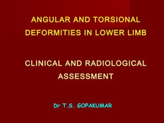
- 1. ANGULAR AND TORSIONAL DEFORMITIES IN LOWER LIMB CLINICAL AND RADIOLOGICAL ASSESSMENT Dr T.S. GOPAKUMAR
- 2. EVOLUTION OF ALIGNMENT IN THE LOWER LIMBS Torsion Fetus MM behind LM Birth same level 1 year LM behind MM Adult 20 degrees External torsion
- 3. Evolution of alignment in the lower limbs
- 6. Every change in the form and function of the bones or function alone is followed by certain definite changes in the external configurations in accordance with mathematical laws. WOLFF LAW
- 7. HEUTER VOLKMANN LAW (1862) Pressure inhibit growth and decreased pressure accelerate the growth of the physis
- 8. ASSESSMENT OF ANGULAR DEFORMITY History Nutritional deficiency Renal diseases Muscle weakness Gastrointestinal problems Family history
- 9. ASSESSMENT OF ANGULAR DEFORMITY Stature Upper segment lower segment ratio Facies Teeth Metaphyseal thickening Hand Nails Changes of rickets Proximal muscle weakness
- 10. CAUSES OF GENU VARUM Metabolic Bone Disease Nutritional Rickets Renal tubular rickets Renal Glomerular rickets Renal Tubular acidosis
- 12. Assymetric growth arrest Blount’s disease Trauma Infection Tumor
- 13. Physiological genu varum Bow leg Medial tibial torsion
- 14. Distance between the femoral condyles Lateral thigh leg angle Foot normal / postural MT varus Foot progression angle Lateral thrust indicate progression Ligamentous stability Torsional profile
- 15. X ray Unnecessary Tibia angulated medially at the jn. Of proximal and middle third Femur angulated in the distal third Medial cortex of tibia and femur thickened and sclerosed Epiphysis,Physis and metaphysis have normal appearance Symmetrical involvement Metaphyseo diaphyseal angle < 11 degrees
- 16. TREATMENT Spontaneous regression Orthopaedic shoes and Orthosis Osteotomy
- 17. Blount’s disease
- 18. TIBIA VARA (BLOUNT’S disease) Growth defect in the proximal medial tibial epiphysis Infantile <3 years Juvenile 3 – 10 years Adolescent > 10 years Manifest 18 – 24 years Obese children Often assymetrical Progressive varus deformity Lateral thrust on standing Siffert Katz sign
- 19. RADIOGRAPHIC FEATURES Varus angulation at epiphseo metaphysealjn Widened and irregular physeal line medially Medially sloping and irregularly ossified epiphysis Prominent beaking of medial metaphysis Lateral subluxation of proximal tibia Normal knee radiograph in a toddler does not exclude Blount’s
- 20. Tibiofemoral angle Metaphyseo diaphyseal angle Epiphyseo metaphyseal angle Langenskiold classification
- 21. Metaphysio diaphyseal angleTibio femoral angle
- 23. Physiological genu varum Blounts disease Invovement Symmetrical Often assymetrical Site of angulation prox &middle third Proximal metphysis Femur Bowed medially Normal except late Lateral thrust Absent Often present Meta Dia angle < 11 Greater than 11 Upper tib Metaphysis Normal Irregular rarifaction Upper tib Epiphysis Normal Sloping Upper tib Physis Normal Narrowed medially Lateral Tib Cortex Gentle curve Straight Med Tib Cortex Gentle curve Sharp angulation
- 24. ADOLESCENT TIBIA VARA 8 Years Males Obese Often Unilateral Black Africans Tibia vara Internal tibial torsion
- 25. X RAY Shape of epiphysis normal Lack of beaking of medial tibial metaphysis Widening of medial tibial epiphyseal plate Widening of lateral distal femoral physis
- 26. Achondroplasia
- 28. Rickets
- 29. Biochemical investigations S. Calcium S. Phosphorus S. Alkaline phosphatase Renal function tests Urine pH Glucose Amino acids 24 hr urine calcium 24 hr urine phosphorus
- 30. X-ray Epiphysis small fragmented Physis wide Metaphysis cupping flaring Diaphysis thinning of cortex
- 31. Post infective genu varum
- 32. GENU VALGUM Awkward gait Easy fatigue due to swinging of legs Shoes collapse medially due to pronated feet Calf and leg pain Patellar mal alignment Obesity due to inactivity Early degenerative arthritis
- 33. ASSESSMENT Inter malleolar distance Lateral tibiofemoral angle Q angle Patellar stability Tibial torsion Flat foot
- 34. CAUSES OF GENU VALGUM Metabolic Bone Disease Nutritional Rickets Renal tubular rickets Renal Glomerular rickets Renal Tubular acidosis
- 35. Assymetric Growth Arrest Trauma Infection Tumor Primary tibia valga
- 36. Bone Dysplasia MED SED Chondroectodermal dysplasia Multiple hereditary exostosis Ollier’s disease
- 37. Endocrine Turners syndrome Congenital Congenital def of fibula Inflammatory Rh arthritis Tuberculosis Paralytic Polio ITB contracture
- 40. Ellis van Creveld syndrome Pyknodysostosis
- 44. TREATMENT Reassurance Stretching of ITB Shoe modification to avoid foot strain Knock knee orthosis Epiphyseal stapling Epiphyseodesis Osteotomy Ilizarov Hemichondrodiactasis
- 45. Genu Recurvatum
- 46. TORSION Twisting of long bone in the longitudinal axis Internal tibial torsion External tibial torsion Femoral antetorsion Femoral retrotorsion Tibial vs Tibiofemoral torsion
- 47. CAUSES OF TOEING IN GAIT Metatarsus varus CTEV Pronated feet Tibia vara Medial tibial torsion Genu valgum (shift center of gravity medially) Congenital tibial deficiency Abnormal femoral antetorsion Spasticity of medial rotators Acetabular anteversion
- 48. TOE OUT GAIT Talipes calcaneovalgus Pes valgus Triceps surae contracture Lateral tibial torsion Cong absence of tibia Abnormal femoral retroversion Paralysis of medial rotators Acetabular retroversion
- 49. Rotational Profile (Staheli) 1. Foot progression angle 2. Medial hip rotation in extension 3. Lateral hip rotation in extension 4. Thigh foot angle 5. Angle of the trans malleolar axis 6. Configuration of the foot
- 50. 1. Foot progression angle Normal average + 10-15 degrees Compensatory tibial torsion may make FPA normal even with excessive femoral torsion
- 51. Medial and lateral hip rotation in extension Medial 40 –60 50 more in females Lateral 25- 65 45 equal in both sexes Femoral anteversion (Staheli) >90 IR 0 ER severe 80- 90 IR 0-10 ER moderate 70- 80 IR 10- 20 ER mild
- 52. Thigh foot angle Patient prone Knee flexed 90 degrees Ankle neutral Angle between the long axis of foot and long axis of the thigh Assessment of tibial torsion Normal +10
- 53. Angle of transmalleolar axis Patient prone Knee flexed 90 degrees Ankle neutral Line joining the center point of medial and lateral malleolus are marked on sole of foot Perpendicular to trans malleolar axis Thigh axis line Mean +15
- 54. Foot deformities Metatarsus varus in toeing Calcaneovalgus out toeing Planovalgus
- 55. Femoral torsion 1 year 40 degrees 2 years 30 degrees (Reduces 1-2 degrees /year) 10 years 20 degrees 15 years 16 degrees Adult 15 +/- 10
- 56. Femoral torsion Clinical features In toeing gait Exaggerated IR in extension of the hip Limitation of ER ER of hip increased in 90 degree flexion of the hip Adaptive changes Hind foot valgus External tibial torsion
- 57. Effect Cosmetic Torsional mal alignment Patellofemoral problems Femoral Torsion
- 58. Femoral Torsion Assessment Ryder method Prone GT palpated Leg is laterally rotated till GT is most prominent The degree of rotation from neutral is the degree of anteversion
- 59. Femoral Torsion Assessment X ray CT MRI USG
- 60. Femoral Torsion Treatment Reassurance No role for shoe modifications Orthosis with twister cables has no role DB splint harmful Avoid reverse tailors position while sitting. Encourage cross leg sitting
- 61. Surgery Child more than 9 years Measured anteversion > 45 degrees(CT/MRI) Clinically severe (IR>90, ER 0) Lateral tibial torsion <35 Functional and cosmetic disability Does not increase incidence of OA of hip/ knee
- 62. Surgery Derotational Osteotomy Trochanteric Supramalleolar Middiaphyseal Ilizarov How much to rotate ?
- 63. TIBIAL TORSION Rotational profile (Staheli) 1. Foot progression angle 2. Medial hip rotation in extension 3. Lateral hip rotation in extension 4. Thigh foot angle 5. Angle of the trans malleolar axis 6. Configuration of the foot
- 64. Xray Nachlas method Hutter and Scott method Rosen and sandick method CT USG TIBIAL TORSION Assessment
- 65. MEDIAL TIBIAL TORSION Idiopathic Cong metatarsus varus Genu varum Femoral anteversion Familial
- 66. CLINICAL PRESENTATION Intoeing gait Bow legs Kites rotation test Staheli’s torsion profile
- 67. LATERAL TIBIAL TORSION Contracture of IT band Idiopathic Congenital
- 68. Patella point laterally Feet point outwards Axis medial to 2nd MT LM posterior to MM Knock knee Ober test ITB IR of hip restricted Femoral antetorsion ER of hip restricted Triceps surae contracture cause toeing out gait CLINICAL PRESENTATION
- 69. External Tibial Torsion Does not correct with growth Contracted ITB /TA DB splint Osteotomy
- 70. Internal Tibial Torsion Spontaneous correction DB splints Corrective casts Osteotomy severe deformity above 8 years
- 71. Thank You