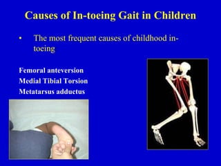
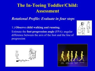
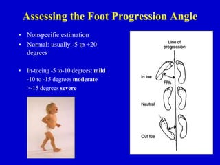
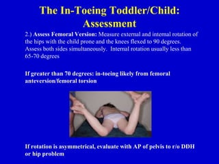
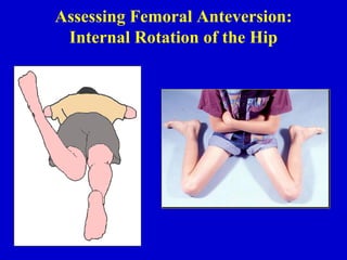
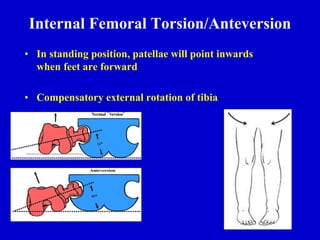
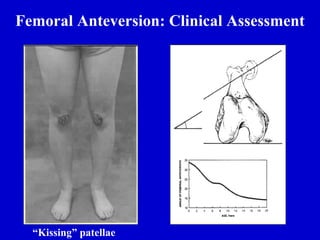
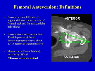
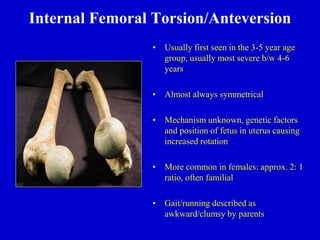
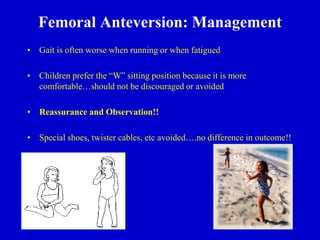
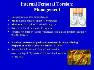
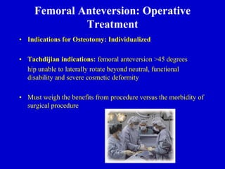
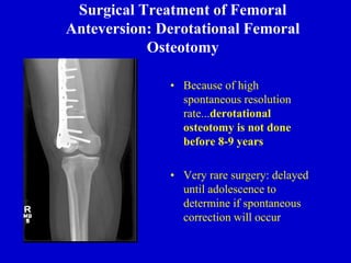
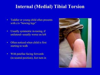
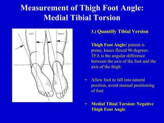
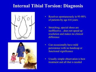
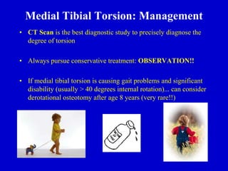
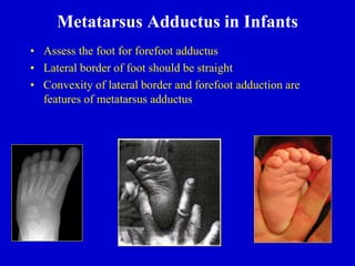
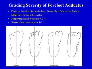
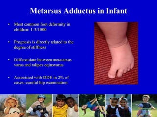
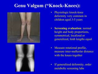
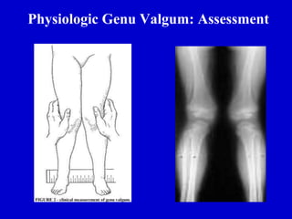
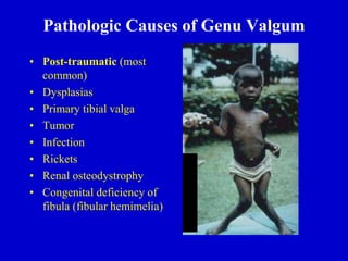
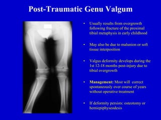
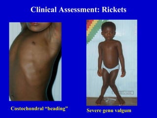
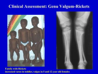
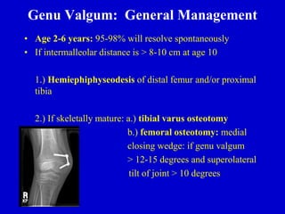
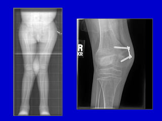
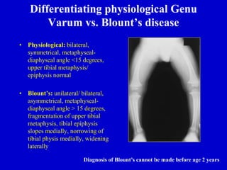
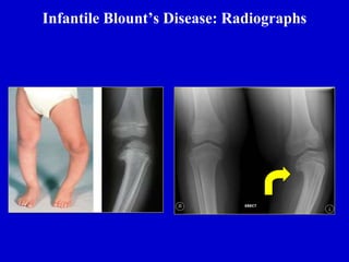
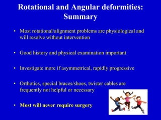
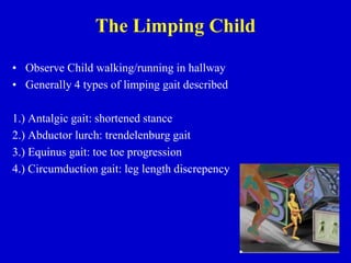
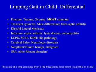
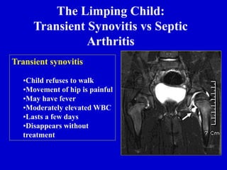
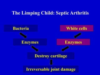
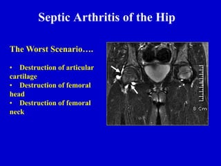
1. In-toeing, Out-toeing, and Limping—Making
Sense of Common Pediatric Gait
Abnormalities
HOSSEIN ASLANI MD
Pediatric orthopedic fellowship
2. Rotational & Angular Deformities in Children
Objectives:
1.) Review common physiologic and pathologic causes of in-toeing/out-toeing gait in
children
2.) Review diagnosis and physical examination techniques used in assessing pediatric
rotational and angular deformities
3.) Review the current management of pediatric rotational and angular deformities in
children
4.) Review the differential diagnosis of limping gait/gait abnormality in children
3. Rotational & Angular Deformities in
Children: Introduction
• Rotational and Angular
Deformities are quite common
in pediatrics
• Very diverse spectrum of
diagnoses: physiologic to
pathologic
• In-toeing/Out-toeing, Genu
varum (bowlegs)/valgum
(knock-knees)
4. The In-toeing Toddler or Child
• History: Inquire about onset, severity, progression, disability,
and previous treatment
• Always assess developmental history: when did child start
walking independently, gross and fine motor skills
• Screening examination to r/o hip dysplasia (DDH), other
neurological problems (cerebral palsy)
5. Causes of In-toeing Gait in Children
• The most frequent causes of childhood in-
toeing:
Femoral anteversion
Medial Tibial Torsion
Metatarsus adductus
6. The In-Toeing Toddler/Child:
Assessment
Rotational Profile: Evaluate in four steps
1.) Observe child walking and running.
Estimate the foot progression angle (FPA): angular
difference between the axis of the foot and the line of
progression
7. Assessing the Foot Progression Angle
• Nonspecific estimation
• Normal: usually -5 tp +20
degrees
• In-toeing -5 to-10 degrees: mild
-10 to -15 degrees moderate
>-15 degrees severe
8. The In-Toeing Toddler/Child:
Assessment
2.) Assess Femoral Version: Measure external and internal rotation of
the hips with the child prone and the knees flexed to 90 degrees.
Assess both sides simultaneously. Internal rotation usually less than
65-70 degrees
If greater than 70 degrees: in-toeing likely from femoral
anteversion/femoral torsion
If rotation is asymmetrical, evaluate with AP of pelvis to r/o DDH
or hip problem
10. Internal Femoral Torsion/Anteversion
• In standing position, patellae will point inwards
when feet are forward
• Compensatory external rotation of tibia
12. Femoral Anteversion: Definitions
• Femoral version defined as the
angular difference between axis of
femoral neck and the transcondylar
axis of knee
• Femoral anteversion ranges from
30-40 degrees at birth and
decreases progressively to about
10-15 degrees at skeletal maturity
• Measurement:X-rays (biplane):
technically difficult
CT--most accurate method
13. Internal Femoral Torsion/Anteversion
• Usually first seen in the 3-5 year age
group, usually most severe b/w 4-6
years
• Almost always symmetrical
• Mechanism unknown, genetic factors
and position of fetus in uterus causing
increased rotation
• More common in females: approx. 2: 1
ratio, often familial
• Gait/running described as
awkward/clumsy by parents
14. Femoral Anteversion: Management
• Gait is often worse when running or when fatigued
• Children prefer the “W” sitting position because it is more
comfortable…should not be discouraged or avoided
• Reassurance and Observation!!
• Special shoes, twister cables, etc avoided….no difference in outcome!!
15. Internal Femoral Torsion:
Management
• Internal femoral torsion/antetorsion
• Mild: internal rotation of hip 70-80 degrees
• Moderate: internal rotation 80-90 degrees
• Severe: internal rotation > 90 degrees
• External hip rotation is usually reduced: total arch of rotation is usually
90-100 degrees
• Resolves spontaneously without treatment in overwhelming
majority of patients: most literature-- 98-99%
• Results from decrease in femoral anterversion
over time (age 8-9 years) and from a lateral rotation
of the tibia
16. Femoral Anteversion: Operative
Treatment
• Indications for Osteotomy: Individualized
• Tachdijian indications: femoral anteversion >45 degrees
hip unable to laterally rotate beyond neutral, functional
disability and severe cosmetic deformity
• Must weigh the benefits from procedure versus the morbidity of
surgical procedure
17. Surgical Treatment of Femoral
Anteversion: Derotational Femoral
Osteotomy
• Because of high
spontaneous resolution
rate...derotational
osteotomy is not done
before 8-9 years
• Very rare surgery: delayed
until adolescence to
determine if spontaneous
correction will occur
18. Internal (Medial) Tibial Torsion
• Toddler or young child often presents
with c/o “bowing legs”
• Usually symmetric in-toeing, if
unilateral--usually worse on left
• Often noticed when child is first
starting to walk
• With patellae facing forwards
(in neutral position), feet turn in
19. Measurement of Thigh Foot Angle:
Medial Tibial Torsion
3.) Quantify Tibial Version
Thigh Foot Angle: patient is
prone, knees flexed 90 degrees:
TFA is the angular difference
between the axis of the foot and the
axis of the thigh
• Allow foot to fall into natural
position, avoid manual positioning
of foot
• Medial Tibial Torsion: Negative
Thigh Foot Angle
20. Internal Tibial Torsion: Diagnosis
• Resolves spontaneously in 95-98%
of patients by age 4-6 years
• Stretching, special shoes are
inefffective…does not speed up
resolution and makes no clinical
difference
• Can occasionally have mild
persistence with no handicap or
functional significance
• Usually simple observation is best
treatment and all that is needed
21. Medial Tibial Torsion: Management
• CT Scan is the best diagnostic study to precisely diagnose the
degree of torsion
• Always pursue conservative treatment: OBSERVATION!!
• If medial tibial torsion is causing gait problems and significant
disability (usually > 40 degrees internal rotation)... can consider
derotational osteotomy after age 8 years (very rare!!)
22. Metatarsus Adductus in Infants
• Assess the foot for forefoot adductus
• Lateral border of foot should be straight
• Convexity of lateral border and forefoot adduction are
features of metatarsus adductus
23. Grading Severity of Forefoot Adductus
• Project a line that bisects the heel. Normally it falls on the 2nd toe
• Mild: falls through the 3rd toe
• Moderate: falls between toes 3-4
• Severe: falls between toes 4-5
24. Metarsus Adductus in Infant
• Most common foot deformity in
children: 1-3/1000
• Prognosis is directly related to the
degree of stiffness
• Differentiate between metatarsus
varus and talipes eqinovarus
• Associated with DDH in 2% of
cases--careful hip examination
25. Metatarsus Adductus: Assessment
• Exact cause is unknown
• Commonly believed to be
caused by intrauterine
positioning or crowding
• No correlation between
gestational age at birth,
maternal age, or birth
order
26. Metatarsus Adductus: Management
• Forefoot can gently be stretched passively with each diaper change
• Occasionally will use serial casting and reverse/straight last shoes to
correct deformity
• Observation and Reassurance: will resolve spontaneously in 90-95%
of patients (tends to persist until age 12-18 months)
27. Physiologic Infantile Out-Toeing
• Out-toeing in early infancy is usually due to a lateral rotation
contracture of the hips
• When infant is positioned upright, the feet will usually turn out
• Resolves spontaneously with ambulation…no treatment is
needed
28. Out-toeing: External Tibial Torsion
• Most common cause of out-toeing in
children
• May worsen over time because of the
normal lateral rotation of the tibia that
occurs with growth
• May be associated with patellofemoral
knee pain
• If combined with femoral anteversion (
knee internally rotated and ankle
externally rotated):“miserable
malalignment syndrome”…inefficient
gait and patellofemoral joint pain
30. Lower Extremity Rotational Profile at
Various Ages
• Normal alignment progresses from 10-15 degrees of varus
at birth to maximum valgus angulation of 10-15 degrees at
3-4 years of age
31. Genu Valgum (“Knock-Knees):
• Physiologic knock-knee
deformity very common in
children aged 3-5 years
• Screening evaluation: normal
height and body proportions,
symmetrical, localized or
generalized, limb lengths equal
• Measure rotational profile,
measure inter-malleolar distance
with the knees together
• If generalized deformity, order
metabolic screening labs
34. Post-Traumatic Genu Valgum
• Usually results from overgrowth
following fracture of the proximal
tibial metaphysis in early childhood
• May also be due to malunion or soft
tissue interposition
• Valgus deformity develops during the
1st 12-18 months post-injury due to
tibial overgrowth
• Management: Most will correct
spontaneously over course of years
without operative treatment
• If deformity persists: osteotomy or
hemiepiphyseodesis
35. Rickets: Diagnosis and Management
• Suspect rickets in child with increasing genu varum/valgum, short
stature, poor nutritional status (vitamin D deficiency)
• Produces generalized genu varum/valgum with bowing of the
diaphysis and distinctive cupping and widening of the epiphysis
• Refer to endocrinologist for medical management: Ca, Phos
supplementation (Vitamin D resistant form possible)
• Correction (if necessary) usually delayed until the end of growth
as recurrence of deformity is quite common
37. Clinical Assessment: Genu Valgum-Rickets
Family with Rickets
increased varus in toddler, valgus in 5 and 12 year old females
38. Genu Valgum: General Management
• Age 2-6 years: 95-98% will resolve spontaneously
• If intermalleolar distance is > 8-10 cm at age 10
1.) Hemiephiphyseodesis of distal femur and/or proximal
tibia
2.) If skeletally mature: a.) tibial varus osteotomy
b.) femoral osteotomy: medial
closing wedge: if genu valgum
> 12-15 degrees and superolateral
tilt of joint > 10 degrees
39.
40. Physiologic Genu Varum: Assessment
• Parents will often note bow leg
deformity, usually recognized
when child starts to walk (12-18
months)
• Commonly bilateral and
symmetric bowing
• Seldom causes functional
disability [X-rays unnecessary
until at least 18 months of age]
• Physiologic bowing usually
spontaneously resolves by the age
of two years
41. Differentiating physiological Genu
Varum vs. Blount’s disease
• Physiological: bilateral,
symmetrical, metaphyseal-
diaphyseal angle <15 degrees,
upper tibial metaphysis/
epiphysis normal
• Blount’s: unilateral/ bilateral,
asymmetrical, metaphyseal-
diaphyseal angle > 15 degrees,
fragmentation of upper tibial
metaphysis, tibial epiphysis
slopes medially, norrowing of
tibial physis medially, widening
laterally
Diagnosis of Blount’s cannot be made before age 2 years
43. Pathological Conditions Causing
Varus Deformity of the Legs
• Metabolic bone disease: Vitamin
D deficiency, Vitamin D refractory
rickets
• Asymmetrical growth arrest or
retardation: Blount’s disease,
Trauma, Infection, Tumor
• Bone dysplasia: metaphyseal
dysplasia
• Congenital
• Neuromuscular
44. Infantile Blount’s Disease:
Epidemiology
• Risk factors: Obesity, African
American,Walking at early age, +
Family history
• Differential Diagnosis:
Physiologic genu varum
(metaphyseal-diaphyseal angle less than
15 degrees)
Rickets
Osteomyelitis,
Trauma, Tumor
Fibrous dysplasia
Metaphyseal chondrodysplasia
45. Blount’s Disease: Classification
• Infantile: (early onset)
onset between 1-3 years, bilateral, usually symmetric, pts often
large for age, etiology is abnormal compression on medial
proximal tibial physis, may feel bony prominence or “beak”
over the medial tibial condyle, often have lateral thrust to gait
Very difficult to differentiate from physiologic varus/
bowlegs in patients < 2 years
Adolescent: (onset over 11 years)
often presents with tenderness/pain over the medial prominence
of the proximal tibia, pts often obese
Prevalence: General population <0.3%, Obese African
American male:2.5%
46. Metaphyseal/Diaphyseal Varus Angle
• Often used to differntiate
physiologic from Blount’s
disease
• If greater than 15-17 degrees,
tibia vara or Blount’s disease
is likely
• Follow radiographs every six
months….physiologic varus
will gradually improve after
age 2 years while Blount’s will
progressively worsen
47. Measuring the Tibial-Femoral Angle
• Line is drawn down center
of tibia and femur….
Intersecting angle is the
tibio-femoral angle
49. Adolescent Blount’s Disease
• Definition: Growth disorder involving
the medial portion of the proximal
tibial growth plate that produces a
localized varus deformity
• More often unilateral, usually seen in
obese individuals, slightly more males
than females, African American,
certain geographic regions
• Definite cause unknown:
biomechanical overload to proximal
tibia physis due to varus alignment
and excessive body weight
50. Adolescent Blount’s: Theorized Cause of
Progressive Varus Deformity
Childhood Varus
Rapid Weight Gain
Medial Growth Plate Injury
Progressive Varus Knee
53. Blount’s Disease: Non-Operative
Treatment
• Non-operative: Observation only until age 2 years
• Anti-Blount’s Brace: Usually used for pts aged 2-3 years with:
1.) metaphyseal/diaphyseal angle > 15-17 degrees
2.) tibial/femoral angle > 15 degrees
3.) brace is designed to provide rotational support, usually worn FT
4.) usually takes 1 year to determine effectiveness of brace
5.) brace is ineffective in adolescents
• If operative correction necessary in infantile Blount’s….results are
better when done before the age of 4 years
54. Blount’s Disease: Operative
Treatment
• For optimum correction and results in infantile tibia vara: Surgical
treatment in early stages is crucial
Avoids sequelae of joint incongruity, limb shortening, and persistent
angulation
• Proximal tibial osteotomy: distal to patellar tendon insertion (avoid
proximal physis (dome, closing or opening wedge)
• Adolescent tibia vara: predominately surgical treatment:
1.) Lateral epiphyseodesis: recommended as initial procedure if more
than one year of growth remaining
2.) High tibial osteotomy with internal fixation ( usually correct to
about 0-5 degrees of varus)
3.) Realignment by external fixation: Ilizarov, Dynamic Axial
External fixator, Taylor Spatial Frame
55. Surgical Correction: Proximal Tibial
Osteotomy
• Demonstration of opening
wedge tibial osteotomy
procedure for correction
of infantile blount’s
disease
• Usually recommend slight
overcorrection into mild
valgus (reverse excessive
compression forces
medially: avoid injury to
physis)
57. Rotational and Angular deformities:
Summary
• Most rotational/alignment problems are physiological and
will resolve without intervention
• Good history and physical examination important
• Investigate more if asymmetrical, rapidly progressive
• Orthotics, special braces/shoes, twister cables are
frequently not helpful or necessary
• Most will never require surgery
58. The Limping Child….
• Relatively large differential diagnosis list
• Obtain good history: VERY important
• Observe child…do they look sick, do they have
fever, will he/she put weight on leg or let you move
extremity?---rule out septic arthritis/infection first!!
• Determine history of trauma, fall, or injury?
• Age of patient, duration of symptoms, onset of
symptoms, family history
59. The Limping Child
• Observe Child walking/running in hallway
• Generally 4 types of limping gait described
1.) Antalgic gait: shortened stance
2.) Abductor lurch: trendelenburg gait
3.) Equinus gait: toe toe progression
4.) Circumduction gait: leg length discrepency
60. Limping Gait in Child: Differential
• Fracture, Trauma, Overuse: MOST common
• Transient synovitis: Must differentiate from septic arthritis
• Discoid Lateral Meniscus
• Infection: septic arthritis, lyme disease, osteomyelitis
• LCPD, SCFE, DDH: Hip pathology
• Cerebral Palsy, Neurologic disorders
• Neoplasm/Tumor: benign, malignant
• JRA, other Rheum disorders
The cause of a limp can range from a life-threatening bone tumor to a pebble in a shoe!
61. The Limping Child:
Transient Synovitis vs Septic
Arthritis
Transient synovitis
•Child refuses to walk
•Movement of hip is painful
•May have fever
•Moderately elevated WBC
•Lasts a few days
•Disappears without
treatment
63. The Worst Scenario….
• Destruction of articular
cartilage
• Destruction of femoral
head
• Destruction of femoral
neck
Septic Arthritis of the Hip
64. Treatment:
1. Kill the bacteria!
• IV Antibiotics
2. Eliminate the white cells
• Early Incision and drainage
3. Don’t delay!!!
The Limping Child: Septic
Arthritis
65. • How to tell the difference?
• Four predictors
– History of fever >101.5
– Refusal to weight-bear
– ESR > 40 mm/hr
– WBC > 12,000
• If in doubt…
– Review in 12 hours
– Do incision and drainage!
The Limping Child: Transient Synovitis
vs. Septic Arthritis