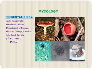
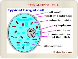
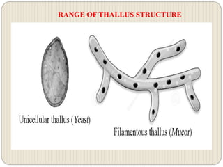
Agaricus is a genus of mushrooms containing both edible and poisonous species, with possibly over 300 members worldwide. The genus includes the common ("button") mushroom (Agaricus bisporus) and the field mushroom (A. campestris), the dominant cultivated mushrooms of the West.
This is an illustrated account for Unit 1 of Coure Course III Mycology and Phytopathology of Bsc Hons Program - Introduction to True fungi including characters, affinities, thallus, cell wall, nutrition and classification
Rhizopus is a genus of common saprophytic fungi on plants and specialized parasites on animals. They are found in a wide variety of organic substances , including "mature fruits and vegetables", jellies, syrups, leather, bread, peanuts, and tobacco.
This is a detailed presentation on Morphology, anatomy and reproduction of Marchantia spp. with high quality pics and eye capturing transitions and animations
1) Strategies and structuresIn Protozoans the method of movement .pdfaptelecom16999
1) Strategies and structures:
In Protozoans the method of movement is determined by the type of organism and the
surrounding environment. Protozoans mainly move by cell extension, flagella or pseudopodia
and cilia, the movement as per the presence of structure can be classified as ciliary, flagellar and
amoeboid movement.
Ciliates : Ciliates form the largest group of protozoa. These organisms vary in size and often live
in watery environments, including oceans, marshes, bays and streams. Ciliates move using tiny
cilia, which are hair-like strands that act as sensors and tiny limbs.
Flagella are longer and less numerous that cilia, they use their long tail like flagella to move.
Amoebas : In these two cytoskeleton get polymerized . This creates a vacancy and cytoplasmice
material flow to cover the vacancy created. When amoeba moves cytoplasm moves to the arm
like extension called pseudopodium. This pseudopodium extends and enlarge and hence this
push the animal body towards that respective direction.
2) A) Flagellates can live as single cells, in colonies, or as parasites.
Commonly live in niche\'s of water.
They conduct photosynthesis and have a cell wall.
They contain flagella for propulsion or to create a current to bring in food.
They can inhabit the reproductive tract, alimentary canal, tissue sites and also the blood stream,
lymph vessels and cerebrospinal canal.
B) Pseudopods : Also called as false feet , are projections that can appear and disappear from the
organism\'s body. These are used for movement and to engulf prey and digest them using
enzymes.
C) Apicomplexa : Unicellular and spore forming, most of them possess a unique form of
organelle that comprises a type of plastid called an apicoplast, and an apical complex structure.
They have apicoplast(non photosynthetic plastid) , mitochondria and nuclear genomes.
Lack of cilia, sexual reproduction, use micropores for feeding, and the production of oocysts
containing sporozoites as the infective form.
They have unique gliding capability which enables them to cross through tissues and enter and
leave their host cells. This gliding ability is made possible by the use of adhesions and small
static myosin motors.
3) Key characteristics of fungi :
Fungi are unicellular or multicellular.
Most of the fungi grow as tubular filaments called hyphae
They are haploid.
Fungus are heterotrophs (they can obtain nutrients by absorption) . They absorb food and secrete
enzymes to digest complex molecules
Propogate by spores
Asexual or sexual reproduction
They can be multinucleated
Fungi are achlorophyllous (lack of cholorphyll pigment)
Both Fungi and protists belong to same kingdom but fungi is different from protist, protists are
able to live in an anaerobic environment without oxygen but fungi need aerobic respiration to
survive.
Protists are unicellular but fungi are multicellular. Protists are autotrophic (make their own
energy) and heterotrophic (rely on outside source to get energy), but fungi a.
Morphology, Classification, Cultivation and Reproduction of FungiKrutika Pardeshi
This presentation is Useful for B. Pharmacy SEM III Students to study the Topic Fungi According to PCI Syllabus.
It Consist of Morpholoy of Fungi, Cultivation , Reproduction and Classification of Fungi.
Described about general characters of fungi which include sexual and asexual reproduction with diagram, so it will be easy for undergraduates to understand the various concepts
Describe in detail about fungi and general characters of fungi and different modifications and reproduction in fungi especially for undergraduate students
This presentation includes all the general characteristics of fungi, types, structure of a fungi, classifications, and reproduction. Different types of fungi and its classification, its reproduction are all included.
The presentation covers all the basic aspects of Kingdom Fungi including its salient features, cell wall structure, nutrition, spore forms, and reproduction.
This presentation explores a brief idea about the structural and functional attributes of nucleotides, the structure and function of genetic materials along with the impact of UV rays and pH upon them.
This presentation intends to explore the communication of the cell within and others for sustainability along the regulation mechanisms by the cellular neural networks and others to sing the song of the life.
Bioenergetics is an important domain in biology. This presentation has explored ATP production and its optimum utilization in biological systems along with certain theories and experiments to give a bird's eye view of this important issue.
This presentation offers the bird's eye view of the cell as the basic structural and functional unit of life. It also addresses the origin of eukaryotic cells from the prokaryotic cell by the endosymbiotic theory.
This presentation has been intended to offer a bird's eye view about the phylogenetic classification of the plant kingdom in general and the Engler and Prantl system in particular with merits and demerits.
This PPT has been made to explore the plant classification in general and the classification as made by Bentham & Hooker for the classification of the flowering plants. It also offers the history of plant classification along with the merits and demerits of this aforesaid classification.
Energy and the biological systems are joined together and no biological world is almost impossible without ATP. This study material intends to explore the beauty of ATP to drive different biological processes.
This PPT offers a bird's eye view of ICBN and its different rules along with regulations for the naming of plants. It also highlights the history of IBC and its contribution to plant taxonomy.
This presentation intends to offer the basic features of plant metabolism along with the different types of mechanisms to regulate and control the metabolic pathways.
This presentation has been designed to give the foundation of taxonomy in general and Plant Taxonomy in particular as a matter of pleasure to explore the diversity of the plant world.
Sex and sexuality are very common words in biology but para-sexuality is a little bit uncommon, several organisms in general and fungi in particular have the pleasure of sexuality to bring variations by beside sex. This PPT explores the beauty of para-sexuality for the academic fraternity.
Sex life in fungi is not less fascinating than in other organisms. Heterosexuality is a matter of pleasure to explore the diversity of sex in fungi along with its cause and consequences. You can find a pleasure to go through the content.
This PowerPoint wants to explore the bird's eye view of the reproduction of bacteria in general and the genetic recombination of bacteria in particular.
This presentation gives the bird's eye view of bacterial nutrition along with some other issues required to understand bacterial diversity as far as nutrition is concerned.
This presentation explores the food value of mushrooms along with the long-term and short-term storage procedures. It also offers a detailed account of the nutrients that remain present in the edible mushrooms.
If you want to explore the role of Cyanobacteria in soil fertility in general & Azolla-Anabena association in particular, you can visit this PowerPoint Presentation.
The increased availability of biomedical data, particularly in the public domain, offers the opportunity to better understand human health and to develop effective therapeutics for a wide range of unmet medical needs. However, data scientists remain stymied by the fact that data remain hard to find and to productively reuse because data and their metadata i) are wholly inaccessible, ii) are in non-standard or incompatible representations, iii) do not conform to community standards, and iv) have unclear or highly restricted terms and conditions that preclude legitimate reuse. These limitations require a rethink on data can be made machine and AI-ready - the key motivation behind the FAIR Guiding Principles. Concurrently, while recent efforts have explored the use of deep learning to fuse disparate data into predictive models for a wide range of biomedical applications, these models often fail even when the correct answer is already known, and fail to explain individual predictions in terms that data scientists can appreciate. These limitations suggest that new methods to produce practical artificial intelligence are still needed.
In this talk, I will discuss our work in (1) building an integrative knowledge infrastructure to prepare FAIR and "AI-ready" data and services along with (2) neurosymbolic AI methods to improve the quality of predictions and to generate plausible explanations. Attention is given to standards, platforms, and methods to wrangle knowledge into simple, but effective semantic and latent representations, and to make these available into standards-compliant and discoverable interfaces that can be used in model building, validation, and explanation. Our work, and those of others in the field, creates a baseline for building trustworthy and easy to deploy AI models in biomedicine.
Bio
Dr. Michel Dumontier is the Distinguished Professor of Data Science at Maastricht University, founder and executive director of the Institute of Data Science, and co-founder of the FAIR (Findable, Accessible, Interoperable and Reusable) data principles. His research explores socio-technological approaches for responsible discovery science, which includes collaborative multi-modal knowledge graphs, privacy-preserving distributed data mining, and AI methods for drug discovery and personalized medicine. His work is supported through the Dutch National Research Agenda, the Netherlands Organisation for Scientific Research, Horizon Europe, the European Open Science Cloud, the US National Institutes of Health, and a Marie-Curie Innovative Training Network. He is the editor-in-chief for the journal Data Science and is internationally recognized for his contributions in bioinformatics, biomedical informatics, and semantic technologies including ontologies and linked data.
Seminar of U.V. Spectroscopy by SAMIR PANDASAMIR PANDA
Spectroscopy is a branch of science dealing the study of interaction of electromagnetic radiation with matter.
Ultraviolet-visible spectroscopy refers to absorption spectroscopy or reflect spectroscopy in the UV-VIS spectral region.
Ultraviolet-visible spectroscopy is an analytical method that can measure the amount of light received by the analyte.
Nutraceutical market, scope and growth: Herbal drug technologyLokesh Patil
As consumer awareness of health and wellness rises, the nutraceutical market—which includes goods like functional meals, drinks, and dietary supplements that provide health advantages beyond basic nutrition—is growing significantly. As healthcare expenses rise, the population ages, and people want natural and preventative health solutions more and more, this industry is increasing quickly. Further driving market expansion are product formulation innovations and the use of cutting-edge technology for customized nutrition. With its worldwide reach, the nutraceutical industry is expected to keep growing and provide significant chances for research and investment in a number of categories, including vitamins, minerals, probiotics, and herbal supplements.
Slide 1: Title Slide
Extrachromosomal Inheritance
Slide 2: Introduction to Extrachromosomal Inheritance
Definition: Extrachromosomal inheritance refers to the transmission of genetic material that is not found within the nucleus.
Key Components: Involves genes located in mitochondria, chloroplasts, and plasmids.
Slide 3: Mitochondrial Inheritance
Mitochondria: Organelles responsible for energy production.
Mitochondrial DNA (mtDNA): Circular DNA molecule found in mitochondria.
Inheritance Pattern: Maternally inherited, meaning it is passed from mothers to all their offspring.
Diseases: Examples include Leber’s hereditary optic neuropathy (LHON) and mitochondrial myopathy.
Slide 4: Chloroplast Inheritance
Chloroplasts: Organelles responsible for photosynthesis in plants.
Chloroplast DNA (cpDNA): Circular DNA molecule found in chloroplasts.
Inheritance Pattern: Often maternally inherited in most plants, but can vary in some species.
Examples: Variegation in plants, where leaf color patterns are determined by chloroplast DNA.
Slide 5: Plasmid Inheritance
Plasmids: Small, circular DNA molecules found in bacteria and some eukaryotes.
Features: Can carry antibiotic resistance genes and can be transferred between cells through processes like conjugation.
Significance: Important in biotechnology for gene cloning and genetic engineering.
Slide 6: Mechanisms of Extrachromosomal Inheritance
Non-Mendelian Patterns: Do not follow Mendel’s laws of inheritance.
Cytoplasmic Segregation: During cell division, organelles like mitochondria and chloroplasts are randomly distributed to daughter cells.
Heteroplasmy: Presence of more than one type of organellar genome within a cell, leading to variation in expression.
Slide 7: Examples of Extrachromosomal Inheritance
Four O’clock Plant (Mirabilis jalapa): Shows variegated leaves due to different cpDNA in leaf cells.
Petite Mutants in Yeast: Result from mutations in mitochondrial DNA affecting respiration.
Slide 8: Importance of Extrachromosomal Inheritance
Evolution: Provides insight into the evolution of eukaryotic cells.
Medicine: Understanding mitochondrial inheritance helps in diagnosing and treating mitochondrial diseases.
Agriculture: Chloroplast inheritance can be used in plant breeding and genetic modification.
Slide 9: Recent Research and Advances
Gene Editing: Techniques like CRISPR-Cas9 are being used to edit mitochondrial and chloroplast DNA.
Therapies: Development of mitochondrial replacement therapy (MRT) for preventing mitochondrial diseases.
Slide 10: Conclusion
Summary: Extrachromosomal inheritance involves the transmission of genetic material outside the nucleus and plays a crucial role in genetics, medicine, and biotechnology.
Future Directions: Continued research and technological advancements hold promise for new treatments and applications.
Slide 11: Questions and Discussion
Invite Audience: Open the floor for any questions or further discussion on the topic.
Multi-source connectivity as the driver of solar wind variability in the heli...Sérgio Sacani
The ambient solar wind that flls the heliosphere originates from multiple
sources in the solar corona and is highly structured. It is often described
as high-speed, relatively homogeneous, plasma streams from coronal
holes and slow-speed, highly variable, streams whose source regions are
under debate. A key goal of ESA/NASA’s Solar Orbiter mission is to identify
solar wind sources and understand what drives the complexity seen in the
heliosphere. By combining magnetic feld modelling and spectroscopic
techniques with high-resolution observations and measurements, we show
that the solar wind variability detected in situ by Solar Orbiter in March
2022 is driven by spatio-temporal changes in the magnetic connectivity to
multiple sources in the solar atmosphere. The magnetic feld footpoints
connected to the spacecraft moved from the boundaries of a coronal hole
to one active region (12961) and then across to another region (12957). This
is refected in the in situ measurements, which show the transition from fast
to highly Alfvénic then to slow solar wind that is disrupted by the arrival of
a coronal mass ejection. Our results describe solar wind variability at 0.5 au
but are applicable to near-Earth observatories.
THE IMPORTANCE OF MARTIAN ATMOSPHERE SAMPLE RETURN.Sérgio Sacani
The return of a sample of near-surface atmosphere from Mars would facilitate answers to several first-order science questions surrounding the formation and evolution of the planet. One of the important aspects of terrestrial planet formation in general is the role that primary atmospheres played in influencing the chemistry and structure of the planets and their antecedents. Studies of the martian atmosphere can be used to investigate the role of a primary atmosphere in its history. Atmosphere samples would also inform our understanding of the near-surface chemistry of the planet, and ultimately the prospects for life. High-precision isotopic analyses of constituent gases are needed to address these questions, requiring that the analyses are made on returned samples rather than in situ.
This pdf is about the Schizophrenia.
For more details visit on YouTube; @SELF-EXPLANATORY;
https://www.youtube.com/channel/UCAiarMZDNhe1A3Rnpr_WkzA/videos
Thanks...!
Introduction:
RNA interference (RNAi) or Post-Transcriptional Gene Silencing (PTGS) is an important biological process for modulating eukaryotic gene expression.
It is highly conserved process of posttranscriptional gene silencing by which double stranded RNA (dsRNA) causes sequence-specific degradation of mRNA sequences.
dsRNA-induced gene silencing (RNAi) is reported in a wide range of eukaryotes ranging from worms, insects, mammals and plants.
This process mediates resistance to both endogenous parasitic and exogenous pathogenic nucleic acids, and regulates the expression of protein-coding genes.
What are small ncRNAs?
micro RNA (miRNA)
short interfering RNA (siRNA)
Properties of small non-coding RNA:
Involved in silencing mRNA transcripts.
Called “small” because they are usually only about 21-24 nucleotides long.
Synthesized by first cutting up longer precursor sequences (like the 61nt one that Lee discovered).
Silence an mRNA by base pairing with some sequence on the mRNA.
Discovery of siRNA?
The first small RNA:
In 1993 Rosalind Lee (Victor Ambros lab) was studying a non- coding gene in C. elegans, lin-4, that was involved in silencing of another gene, lin-14, at the appropriate time in the
development of the worm C. elegans.
Two small transcripts of lin-4 (22nt and 61nt) were found to be complementary to a sequence in the 3' UTR of lin-14.
Because lin-4 encoded no protein, she deduced that it must be these transcripts that are causing the silencing by RNA-RNA interactions.
Types of RNAi ( non coding RNA)
MiRNA
Length (23-25 nt)
Trans acting
Binds with target MRNA in mismatch
Translation inhibition
Si RNA
Length 21 nt.
Cis acting
Bind with target Mrna in perfect complementary sequence
Piwi-RNA
Length ; 25 to 36 nt.
Expressed in Germ Cells
Regulates trnasposomes activity
MECHANISM OF RNAI:
First the double-stranded RNA teams up with a protein complex named Dicer, which cuts the long RNA into short pieces.
Then another protein complex called RISC (RNA-induced silencing complex) discards one of the two RNA strands.
The RISC-docked, single-stranded RNA then pairs with the homologous mRNA and destroys it.
THE RISC COMPLEX:
RISC is large(>500kD) RNA multi- protein Binding complex which triggers MRNA degradation in response to MRNA
Unwinding of double stranded Si RNA by ATP independent Helicase
Active component of RISC is Ago proteins( ENDONUCLEASE) which cleave target MRNA.
DICER: endonuclease (RNase Family III)
Argonaute: Central Component of the RNA-Induced Silencing Complex (RISC)
One strand of the dsRNA produced by Dicer is retained in the RISC complex in association with Argonaute
ARGONAUTE PROTEIN :
1.PAZ(PIWI/Argonaute/ Zwille)- Recognition of target MRNA
2.PIWI (p-element induced wimpy Testis)- breaks Phosphodiester bond of mRNA.)RNAse H activity.
MiRNA:
The Double-stranded RNAs are naturally produced in eukaryotic cells during development, and they have a key role in regulating gene expression .
Observation of Io’s Resurfacing via Plume Deposition Using Ground-based Adapt...Sérgio Sacani
Since volcanic activity was first discovered on Io from Voyager images in 1979, changes
on Io’s surface have been monitored from both spacecraft and ground-based telescopes.
Here, we present the highest spatial resolution images of Io ever obtained from a groundbased telescope. These images, acquired by the SHARK-VIS instrument on the Large
Binocular Telescope, show evidence of a major resurfacing event on Io’s trailing hemisphere. When compared to the most recent spacecraft images, the SHARK-VIS images
show that a plume deposit from a powerful eruption at Pillan Patera has covered part
of the long-lived Pele plume deposit. Although this type of resurfacing event may be common on Io, few have been detected due to the rarity of spacecraft visits and the previously low spatial resolution available from Earth-based telescopes. The SHARK-VIS instrument ushers in a new era of high resolution imaging of Io’s surface using adaptive
optics at visible wavelengths.
Professional air quality monitoring systems provide immediate, on-site data for analysis, compliance, and decision-making.
Monitor common gases, weather parameters, particulates.
In silico drugs analogue design: novobiocin analogues.pptx
Fungi- Cell Wall & Thallus Structure.pdf
1. MYCOLOGY
PRESENTATION BY:
Dr. N. Sannigrahi,
Associate Professor,
Department of Botany,
Nistarini College, Purulia,
D.B. Road, Purulia
( W.B), 723101,
INDIA.
2. CONTENTS
Fungi as cell,
Cell components,
Thallus organization,
Thallus modifications,
Special structure of thallus,
Cell wall-Structure & Function,
Ultra structure of cell wall & Composition,
Conclusions.
3. FUNGAL CELL
Fungi as ubiquitous in the form of saprophytes, parasites or symbionts
is the eukaryotic in nature with wide diversity as far as the somatic
structure is concerned. The fungi enjoys a wide range as far as
reproduction is concerned and the cellular organizational beauty has
made it unique one in this respect.
Eukaryotic with distinct cell wall. Cytoplasm & nucleus,
Uninucleate or multinucleate as coenocytes,
Heterokaryotic or homokariotic in nature,
Nucleus contains one or more nucleoli,
Cytoplasm colorless and granular to reticulate in appearance,
Cytoplasm contains many big but irregular vacuoles whereas the
higher members contain single large vacuole found in central position,
Cells contain mitochondria and complex system of internal membranes
including ER, Golgi apparatus,
Cell membrane contain ergo sterol that replace the cholesterol in animal
cells.
5. THALLUS ORGANISATION
Unicellular Thallus :
In some of the lower fungi such as the chytrids, the thallus is more or less a
spherical, single-celled structure . At the time of reproduction, it becomes a
reproductive unit. The latter produces the asexual or sexual cells. Such fungi
are called holocarpic. In them, the vegetative and reproductive stages do not
occur together in the same thallus.
Plasmodiophora has a vegetative phase consisting of a naked, multi-
nucleate, amoeboid mass of protoplasm. It is termed Plasmodium. The
protoplast of the diploid Plasmodium cleaves to form the resting spores. The
yeasts, which are related to the filamentous forms, also have a unicellular
thallus. In the unicellular holocarpic forms (Synchytrium) the mycelium is
absent.
Filamentous Thallus :
The vast majority of the fungi have a filamentous thallus. It originates
through the germination of a spore. The spore germinates as it lands on a
suitable substratum where other conditions of life are also favorable. In
some species, the spore, on germination, produces only a short, tubular
structure of limited growth.
7. RANGE OF THALLUS STRUCTURE
It constitutes the thallus and is technically called a hypha. The spores of
most of the fungi, however, give rise to a fluffy thallus consisting of a
cottony mass of fine, branched filaments. These long, fine filaments are
called the hyphae (sing, hypha). Some of these hyphae, at a certain stage of
maturity, extend into the air and bear the reproductive bodies.
The rest spread over or within the substratum and continue the normal
activities. Such fungi are called eucarpic. Collectively the hyphae comprise
the vegetative body (thallus) of a fungus which is called the mycelium. The
hypha is thus a structural unit of the mycelium. It consists of a thin,
transparent wall filled or lined with a layer of cytoplasm.
The medium upon which the mycelium grows is known as substratum. The
mycelium is the food procuring structure in the life cycle. It carries on the
general activities of a plant cell such as absorption, digestion, respiration,
excretion and growth but not photosynthesis. The hyphae constituting the
mycelium branch, spread in all directions within or over the substratum to
form a loose and ramifying network.
8. RANGE OF THALLUS STRUCTURE
The hyphae are usually colourless particularly those embedded in the
substratum. The aerial hyphae in some fungi become colored. Black,
orange, yellow, red, blue and brown are the usual tints. The color is usually
confined to the hyphal wall.
Even when the pigments are present in the protoplasmic contents, they do
not form an integral part of the living matter. The pigments play no role in
the physiology of the fungus. The growth in length takes place at the tips of
the hyphae and is thus termed apical.
The thallus may be grouped into- holocarpic or eucarpic; When the thallus as a
whole is vegetative as well as reproductive in function- holocarpic but when the
portion of the hyphae of the thallus takes part in reproduction-eucarpic.
Hyphae- aerial or substrate , both are branched; substrate hyphae for nutrients but
aerial hyphae for reproductive structures, look black, red, blue , green , brown in
color diversity;
Hyphae may be modified- Prosenchyma, Pseudo parenchyma, Rhizomorph,
Sclerotia, Sporophore, Stroma etc depending on species diversity.
9. KINDS OF MYCELIUM
The fungus mycelium in fact, is a multinucleate structure lacking
complete internal cell boundaries.
i. Aseptate Mycelium- In algal fungi, it is multinucleate, aseptate,
grows terminally by the apical elongation of the hyphae accompanied
by increase in the number of nuclei by nuclear division, becomes
coenocytes.
Ii. Septate Mycelium- Develop internal cross walls that divide the
hyphae into segments at regular intervals behind the hyphal tip,
segments uninucleate or multinucleate. Formation of septa is always
preceded by the division of nucleus, each has a central pore or rarely
more than one pore although complete partitions do not occur during
vegetative phase.
Mostly found in the members of Ascomycetes & Basidiomycetes,
In Basidiomycets, further elaboration of the septum to produce a more
complex pore called dolipore and it is covered by round bracket like
structure – Parenthesome.
10. AGGREGATION OF HYPHAE
Different types of modifications of hyphae mycelium are found as
follows:
PLECTENCHYMA-A kind of false tissue formed by the aggregation of
hyphae, may be two types-
A. Prosenchyma - Rather loosely woven hyphae, hyphae do not lose
their identity, run more or less parallel to each other composed of
elongated cells,
B. Pseudoparenchyma - The hyphae become woven and intertwined
into a compact mass, hyphal mass appears to be continuous structure
consisting oval cells , resemblance with parenchyma tissue of higher
cells
RHIZOMORPH- Interwoven hypae to form rope like structure,
Tightly packed and individual hyphae lose their individual identity,
Outer hard black external layer of the rhizomporph called rind,,
Each rhizomorph contains a growing tip,
Rhizomorphs are perennating structure to withstand adverse conditions.
12. AGGREGATION OF HYPHAE
SCLEROTIUM- Specially modified mycelium for storage,
perennation and vegetative propagation,
Compact, rounded. Cushion shaped, cylindrical or irregular object,
Dense mass of thick walled short hyphae to form
pseudoparenchymatous structure,
polyhedral shape,
Outer surface dark brown black and crest like,
It may be pin head to large with several inches in diameter.
SPOROPHORES- Spore bearing modified into group to form
pycnidia, sporodochia, hymenia, acervuli etc,
Aerial, erect and arise from the prostrate hyphae,
Branched or unbranched with sporangia or conidia with them,
Spore bearing sporophores are sporangiosphores and conidia bearing
called conidiophores.
13. AGGREGATION OF HYPHAE
STROMATA: The fungal tissue forms a mass of
pseudoparenchymatous tissue like flat or cushion shape structure,
Several fructifications to form reproductive structures,
PESUDOSCLEROTIUM
The sclerotium like bodies are formed at the base of the fruit bodies of
higher fungi,
In Polyporous basilapiloides formed below the soil surface
Composed of sand particles surrounded by hyphal aggregations
APPRESORIUM
Common in parasitic fungi mostly found in ectoparasites, Terminal
simple or lobed swollen structure of germ tubes or infection hyphal
Adheres to the surface of the host and helps in the penetration of the
hyphae of the pathogen,
Found in the parasitic fungi of the orders Erysiphales.
15. AGGREGATION OF HYPHAE
HAUSTORIUM
Mostly found in the intercellular absorbing structure of obligate
parasites,
Usually produced in the fungi in which intercellular mycelium formed,
Vary in shape , may be knob shaped or branched finger shaped,
Secrete certain enzymes which hydrolyses the proteins and
carbohydrates of the host cell and absorb the nutrients from the host
cell without killing them,
Provide greater surface area for the exchange of materials.
In addition to these above kind of modifications, different other
specialized modifications are formed in order to address the stress and
other issues appear in course of their survival and reproductive
potential.
17. CELL WALL COMPOSITION
The fungal cell wall is located outside the plasma membrane and is the cell
compartment that mediates all the relationships of the cell with the
environment.
It protects the contents of the cell, gives rigidity and defines the cellular
structure.
The cell wall is a skeleton with high plasticity that protects the cell from
different stresses, among which osmotic changes stand out.
The cell wall allows interaction with the external environment since some of
its proteins are adhesions and receptors.
Since, some components have a high immunogenic capacity, certain wall
components can drive the host’s immune response to promote fungus
growth and dissemination.
The cell wall is a characteristic structure of fungi and is composed mainly
of glucans, chitin and glycoprotein.
19. CELL WALL COMPOSITION
The cell wall is a specific and complex cellular organelle composed of
glucans, chitin, chitosan, and glycosylated proteins. Proteins are generally
associated with polysaccharides resulting in glycoprotein. Together, these
components contribute to the cell wall rigidity. The synthesis and
maintenance of cell wall involves a large number of biosynthetic and
signaling pathways (Casadevall and Perfect, 1998).
The cell wall is structured in different layers where the innermost layer is a
more conserved structure on which the remaining layers are deposited and
can vary between different species of fungi.
Glucans is the most important structural polysaccharide of the fungal cell
wall and represents 50–60% of the dry weight of this structure. Most
polymers of glucans are composed of 1,3 linkage glucose units (65–90%),
although there are also glucans with β-1,6 (in Candida but not in
Aspergillus), β-1,4, α-1,3 and α-1,4 links. The β-1,3-D-glucan is the most
important structural component of the wall, to which other components of
this structure are covalently linked. The β-1,3-D-glucan is synthesized by a
complex of enzymes located in the plasma membrane called glucans
synthases.
20. CELL WALL COMPOSITION
The chitin content of the fungal wall varies according to the morphological
phase of the fungus. It represents 1–2% of the dry weight of yeast cell wall
while in filamentous fungi, it can reach up to 10–20%. Chitin is synthesized
from n- acetyl glucosamine by the enzyme chitin synthase, which deposits
chitin polymers in the extracellular space next to the cytoplasmic
membrane.
Proteins compose 30–50% of the dry weight of fungal wall in yeast and 20–
30% of the dry weight of the wall of the filamentous fungi. Most proteins
are associated to carbohydrates by O or N linkages resulting in
glycoprotein. Cell wall proteins have different functions including
participation in the maintenance of the cellular shape, adhesion processes,
cellular protection against different substances, absorption of molecules,
signal transmission, and synthesis and reorganization of wall components
(Bowman and Free, 2006; Ponton, 2008).
Lipids are found in small amounts, prevent to desiccation of cells,
Presence of pigments and salts; Melanin is most abundant, important to
protect the hyphen, spores and help in pathogenesis , attachment to the
surfaces
21. CELL WALL COMPOSITION
Enzymes cross linking the fibrils in the wall are released through the plasma
membrane,
Wall constrictions take place in the apical cap; the synthesis of chitin and
glucans take place within in 1 mm. in the apex; the rigidity of the wall; is
done by cross linking of polymers,
The wall contains hydrophobins, glomalins ; Hydrophobins constitute 10%
of the total wall proteins, the amphoetric nature provides an array of extra
ordinary functions and play role biotechnology.
Glycoprotein includes mannoprotein, galactoprotein, xyloproteins etc.
The wide diversity of the cell wall composition observed as far as the
diversity of the fungal kingdom as stated below.
22. COMMON WALL CONSTITUENTS IN EACH DIVISION
DIVISION FIBROUS
COMPONENTS
GEL LIKE POLYMERS
BASIDIOMYCOTINA Chitin ,β(1-3),β (1-6)
Glucans
Xylomannoproteins,α (1-3)
Glucans
ASCOMYCOTINA Chitin ,β(1-3),β (1-6)
Lucan
Galactomannoproteins, α
(1-3) Glucans
ZYGOMYCOTINA Chitin , Chitosan Polygluchonic acid,
Glucurosomannoproteins,
Polyphosphate
CHYTRIDIOMYCOTA Chitin, Glucans Glucans
23. REVIEW
Fungi are heterotrophic either parasites, saprophytes or symbionts,
Unicellular or multicellular, holocarpic or eucarpic with diverse structural
peculiarity as far as thallus is concerned,
Eukaryotic with almost all kind of organizational beauty observed,
Cell wall is diverse type with a fundamental framework in all the aspects,
The reproduction takes place by vegetative, asexual or sexual means following
the route of plasmogamy, karyogamy & meiosis with haplontic life cycle
pattern.
Play a very significant role in the sustainability of environment and offers raw
materials for the industrial wheel,
Play a pivotal role for the deterioration of the productivity of crops both
qualitative and quantitative.
24. THANKS A LOT FOR YOUR CONSIDERATION
References:
Google for images,
Microbiology & Phycology - Mishra & Dash,
Brook Microbiology of Microorganisms-Madigan, Martinko, Bender,
Buckley & Stahl,
A Textbook of Microbiology- Chakraborty,
Microbiology- Pelzer, Chan & Krieg,
Botany for degree students- Vasistha & Sinha,
Introduction to Mycology and Phytopathology - Mishra & Dash,
Different WebPages to enrich content.
Disclaimer: This PPT has been designed to enrich the open source
of learning. The author does not claim any kind of financial pleasure