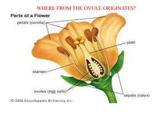
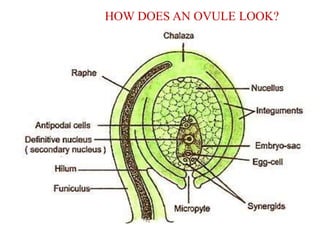
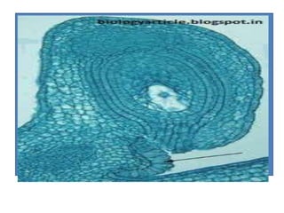
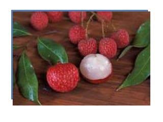
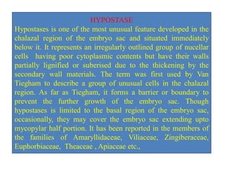
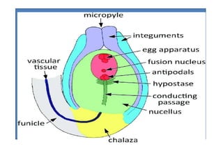
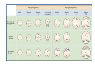
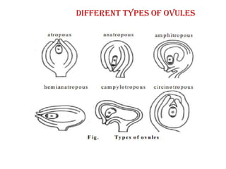
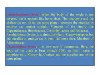
OVULE:
EXCEPTIONAL BEAUTY
Welcome to
Ovule-Structure, Types & Specialty
Dr. N. Sannigrahi, Associate Professor,
Department of Botany,
Nistarini College, Purulia, 723101(W.B) India
MYSTREY OF BIOLOGY
Plants are treasure of all the life forms across the globe. The
beauty of the plants and its reproduction is an awesome appetite
for the natural science and scientists. Irrespective of the nature
except some cases, the plants life is comprising of the two
generations- Sporophytic generation & gametophyte generations.
The sporophytic generations starts from the single cell , zygote.
The zygotes undergoes division mitotically to form embryo and
through a series of biochemical and morphological changes, the
embryo along with the other requisites turn into seed. Seed is the
enigma of life , undergoes germination to promise a baby plants
enjoying the different natural attributes as required. The
sporophytic generations subject to the completion of vegetative
phase undergoes reproductive stage.
The reproductive stages starts with a beauty of the flower- an
outcome of the genes and the different environmental attributes.
The flower is the most attractive part of the plant life
irrespective of its diversity. The flower comprising of the
accessory and essential whorls. The stamen-the male part & the
carpel-the female parts undergo a mutual relationship to promise
the birth of another baby plant. The male gametophyte
generation starts with the haploid microspore and the female
gametophytic generations develop from the megaspore
popularly known as Ovule. Ovule bears a number of characters
and this is always a pleasure to the biologists. Let us examine
the ovule and its diversity and how the ovule brings the
transformation for another zygote to initiate the another
sporophytic generation.
OVULE
Ovule, the developmental precursor of seeds is the organ of prime
importance in angiosperm flowers and it can be traced back in
evolution almost 400 million years ago to the earliest seed plants as
far as phylogeny is concerned. Having diverse position in ovary,
nucellus thickness, number & thickness of integuments, degree and
direction of curvature and histological differentiations have drawn
the importance of plant biologists.
The ovule develops as multicellular placental outgrowth including
the epidermal & number of hypodermal cells. With further
development, this gives to the nucellus and one or two integuments
from its basal region.
Angiosperm ovules basically consists of a nucellus and two
integuments and sessile on the placenta or attached by its stal
WHERE FROM THE OVULE ORIGINATES?
Most commonly a vascular strands extends from the placenta
through the funiculus to the chalaza, i.e. the area below the
base of the nucellus where the integuments depart. The
funiculus and the chalaza are intercalary structure and thus
well demarcated than the nucellus and integuments. The
nucellus represents the mega sporangium, in which the
meiocytes undergoes meiosis forming four megaspores,
typically only one of which develops into embryo sac
representing the megagametophyte.The embryo sac contains
basically four or eight nuclei, organized into four or seven
cells, depending on whether two or three round mitotic
divisions in the developing embryo(Maheswari,
1950;Friedman, 2006).
HOW DOES AN OVULE LOOK?
COMPONENTS OF OVULE
A typical angiosperm ovule consists of the followings:
Funicle - Stalk of the ovule that connects ovule to the
placenta, short & multicellular.
Hilum - Point where funicle connects the main body of the
ovule.
Integuments-Layers that surround the ovule-outer & inner
integument’ main function to protect the inner tissues.
Micropyle- Opening which present at the top of the
integuments called micropyle. It allows entry of the pollen
tube into the ovule.
COMPONENTS OF OVULE
Nucellus- Integuments surrounds parenchymatous tissues
which constitutes the main body of the ovule called nucellus.
The nucellus is nutritive in function to the developing embryos.
Chalaza- The swollen part of the ovule present opposite to
micropyle is called chalaza.
Embryo sac-It is present in the nucellus and it contains female
gamete called egg.
Nutrients from the plants travel through phloem of the vascular
system to the funiculus and the outer integument and from there
apoplastically and symplastically through the nucellus inside
the ovule.
ENDOTHELIUM
The endothelium is an additional cell layer, differentiating from the
inner epidermis of the ovule integument. In tomato (Solanum
lycopersicum L.), after fertilization, the endothelium separates from
integument and becomes an independent tissue developing next to
the growing embryo sac. In the absence of fertilization, the
endothelium may proliferate and form pseudo-embryo. Endothelial
cells in normally developing ovules change their structural and
functional specialization from meristematic to secretary and back to
meristematic, and proliferate until seeds fully mature. The secretary
activity of the endothelium is necessary for the lysis of dying cells
of the integument and provides the space for the growth of the new
sporophyte.
OBTURATOR
Any ovular structure which helps in directing the growth of pollen
tube towards the micropyle is generally referred to as obturator.
Obturators exhibit great variation in their origin, morphology,
anatomy and extent of development. They may originate from
placenta or funiculus or both. Within the ovary, the obturator
bridges the pathway of the pollen tube from the style to the
ovule. In several families of angiosperms a characteristic placental
outgrowth is present near the ovule and is referred to as obturator.
This obturator grows into the space between the nucellus and the
integuments and also between the ovary and ovary wall. An
obturator is said to be helping in directing the growth of pollen
tube towards micropyle. Obturator exhibits a great amount of
diversity in its origin, morphology, anatomy and mode of
development.
CARUNCLE
The caruncle is a structure present in the micropylar region of
Euphorbiaceae seeds. This structure has the ecological function of
promoting seed dispersal by ants (myrmecochory), but it is
debated whether it also has an agronomical importance
influencing seed germination. The caruncle is the spongy
outgrowth of the integument near micropyle. The main
function of caruncle is to absorb water which is necessary for
germination. The example of seed which contains caruncle is
castor seeds. But recent investigation rejects the role of caruncle
in the seed germination as far as water absorption is concerned.
ARIL
The term "aril" is sometimes applied to any fleshy appendage
of the seed in flowering plants, such as the mace of the nutmeg
seed, lichi etc. Arils and arillodes are often edible enticements
that encourage animals to transport the seed, thereby assisting in
seed dispersal. The seeds perform important functions of the
plants which are nourishment of embryo, dispersal and travels to
new places and dormancy during unfavorable conditions. Seeds
are defined as the products of sexual reproduction which are a
source of genetic variability which can be induced in the plants.
HYPOSTASE
Hypostases is one of the most unusual feature developed in the
chalazal region of the embryo sac and situated immediately
below it. It represents an irregularly outlined group of nucellar
cells having poor cytoplasmic contents but have their walls
partially lignified or suberised due to the thickening by the
secondary wall materials. The term was first used by Van
Tiegham to describe a group of unusual cells in the chalazal
region. As far as Tiegham, it forms a barrier or boundary to
prevent the further growth of the embryo sac. Though
hypostases is limited to the basal region of the embryo sac,
occasionally, they may cover the embryo sac extending upto
mycopylar half portion. It has been reported in the members of
the families of Amaryllidaceae, Viliaceae, Zingiberaceae,
Euphorbiaceae, Theaceae , Apiaceae etc.,
Female gametophyte– megasporogenesis (monosporic, bisporic
and tetrasporic)
Depending on the number of megaspores taking part in the
development, the embryo sac (Female gametophyte) may be
three types as follows:
MONOSPORIC: Out of the four megaspores developed by the
meiotic cell division of the megaspore mother cells, if only one
cell takes part in the development of the female gametophyte, it is
called monosporic development of embryo sac.
BISPORIC: If out of the four megaspores, only two megaspores
nuclei takes part in the development of the embryo sac, it is
called bisporic .
TETRASPORIC: However, all the four megaspores nuclei take
part in the development of the embryo sac, it is called tetrasporic
development of female gametophyte.
MEGAGAMETOGENESIS (POLYGONUM TYPE)
i. The functional megaspore, the first cell of the female divides
by three successive divisions to form an eight –nucleate
gametophyte or embryo sac ,
ii. Here, the nuclei of the functional megaspore divides to form
two nuclei-the primary micropylar and primary chalazal nuclei,
iii. The nuclei again divide to form four nuclei,
iv. Each of the nuclei again divide to form eight cell stage to form
the complete octate embryo sac,
v. The female gametophyte increases in size , one nuclei from
each end or pole passes inwards and the two polar nuclei fuse
together somewhere in the middle of the embryo sac forming the
secondary nucleus (2N),
vi.The remaining three nuclei at the micropylar end , each
surrounded by a very thin wall, form egg apparatus
vi. The egg apparatus consists of two synergids and an egg cell.
The other three nuclei at the opposite chalazal end, lying in a
group, often surrounded by very thin walls, form the antipodal
cells.
vii. At the end of the development, the embryo sac getting ready
for the execution of the other biological process to be taken into
account followed by the entry of the pollen tube for the
fertilization to lay the foundation of the first sporophytic cell.
This kind of microspore development is monosporic as single
microspore out of the four derived takes part in the development
of the embryo sac. This is also treated as Polygonum type of
embryo sac development as it was first found to occur among the
members of Polygonaceae.
TYPES OF OVULE
The ovules are characterized by a considerable diversity in
shape, degree of development and structure. Mirbel (1929) for
the first time classified ovules under four groups- Orthotropus,
Anatropus, Campylotropous & Amphitropous. But later on
another two types added making the ovules altogether six(6)
types as follows:
Orthotropous or atropous
Anatropous
Hemi-anatropous or Hemitropous
Campylotropous
Amphitropous
Circinotropous
Different types of ovules
TYPES OF OVULE
As stated earlier, Ovules may have diversity,. When only one
integument present called unitegmic, if the ovule contains two
integuments called bitegmic and very rarely three integuments
called tritegmic is present in plants like Asphodelus. In some
plants like Santalum, no integument called ategmic is also found
to occur.
On the basis of the position of micropyle, with respect to
funiculus, Ovules are classified under six broad categories as
stated earlier. The details are given below.
Orthotropous Ovule: It is atropous or straight where the
micropyle, Chalaza & funiculus , all are in the same line. E.g.
Cycas, members of Polygonaceae and Piperaceae.
Anatropous Ovule: In 80% genera, this is very common type.In
this ovule, the funicle is long , whole body is inverted through
180⁰ , As a result, micropyle comes close to the funicle. Most
common almost monocots and dicots like the members of
Asteraceae , Solanaceae etc.
Hemianatropous or Hemitropous Ovule: The body of the
ovule is inverted through 90⁰. As a result the funicle comes to
lie at right angle to the nucellus.Micropyle and the chalaza lie
in the same plane. Members of primitive family
Rananunculaceae like Ranunculus etc.
Campylotropous Ovule: When the body of the ovule is not
inverted but it appears like horse shoe. The micropyle and the
chalaza do not lie on the same plane , however the nucellus or
embryo sac remain straight. Members of the family like
Capparidaceae, Brassicaceae, Caryophyllaceae and Fabaceae.
Amphitropous Ovule: It is almost similar to Campylotropous but
the nucellus or embryo sac is bent like horse shoe. Members of
Alismataceae.
Circinotropous Ovule; It is very rare in occurrence. Here, the
body of the ovule is bent through 360⁰, so that it takes a
complete turn. Micropyle, Chalaza and the nucellus are on the
same plane.
Your Patience deserves appreciation. Thanks a lot
REFERENCES:
Encyclopedia Britannica,
An introduction to the Embryology of angiosperms by P.
Maheswari,
A text book of Botany by Hait, Bhattacharya, Ghosh,
Advanced Plant taxonomy by A.K.Mondal,
Different websites from Google for images and contents.
DISCLAIMER:
This presentation has been made to enrich open source of
information without any financial interest. The presenter
acknowledges Google for images and other open sources
of knowledge to develop this PPT.

Megaspore-Structure & Speciality

  • 1.
  • 2.
    Welcome to Ovule-Structure, Types& Specialty Dr. N. Sannigrahi, Associate Professor, Department of Botany, Nistarini College, Purulia, 723101(W.B) India
  • 3.
    MYSTREY OF BIOLOGY Plantsare treasure of all the life forms across the globe. The beauty of the plants and its reproduction is an awesome appetite for the natural science and scientists. Irrespective of the nature except some cases, the plants life is comprising of the two generations- Sporophytic generation & gametophyte generations. The sporophytic generations starts from the single cell , zygote. The zygotes undergoes division mitotically to form embryo and through a series of biochemical and morphological changes, the embryo along with the other requisites turn into seed. Seed is the enigma of life , undergoes germination to promise a baby plants enjoying the different natural attributes as required. The sporophytic generations subject to the completion of vegetative phase undergoes reproductive stage.
  • 4.
    The reproductive stagesstarts with a beauty of the flower- an outcome of the genes and the different environmental attributes. The flower is the most attractive part of the plant life irrespective of its diversity. The flower comprising of the accessory and essential whorls. The stamen-the male part & the carpel-the female parts undergo a mutual relationship to promise the birth of another baby plant. The male gametophyte generation starts with the haploid microspore and the female gametophytic generations develop from the megaspore popularly known as Ovule. Ovule bears a number of characters and this is always a pleasure to the biologists. Let us examine the ovule and its diversity and how the ovule brings the transformation for another zygote to initiate the another sporophytic generation.
  • 5.
    OVULE Ovule, the developmentalprecursor of seeds is the organ of prime importance in angiosperm flowers and it can be traced back in evolution almost 400 million years ago to the earliest seed plants as far as phylogeny is concerned. Having diverse position in ovary, nucellus thickness, number & thickness of integuments, degree and direction of curvature and histological differentiations have drawn the importance of plant biologists. The ovule develops as multicellular placental outgrowth including the epidermal & number of hypodermal cells. With further development, this gives to the nucellus and one or two integuments from its basal region. Angiosperm ovules basically consists of a nucellus and two integuments and sessile on the placenta or attached by its stal
  • 6.
    WHERE FROM THEOVULE ORIGINATES?
  • 7.
    Most commonly avascular strands extends from the placenta through the funiculus to the chalaza, i.e. the area below the base of the nucellus where the integuments depart. The funiculus and the chalaza are intercalary structure and thus well demarcated than the nucellus and integuments. The nucellus represents the mega sporangium, in which the meiocytes undergoes meiosis forming four megaspores, typically only one of which develops into embryo sac representing the megagametophyte.The embryo sac contains basically four or eight nuclei, organized into four or seven cells, depending on whether two or three round mitotic divisions in the developing embryo(Maheswari, 1950;Friedman, 2006).
  • 8.
    HOW DOES ANOVULE LOOK?
  • 9.
    COMPONENTS OF OVULE Atypical angiosperm ovule consists of the followings: Funicle - Stalk of the ovule that connects ovule to the placenta, short & multicellular. Hilum - Point where funicle connects the main body of the ovule. Integuments-Layers that surround the ovule-outer & inner integument’ main function to protect the inner tissues. Micropyle- Opening which present at the top of the integuments called micropyle. It allows entry of the pollen tube into the ovule.
  • 10.
    COMPONENTS OF OVULE Nucellus-Integuments surrounds parenchymatous tissues which constitutes the main body of the ovule called nucellus. The nucellus is nutritive in function to the developing embryos. Chalaza- The swollen part of the ovule present opposite to micropyle is called chalaza. Embryo sac-It is present in the nucellus and it contains female gamete called egg. Nutrients from the plants travel through phloem of the vascular system to the funiculus and the outer integument and from there apoplastically and symplastically through the nucellus inside the ovule.
  • 11.
    ENDOTHELIUM The endothelium isan additional cell layer, differentiating from the inner epidermis of the ovule integument. In tomato (Solanum lycopersicum L.), after fertilization, the endothelium separates from integument and becomes an independent tissue developing next to the growing embryo sac. In the absence of fertilization, the endothelium may proliferate and form pseudo-embryo. Endothelial cells in normally developing ovules change their structural and functional specialization from meristematic to secretary and back to meristematic, and proliferate until seeds fully mature. The secretary activity of the endothelium is necessary for the lysis of dying cells of the integument and provides the space for the growth of the new sporophyte.
  • 12.
    OBTURATOR Any ovular structurewhich helps in directing the growth of pollen tube towards the micropyle is generally referred to as obturator. Obturators exhibit great variation in their origin, morphology, anatomy and extent of development. They may originate from placenta or funiculus or both. Within the ovary, the obturator bridges the pathway of the pollen tube from the style to the ovule. In several families of angiosperms a characteristic placental outgrowth is present near the ovule and is referred to as obturator. This obturator grows into the space between the nucellus and the integuments and also between the ovary and ovary wall. An obturator is said to be helping in directing the growth of pollen tube towards micropyle. Obturator exhibits a great amount of diversity in its origin, morphology, anatomy and mode of development.
  • 14.
    CARUNCLE The caruncle isa structure present in the micropylar region of Euphorbiaceae seeds. This structure has the ecological function of promoting seed dispersal by ants (myrmecochory), but it is debated whether it also has an agronomical importance influencing seed germination. The caruncle is the spongy outgrowth of the integument near micropyle. The main function of caruncle is to absorb water which is necessary for germination. The example of seed which contains caruncle is castor seeds. But recent investigation rejects the role of caruncle in the seed germination as far as water absorption is concerned.
  • 16.
    ARIL The term "aril"is sometimes applied to any fleshy appendage of the seed in flowering plants, such as the mace of the nutmeg seed, lichi etc. Arils and arillodes are often edible enticements that encourage animals to transport the seed, thereby assisting in seed dispersal. The seeds perform important functions of the plants which are nourishment of embryo, dispersal and travels to new places and dormancy during unfavorable conditions. Seeds are defined as the products of sexual reproduction which are a source of genetic variability which can be induced in the plants.
  • 18.
    HYPOSTASE Hypostases is oneof the most unusual feature developed in the chalazal region of the embryo sac and situated immediately below it. It represents an irregularly outlined group of nucellar cells having poor cytoplasmic contents but have their walls partially lignified or suberised due to the thickening by the secondary wall materials. The term was first used by Van Tiegham to describe a group of unusual cells in the chalazal region. As far as Tiegham, it forms a barrier or boundary to prevent the further growth of the embryo sac. Though hypostases is limited to the basal region of the embryo sac, occasionally, they may cover the embryo sac extending upto mycopylar half portion. It has been reported in the members of the families of Amaryllidaceae, Viliaceae, Zingiberaceae, Euphorbiaceae, Theaceae , Apiaceae etc.,
  • 20.
    Female gametophyte– megasporogenesis(monosporic, bisporic and tetrasporic) Depending on the number of megaspores taking part in the development, the embryo sac (Female gametophyte) may be three types as follows: MONOSPORIC: Out of the four megaspores developed by the meiotic cell division of the megaspore mother cells, if only one cell takes part in the development of the female gametophyte, it is called monosporic development of embryo sac. BISPORIC: If out of the four megaspores, only two megaspores nuclei takes part in the development of the embryo sac, it is called bisporic . TETRASPORIC: However, all the four megaspores nuclei take part in the development of the embryo sac, it is called tetrasporic development of female gametophyte.
  • 22.
    MEGAGAMETOGENESIS (POLYGONUM TYPE) i.The functional megaspore, the first cell of the female divides by three successive divisions to form an eight –nucleate gametophyte or embryo sac , ii. Here, the nuclei of the functional megaspore divides to form two nuclei-the primary micropylar and primary chalazal nuclei, iii. The nuclei again divide to form four nuclei, iv. Each of the nuclei again divide to form eight cell stage to form the complete octate embryo sac, v. The female gametophyte increases in size , one nuclei from each end or pole passes inwards and the two polar nuclei fuse together somewhere in the middle of the embryo sac forming the secondary nucleus (2N), vi.The remaining three nuclei at the micropylar end , each surrounded by a very thin wall, form egg apparatus
  • 24.
    vi. The eggapparatus consists of two synergids and an egg cell. The other three nuclei at the opposite chalazal end, lying in a group, often surrounded by very thin walls, form the antipodal cells. vii. At the end of the development, the embryo sac getting ready for the execution of the other biological process to be taken into account followed by the entry of the pollen tube for the fertilization to lay the foundation of the first sporophytic cell. This kind of microspore development is monosporic as single microspore out of the four derived takes part in the development of the embryo sac. This is also treated as Polygonum type of embryo sac development as it was first found to occur among the members of Polygonaceae.
  • 25.
    TYPES OF OVULE Theovules are characterized by a considerable diversity in shape, degree of development and structure. Mirbel (1929) for the first time classified ovules under four groups- Orthotropus, Anatropus, Campylotropous & Amphitropous. But later on another two types added making the ovules altogether six(6) types as follows: Orthotropous or atropous Anatropous Hemi-anatropous or Hemitropous Campylotropous Amphitropous Circinotropous
  • 26.
  • 27.
    TYPES OF OVULE Asstated earlier, Ovules may have diversity,. When only one integument present called unitegmic, if the ovule contains two integuments called bitegmic and very rarely three integuments called tritegmic is present in plants like Asphodelus. In some plants like Santalum, no integument called ategmic is also found to occur. On the basis of the position of micropyle, with respect to funiculus, Ovules are classified under six broad categories as stated earlier. The details are given below.
  • 28.
    Orthotropous Ovule: Itis atropous or straight where the micropyle, Chalaza & funiculus , all are in the same line. E.g. Cycas, members of Polygonaceae and Piperaceae. Anatropous Ovule: In 80% genera, this is very common type.In this ovule, the funicle is long , whole body is inverted through 180⁰ , As a result, micropyle comes close to the funicle. Most common almost monocots and dicots like the members of Asteraceae , Solanaceae etc. Hemianatropous or Hemitropous Ovule: The body of the ovule is inverted through 90⁰. As a result the funicle comes to lie at right angle to the nucellus.Micropyle and the chalaza lie in the same plane. Members of primitive family Rananunculaceae like Ranunculus etc.
  • 29.
    Campylotropous Ovule: Whenthe body of the ovule is not inverted but it appears like horse shoe. The micropyle and the chalaza do not lie on the same plane , however the nucellus or embryo sac remain straight. Members of the family like Capparidaceae, Brassicaceae, Caryophyllaceae and Fabaceae. Amphitropous Ovule: It is almost similar to Campylotropous but the nucellus or embryo sac is bent like horse shoe. Members of Alismataceae. Circinotropous Ovule; It is very rare in occurrence. Here, the body of the ovule is bent through 360⁰, so that it takes a complete turn. Micropyle, Chalaza and the nucellus are on the same plane.
  • 30.
    Your Patience deservesappreciation. Thanks a lot
  • 31.
    REFERENCES: Encyclopedia Britannica, An introductionto the Embryology of angiosperms by P. Maheswari, A text book of Botany by Hait, Bhattacharya, Ghosh, Advanced Plant taxonomy by A.K.Mondal, Different websites from Google for images and contents. DISCLAIMER: This presentation has been made to enrich open source of information without any financial interest. The presenter acknowledges Google for images and other open sources of knowledge to develop this PPT.