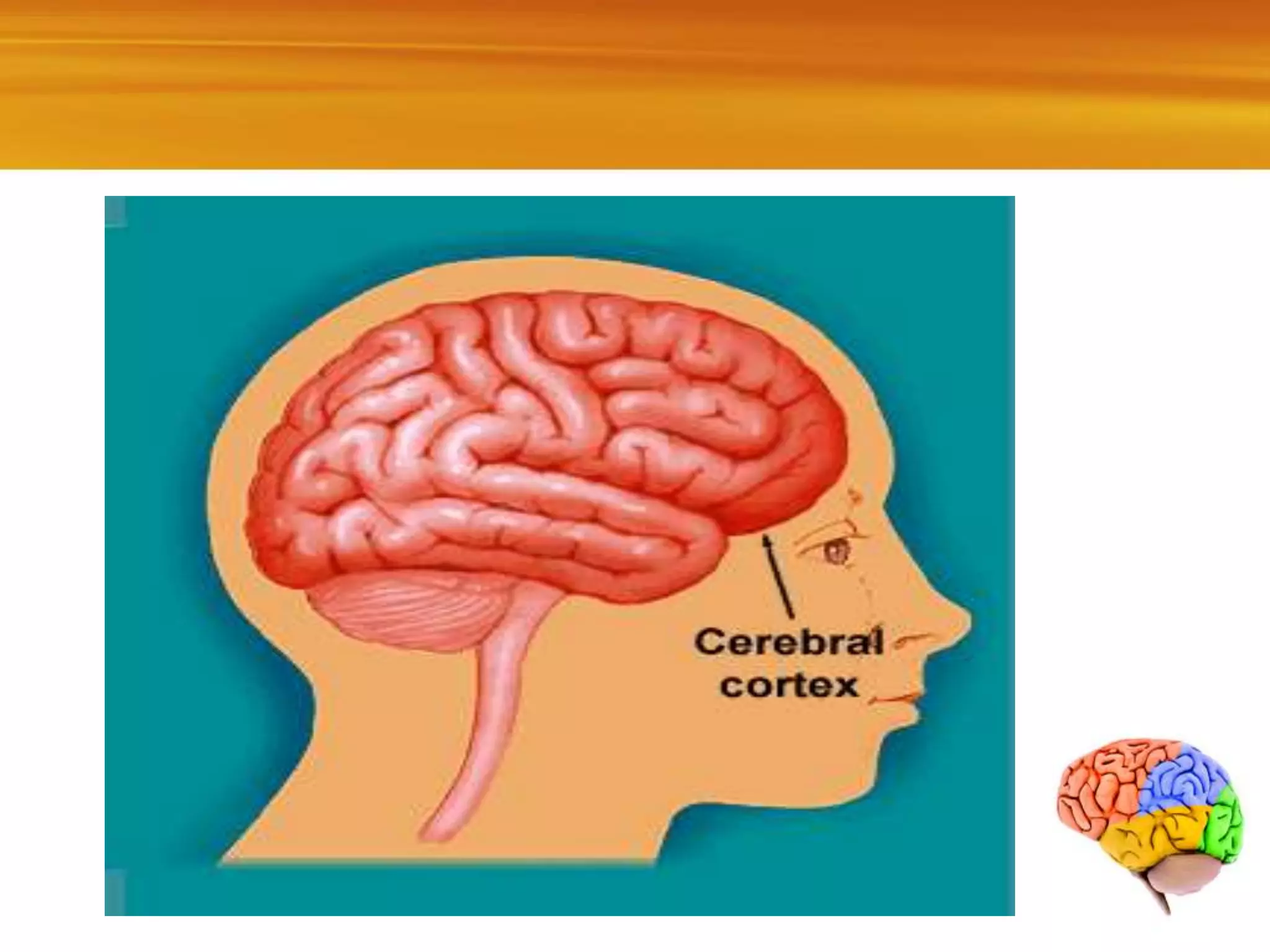
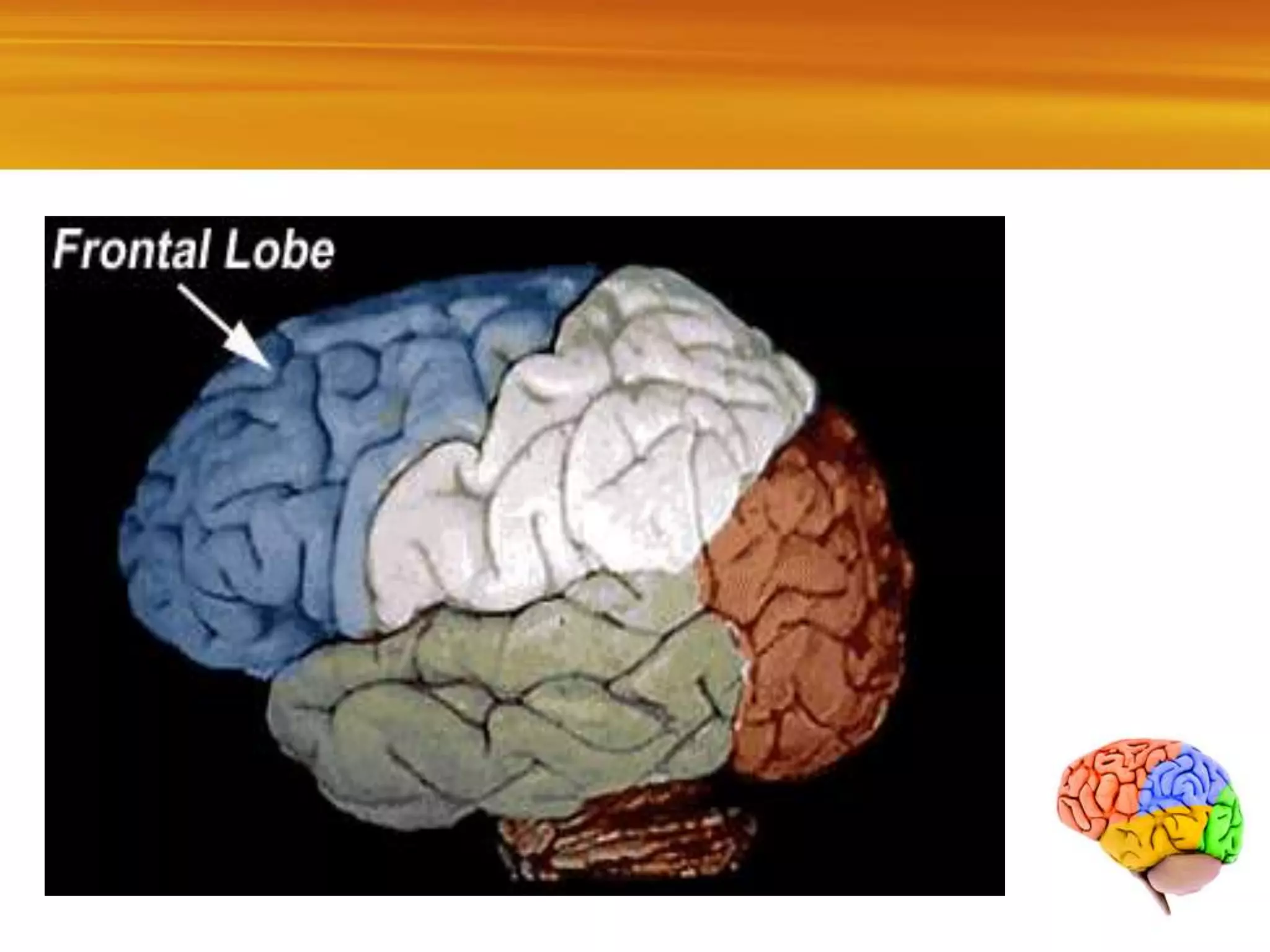
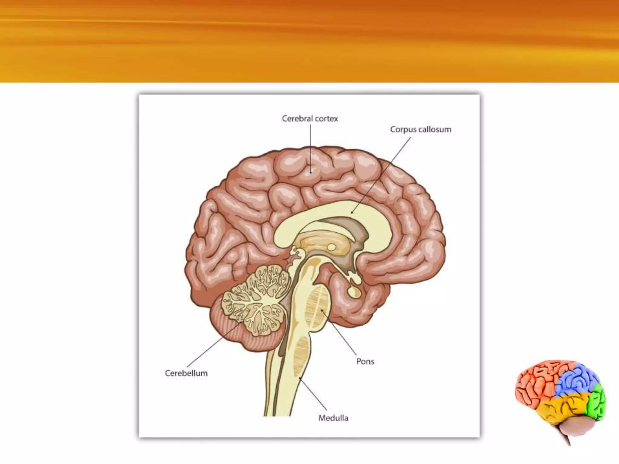
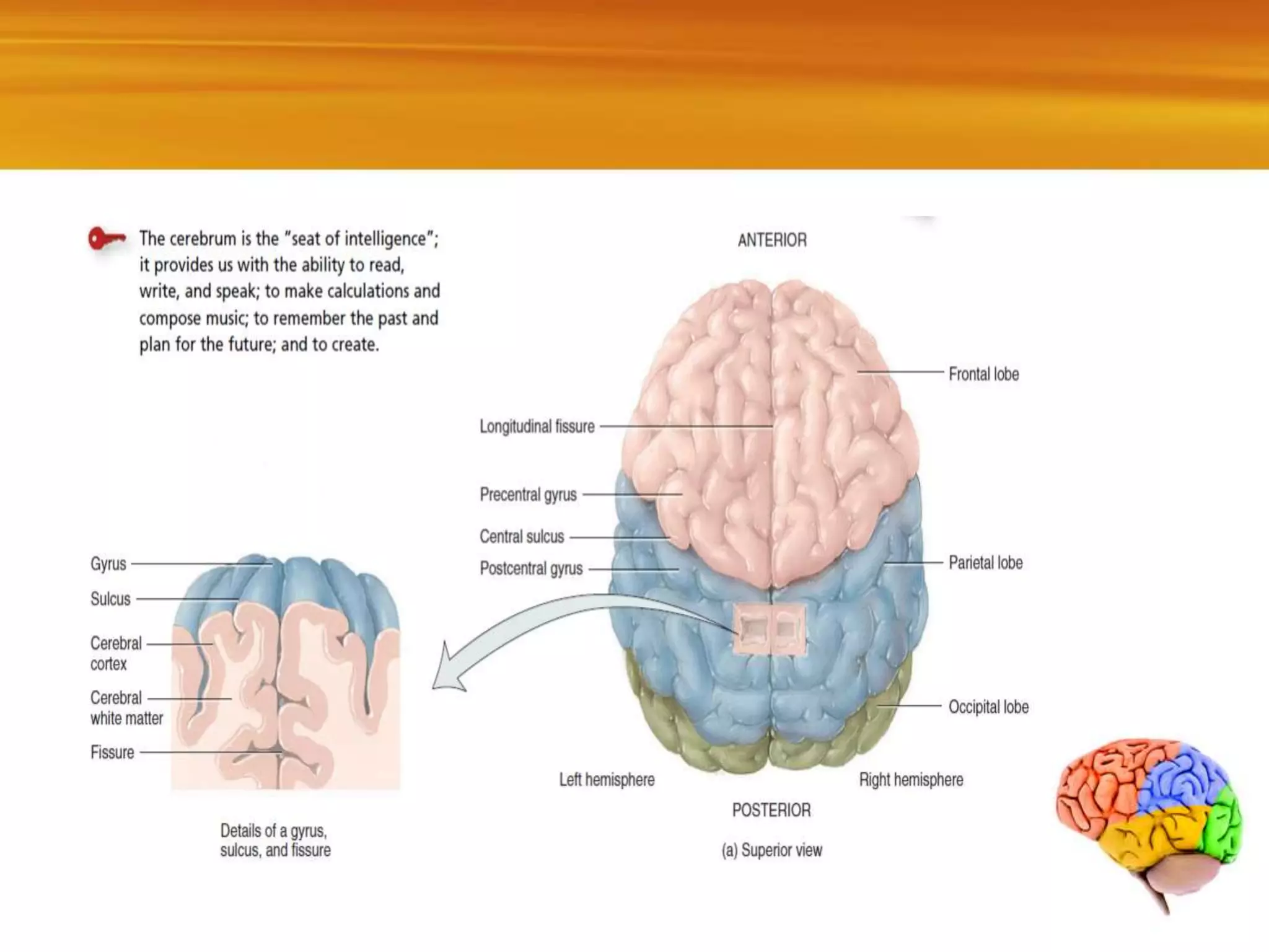
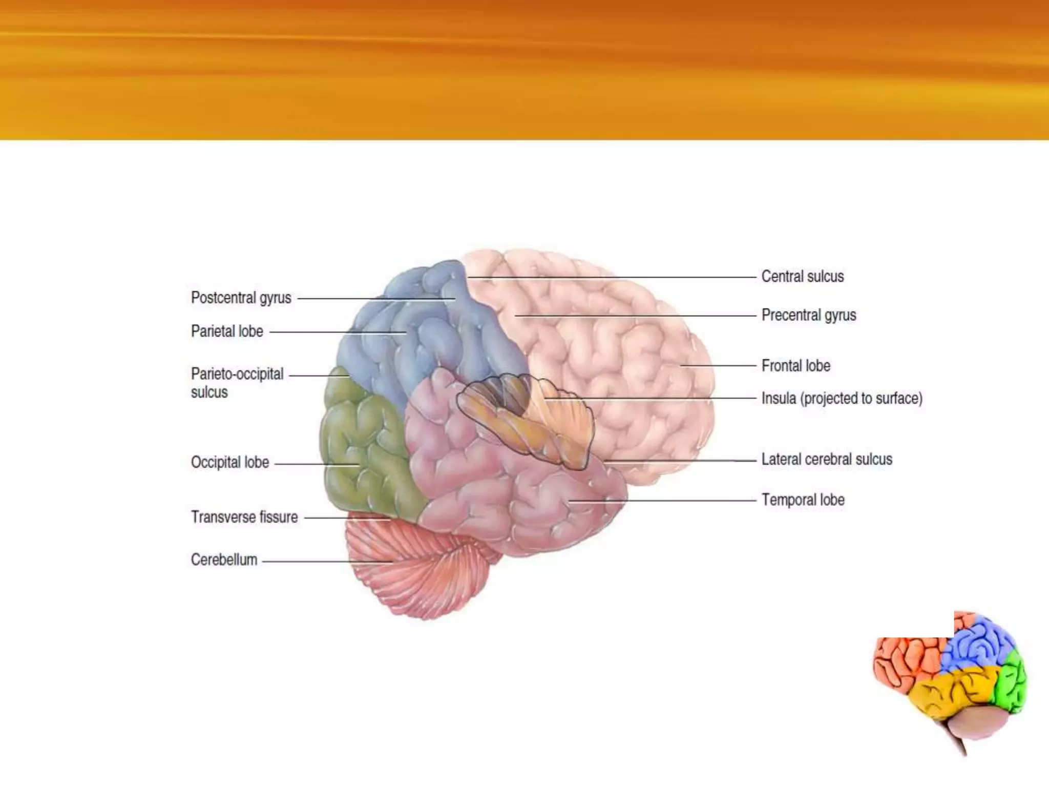
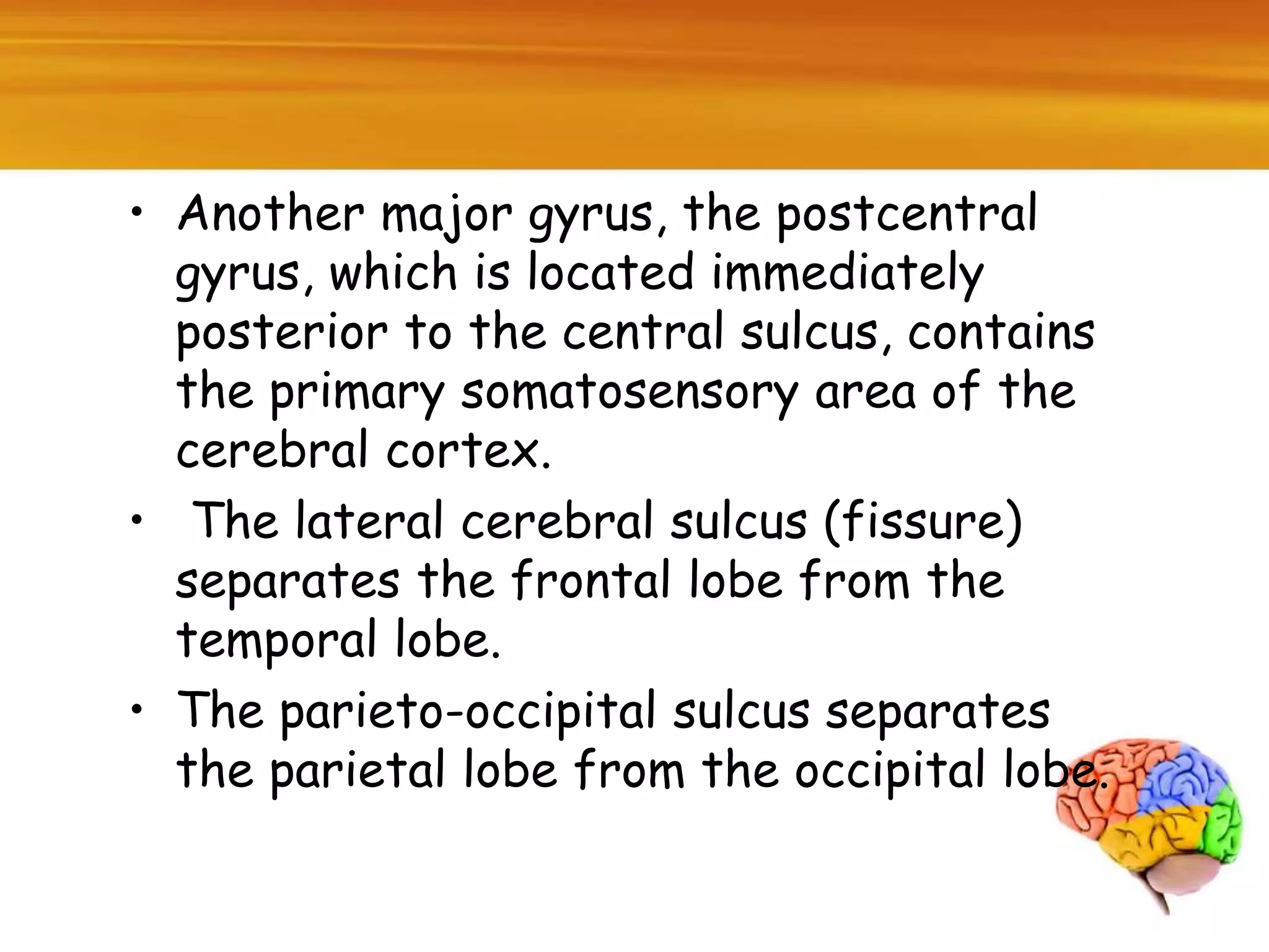
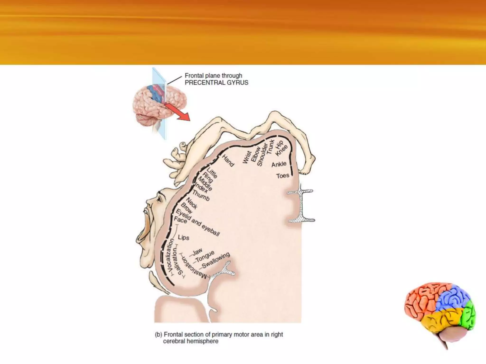
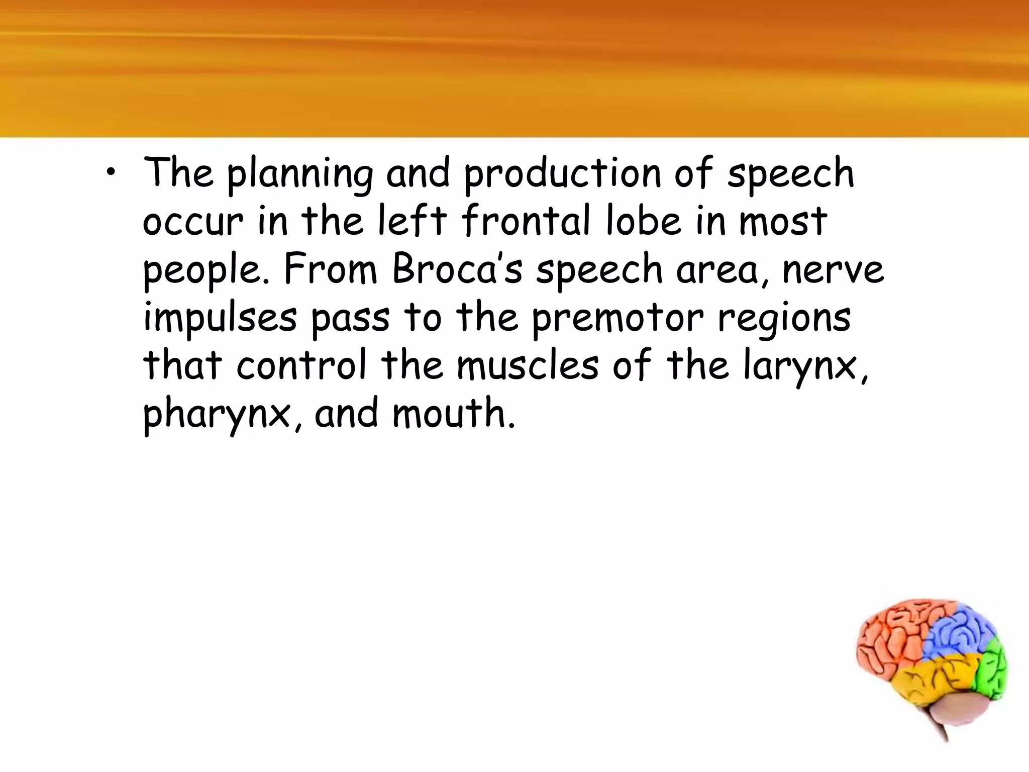
FUNCTIONAL
ORGANIATION OF
THE CEREBRA CORTEX
FATIMA WAHID MANGRIO
fatimawahid1234@gmail.com
Cerebral Cortex
• The cerebral cortex is a region of gray
matter that forms the outer rim of
the cerebrum.
• The cerebral cortex contains billions
of neurons arranged in layers.
• During embryonic development, when
brain size increases rapidly, the gray
matter of the cortex enlarges much
faster than the deeper white matter.
As a result, the cortical region rolls
and folds on itself. The folds are
called gyri or convolutions.
• The deepest grooves between folds are
known as fissures; the shallower grooves
between folds are termed sulci.
• The most prominent fissure, the
longitudinal fissure, separates the
cerebrum into right and left halves called
cerebral hemispheres.
• The cerebral hemispheres are connected
internally by the corpus callosum a broad
band of white matter containing axons
that extend between the hemispheres
Lobes of the Cerebrum
• Each cerebral hemisphere can be further
subdivided into several lobes.
• The central sulcus separates the frontal
lobe from the parietal lobe. A major
gyrus, the precentral gyrus—located
immediately anterior to the central
sulcus contains the primary motor area of
the cerebral cortex.
• Another major gyrus, the postcentral
gyrus, which is located immediately
posterior to the central sulcus, contains
the primary somatosensory area of the
cerebral cortex.
• The lateral cerebral sulcus (fissure)
separates the frontal lobe from the
temporal lobe.
• The parieto-occipital sulcus separates
the parietal lobe from the occipital lobe.
Functional Organization of the Cerebral Cortex
• Specific types of sensory, motor, and
integrative signals are processed in certain
regions of the cerebral cortex.
• Generally, sensory areas receive sensory
information and are involved in perception,
the conscious awareness of a sensation; motor
areas control the execution of voluntary
movements; and association areas deal with
more complex integrative functions such as
memory, emotions, reasoning, will, judgment,
personality traits, and intelligence.
Sensory Areas of The Cerebral Cortex
• Sensory impulses arrive mainly in the
posterior half of both cerebral
hemispheres, in regions behind the
central sulci. In the cerebral cortex,
primary sensory areas receive sensory
information that has been relayed from
peripheral sensory receptors through
lower regions of the brain.
• Sensory association areas often are
adjacent to the primary areas. They
usually receive input both from the
primary areas and from other brain
regions. Sensory association areas
integrate sensory experiences to
generate meaningful patterns of
recognition and awareness.
Some Important sensory Areas of the Cerebral
Cortex
• The primary somatosensory area (areas 1, 2,
and 3) is located in the postcentral gyrus of
parietal lobe.
• The primary somatosensory area receives
nerve impulses for touch, pressure,
vibration, itch, tickle, temperature (coldness
and warmth), pain, and proprioception (joint
and muscle position) and is involved in the
perception of these somatic sensations.
Sensory Homunculus
• A “map” of the entire body is present in the
primary somatosensory area: Each point
within the area receives impulses from a
specific part of the body.
• The size of the cortical area receiving
impulses from a particular part of the body
depends on the number of receptors present
there rather than on the size of the body
part.
• For example, a larger region of the
somatosensory area receives impulses from
the lips and fingertips than from the thorax
or hip. This distorted somatic sensory map of
the body is known as the sensory homunculus.
The primary somatosensory area allows you
to pinpoint where somatic sensations
originate, so that you know exactly where on
your body to swat that mosquito.
Sensory Homunculus
Primary visual area
• The primary visual area (area 17),
located at the posterior tip of the
occipital lobe receives visual
information and is involved in visual
perception.
Primary auditory area
• The primary auditory area
(areas 41 and 42), located in the
superior part of the temporal
lobe, receives information for
sound and is involved in auditory
perception.
Primary gustatory area
• The primary gustatory area
(area 43), located at the base of
the postcentral gyrus in the
parietal cortex, receives
impulses for taste and is involved
in gustatory perception and
taste discrimination
Primary olfactory area
• The primary olfactory area (area 28),
located in the temporal lobe on the
medial aspect (and thus not visible)
receives impulses for smell and is
involved in olfactory perception.
Motor Areas of the Cerebral Cortex
• Motor output from the cerebral cortex
flows mainly from the anterior part of
each hemisphere. Among the most
important motor areas are the following
Primary motor area
• The primary motor area (area 4) is
located in the precentral gyrus of the
frontal lobe. As is true for the primary
somatosensory area, a “map” of the
entire body is present in the primary
motor area: Each region within the area
controls voluntary contractions of
specific muscles or groups of muscles.
• Electrical stimulation of any point in the
primary motor area causes contraction of
specific skeletal muscle fibers on the
opposite side of the body. Different muscles
are represented unequally in the primary
motor area.
• More cortical area is devoted to those
muscles involved in skilled, complex, or
delicate movement. For instance, the
cortical region devoted to muscles that
move the fingers is much larger than the
region for muscles that move the toes.
This distorted muscle map of the body is
called the motor homunculus.
BROCA’S SPEECH AREA
• Broca’s speech area (areas 44 and 45) is
located in the frontal lobe.
• Speaking and understanding language are
complex activities that involve several
sensory, association, and motor areas of
the cortex. In about 97% of the
population, these language areas are
localized in the left hemisphere.
• The planning and production of speech
occur in the left frontal lobe in most
people. From Broca’s speech area, nerve
impulses pass to the premotor regions
that control the muscles of the larynx,
pharynx, and mouth.
• The impulses from the premotor area
result in specific, coordinated muscle
contractions. Simultaneously, impulses
propagate from Broca’s speech area to
the primary motor area. From here,
impulses also control the breathing
muscles to regulate the proper flow of
air past the vocal cords.
• The coordinated contractions of your
speech and breathing muscles enable you
to speak your thoughts. People who
suffer a cerebrovascular accident (CVA)
or stroke in this area can still have clear
thoughts but are unable to form words, a
phenomenon referred to as non fluent
aphasia.
Functional organization of the cerebral cortex i sensory & motor cortex

Functional organization of the cerebral cortex i sensory & motor cortex

  • 1.
    FUNCTIONAL ORGANIATION OF THE CEREBRACORTEX FATIMA WAHID MANGRIO fatimawahid1234@gmail.com
  • 3.
    Cerebral Cortex • Thecerebral cortex is a region of gray matter that forms the outer rim of the cerebrum. • The cerebral cortex contains billions of neurons arranged in layers. • During embryonic development, when brain size increases rapidly, the gray matter of the cortex enlarges much faster than the deeper white matter. As a result, the cortical region rolls and folds on itself. The folds are called gyri or convolutions.
  • 7.
    • The deepestgrooves between folds are known as fissures; the shallower grooves between folds are termed sulci. • The most prominent fissure, the longitudinal fissure, separates the cerebrum into right and left halves called cerebral hemispheres. • The cerebral hemispheres are connected internally by the corpus callosum a broad band of white matter containing axons that extend between the hemispheres
  • 10.
    Lobes of theCerebrum • Each cerebral hemisphere can be further subdivided into several lobes. • The central sulcus separates the frontal lobe from the parietal lobe. A major gyrus, the precentral gyrus—located immediately anterior to the central sulcus contains the primary motor area of the cerebral cortex.
  • 11.
    • Another majorgyrus, the postcentral gyrus, which is located immediately posterior to the central sulcus, contains the primary somatosensory area of the cerebral cortex. • The lateral cerebral sulcus (fissure) separates the frontal lobe from the temporal lobe. • The parieto-occipital sulcus separates the parietal lobe from the occipital lobe.
  • 12.
    Functional Organization ofthe Cerebral Cortex • Specific types of sensory, motor, and integrative signals are processed in certain regions of the cerebral cortex. • Generally, sensory areas receive sensory information and are involved in perception, the conscious awareness of a sensation; motor areas control the execution of voluntary movements; and association areas deal with more complex integrative functions such as memory, emotions, reasoning, will, judgment, personality traits, and intelligence.
  • 14.
    Sensory Areas ofThe Cerebral Cortex • Sensory impulses arrive mainly in the posterior half of both cerebral hemispheres, in regions behind the central sulci. In the cerebral cortex, primary sensory areas receive sensory information that has been relayed from peripheral sensory receptors through lower regions of the brain.
  • 15.
    • Sensory associationareas often are adjacent to the primary areas. They usually receive input both from the primary areas and from other brain regions. Sensory association areas integrate sensory experiences to generate meaningful patterns of recognition and awareness.
  • 16.
    Some Important sensoryAreas of the Cerebral Cortex • The primary somatosensory area (areas 1, 2, and 3) is located in the postcentral gyrus of parietal lobe. • The primary somatosensory area receives nerve impulses for touch, pressure, vibration, itch, tickle, temperature (coldness and warmth), pain, and proprioception (joint and muscle position) and is involved in the perception of these somatic sensations.
  • 17.
    Sensory Homunculus • A“map” of the entire body is present in the primary somatosensory area: Each point within the area receives impulses from a specific part of the body. • The size of the cortical area receiving impulses from a particular part of the body depends on the number of receptors present there rather than on the size of the body part.
  • 18.
    • For example,a larger region of the somatosensory area receives impulses from the lips and fingertips than from the thorax or hip. This distorted somatic sensory map of the body is known as the sensory homunculus. The primary somatosensory area allows you to pinpoint where somatic sensations originate, so that you know exactly where on your body to swat that mosquito.
  • 19.
  • 20.
    Primary visual area •The primary visual area (area 17), located at the posterior tip of the occipital lobe receives visual information and is involved in visual perception.
  • 21.
    Primary auditory area •The primary auditory area (areas 41 and 42), located in the superior part of the temporal lobe, receives information for sound and is involved in auditory perception.
  • 22.
    Primary gustatory area •The primary gustatory area (area 43), located at the base of the postcentral gyrus in the parietal cortex, receives impulses for taste and is involved in gustatory perception and taste discrimination
  • 23.
    Primary olfactory area •The primary olfactory area (area 28), located in the temporal lobe on the medial aspect (and thus not visible) receives impulses for smell and is involved in olfactory perception.
  • 24.
    Motor Areas ofthe Cerebral Cortex • Motor output from the cerebral cortex flows mainly from the anterior part of each hemisphere. Among the most important motor areas are the following
  • 25.
    Primary motor area •The primary motor area (area 4) is located in the precentral gyrus of the frontal lobe. As is true for the primary somatosensory area, a “map” of the entire body is present in the primary motor area: Each region within the area controls voluntary contractions of specific muscles or groups of muscles.
  • 27.
    • Electrical stimulationof any point in the primary motor area causes contraction of specific skeletal muscle fibers on the opposite side of the body. Different muscles are represented unequally in the primary motor area.
  • 28.
    • More corticalarea is devoted to those muscles involved in skilled, complex, or delicate movement. For instance, the cortical region devoted to muscles that move the fingers is much larger than the region for muscles that move the toes. This distorted muscle map of the body is called the motor homunculus.
  • 29.
    BROCA’S SPEECH AREA •Broca’s speech area (areas 44 and 45) is located in the frontal lobe. • Speaking and understanding language are complex activities that involve several sensory, association, and motor areas of the cortex. In about 97% of the population, these language areas are localized in the left hemisphere.
  • 30.
    • The planningand production of speech occur in the left frontal lobe in most people. From Broca’s speech area, nerve impulses pass to the premotor regions that control the muscles of the larynx, pharynx, and mouth.
  • 31.
    • The impulsesfrom the premotor area result in specific, coordinated muscle contractions. Simultaneously, impulses propagate from Broca’s speech area to the primary motor area. From here, impulses also control the breathing muscles to regulate the proper flow of air past the vocal cords.
  • 32.
    • The coordinatedcontractions of your speech and breathing muscles enable you to speak your thoughts. People who suffer a cerebrovascular accident (CVA) or stroke in this area can still have clear thoughts but are unable to form words, a phenomenon referred to as non fluent aphasia.