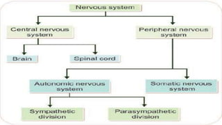
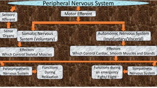
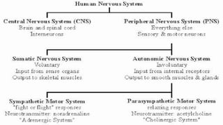
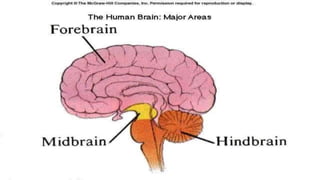
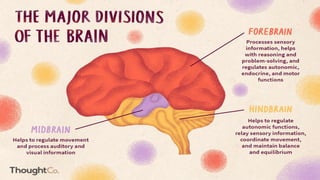
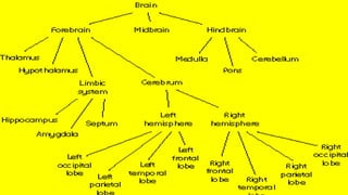
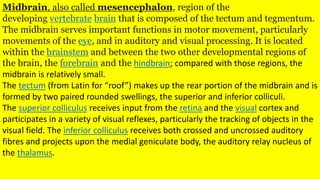
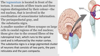
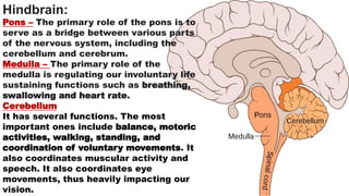
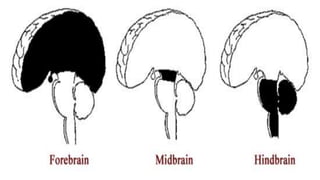
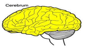
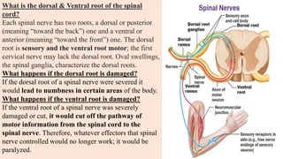
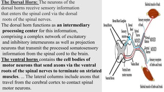
Nervous –System: Classification
Dr Neeta Gupta
Associate Professor
Certified Practitioner of EFT & REBT
(London) & CBT (Scotland) T
DAV PG College
Dehradun
.
Peripheral Nervous System
Sense
Organs
Sensory
Afferent
Motor Efferent
Autonomic Nervous System
(Involuntary/Visceral)
Somatic Nervous
System (Voluntary)
Effectors
Which Control Skeletal Muscles
Effectors
Which Control Cardiac, Smooth Muscles and Glands
Functions
During
Relaxation
Sympathetic
Nervous System
Functions during
an emergency
Fight/ Flight
Parasympathetic
Nervous System
Cranial nerves and spinal nerves are two types of
nerves of the peripheral nervous system. The key
difference between cranial and spinal nerves is that
the cranial nerves come from the brain while the
spinal nerves come from the spinal cord. Spinal
nerves are those that emerge directly from segments of
the spinal cord. Cranial nerve transfers information
between the brain and the other parts of the body.
Cranial nerves arise from the brain.
The nerves Conduct impulses toward
or away from the central nervous
mechanism. In humans
12 pairs of the cranial nerves,
are attached to the brain, and,
as a rule, 31 pairs of the spinal nerves, are attached to the
spinal cord.
The autonomic nervous system:
is also called the visceral nervous
system because it controls smooth
muscle, cardiac muscle, and
glands, which make up the viscera
of the body. ... These inputs elicit
reflex responses through the
efferent autonomic nerves.
Hypothalamus: is located in the floor of the third
ventricle and is the master control of the
autonomic system. It plays a role in controlling
behaviours such as hunger, thirst, sleep, and
sexual response. It also regulates body
temperature, blood pressure, emotions, and
secretion of hormones.
Thalamus: serves as a relay station for almost all
information that comes and goes to the cortex. It
plays a role in pain sensation, attention,
alertness and memory.
Limbic system: is the center of our emotions,
learning, and memory. Included in this system
are the cingulate gyri, hypothalamus, amygdala
(emotional reactions) and hippocampus
(memory). The septum is considered a part of the
limbic system, mediating the connection between
the cortex and subcortical limbic nuclei.
Cerebrum: is the largest part of the brain and is
composed of right and left hemispheres. It performs
higher functions like interpreting touch, vision and
hearing, as well as speech, reasoning, emotions,
learning, and fine control of movement.
The cerebrum is divided into two halves: the right and
left hemispheres .They are joined by a bundle of
fibers called the corpus callosum that transmits
messages from one side to the other. Each
hemisphere controls the opposite side of the body. If
a stroke occurs on the right side of the brain, your left
arm or leg may be weak or paralyzed.
The right hemisphere controls creativity, spatial
ability, artistic, and musical skills. The left hemisphere
is dominant in hand use and language in about 92% of
people.
The cerebral hemispheres have distinct
fissures, which divide the brain into lobes.
Each hemisphere has 4 lobes: frontal,
temporal, parietal, and occipital .
Frontal lobe
•Personality, behavior, emotions
•Judgment, planning, problem solving
•Speech: speaking and writing (Broca’s area)
•Body movement (motor strip)
•Intelligence, concentration, self awareness
Parietal lobe
•Interprets language, words
•Sense of touch, pain, temperature (sensory strip)
•Interprets signals from vision, hearing, motor,
sensory and memory
•Spatial and visual perception
Occipital lobe
•Interprets vision (color, light, movement)
Temporal lobe
•Understanding language (Wernicke’s area)
•Hearing
.
Midbrain, also called mesencephalon, region of the
developing vertebrate brain that is composed of the tectum and tegmentum.
The midbrain serves important functions in motor movement, particularly
movements of the eye, and in auditory and visual processing. It is located
within the brainstem and between the two other developmental regions of
the brain, the forebrain and the hindbrain; compared with those regions, the
midbrain is relatively small.
The tectum (from Latin for “roof”) makes up the rear portion of the midbrain and is
formed by two paired rounded swellings, the superior and inferior colliculi.
The superior colliculus receives input from the retina and the visual cortex and
participates in a variety of visual reflexes, particularly the tracking of objects in the
visual field. The inferior colliculus receives both crossed and uncrossed auditory
fibres and projects upon the medial geniculate body, the auditory relay nucleus of
the thalamus.
The tegmentum is located in front of the
tectum. It consists of fibre tracts and three
regions distinguished by their colour—the
red nucleus, that is involved in the
coordination of sensorimotor information.
The periaqueductal gray, and
the substantia nigra.
A smaller number of fibres synapse on large
cells in caudal regions of the red nucleus;
those give rise to the crossed fibres of the
rubrospinal tract, which runs to the spinal
cord and is influenced by the motor cortex.
The substantia nigra is a large pigmented cluster
of neurons that consists of two parts, the pars
reticulata and the pars compacta.
Hindbrain:
Pons – The primary role of the pons is to
serve as a bridge between various parts
of the nervous system, including the
cerebellum and cerebrum.
Medulla – The primary role of the
medulla is regulating our involuntary life
sustaining functions such as breathing,
swallowing and heart rate.
Cerebellum
It has several functions. The most
important ones include balance, motoric
activities, walking, standing, and
coordination of voluntary movements. It
also coordinates muscular activity and
speech. It also coordinates eye
movements, thus heavily impacting our
vision.
Spinal Cord;
The spinal cord is a long, fragile tubelike structure that
begins at the end of the brain stem and continues down
almost to the bottom of the spine. The spinal cord
consists of bundles of nerve axons forming pathways
that carry incoming and outgoing messages between
the brain and the rest of the body. Like the brain, the
spinal cord is covered by three layers of tissue
(meninges). The spinal cord and meninges are
contained in the spinal canal, which runs through the
center of the spine.
.
In most adults, the spine is composed
of 33 individual back bones
(vertebrae). Just as the skull protects
the brain, vertebrae protect the
spinal cord. The vertebrae are
separated by disks made of cartilage,
which act as cushions, reducing the
forces on the spine generated by
movements such as walking and
jumping. The vertebrae and disks of
cartilage extend the length of the
spine and together form the vertebral
column, also called the spinal
column.
What is the dorsal & Ventral root of the spinal
cord?
Each spinal nerve has two roots, a dorsal or posterior
(meaning “toward the back”) one and a ventral or
anterior (meaning “toward the front”) one. The dorsal
root is sensory and the ventral root motor; the first
cervical nerve may lack the dorsal root. Oval swellings,
the spinal ganglia, characterize the dorsal roots.
What happens if the dorsal root is damaged?
If the dorsal root of a spinal nerve were severed it
would lead to numbness in certain areas of the body.
What happens if the ventral root is damaged?
If the ventral root of a spinal nerve was severely
damaged or cut, it would cut off the pathway of
motor information from the spinal cord to the
spinal nerve. Therefore, whatever effectors that spinal
nerve controlled would no longer work; it would be
paralyzed.
The Dorsal Horn: The neurons of the
dorsal horns receive sensory information
that enters the spinal cord via the dorsal
roots of the spinal nerves.
The dorsal horn functions as an intermediary
processing center for this information,
comprising a complex network of excitatory
and inhibitory interneurons as well as projection
neurons that transmit the processed somatosensory
information from the spinal cord to the brain.
The ventral horns contains the cell bodies of
motor neurons that send axons via the ventral
roots of the spinal nerves to terminate on striated
muscles. ... The lateral columns include axons that
travel from the cerebral cortex to contact spinal
motor neurons.
Mechanism of Simple Reflex
Spinal Reflexes are very simlpe and automatic behaviour that
occur without conscious, voluntary action of the brain.
When you are exposed to flame, receptor cells in your fingertips
respond to the flame, sending neural impulses racing along
sensory neurons, through a dorsal root and into the spinal cord.
Impulses travel to interneurons and go right back out of the
spinal cord (through ventral root) on motor neurons to your
arms and hand where muscles are stimulated to contract and
your hand jerks back from the flame. So this is the simple relex
impulse.
REFERENCES:
https://4.bp.blogspot.com/-
YyolCPZX1Aw/VuPhJUwHoFI/AAAAAAAABug/Kt30PLfhmCIav7Pj6Up8WBlgzoxXe3uFg/s640/organization_of_nervous_system.JPG
https://qbi.uq.edu.au/files/38434/Nervous_System.jpg
https://lh3.googleusercontent.com/proxy/ftC0-vMqCwOhR3-
F6eIpDWgyGs96tfuQDmh8fa3yZAJ_JJp0J_ZkVrg_YfWNgXV_z7fKrmTkDOBzsBc8h9b2ow
https://www.thoughtco.com/thmb/EUnrG_GGF5svvLmUAiBlyrihi_M=/1500x1000/filters:fill(auto,1)/divisions-of-the-brain-
4032899_final1-5c795dddc9e77c00012f81d8.png
https://www.scienceabc.com/wp-content/uploads/2018/06/Cerebrum1.jpg
https://lh4.ggpht.com/-
LuzjIzjUvFs/Uym2VcSh1RI/AAAAAAAABDI/tunSDFTJh84/Brain%252527s%252520three%252520parts%25255B4%25255D.gif?img
max=800
https://mayfieldclinic.com/pe-anatbrain.htm
https://i.pinimg.com/originals/6f/99/df/6f99dfb8c638343c8c8849e580377d97.gif
https://faculty.washington.edu/chudler/gif/lobeani.gif
https://64.media.tumblr.com/0f7befbf5b9c043af0a481a3181a96ec/tumblr_inline_oldpdyGzHf1sndsvm_540.gifv
https://static.wikia.nocookie.net/brain-for-ai/images/b/bd/Tectum.png/revision/latest?cb=20170613125935
https://i.makeagif.com/media/6-03-2017/y_7E4a.gif
https://upload.wikimedia.org/wikipedia/commons/thumb/d/d2/Spinal_nerve.svg/1200px-Spinal_nerve.svg.png
http://31.media.tumblr.com/91040c8752ba3cfe8a2aa95d1c3b832d/tumblr_nk927ftzex1skn1oxo1_500.gif
https://i.makeagif.com/media/5-12-2018/cuxkW2.gif
https://www.msdmanuals.com/en-in/home/brain,-spinal-cord,-and-nerve-disorders/biology-of-the-nervous-system/spinal-cord
https://thumbs.gfycat.com/FearlessNaturalFireant-size_restricted.gif
Nervous System: Classification

Nervous System: Classification

  • 1.
    Nervous –System: Classification DrNeeta Gupta Associate Professor Certified Practitioner of EFT & REBT (London) & CBT (Scotland) T DAV PG College Dehradun
  • 3.
    . Peripheral Nervous System Sense Organs Sensory Afferent MotorEfferent Autonomic Nervous System (Involuntary/Visceral) Somatic Nervous System (Voluntary) Effectors Which Control Skeletal Muscles Effectors Which Control Cardiac, Smooth Muscles and Glands Functions During Relaxation Sympathetic Nervous System Functions during an emergency Fight/ Flight Parasympathetic Nervous System
  • 4.
    Cranial nerves andspinal nerves are two types of nerves of the peripheral nervous system. The key difference between cranial and spinal nerves is that the cranial nerves come from the brain while the spinal nerves come from the spinal cord. Spinal nerves are those that emerge directly from segments of the spinal cord. Cranial nerve transfers information between the brain and the other parts of the body. Cranial nerves arise from the brain. The nerves Conduct impulses toward or away from the central nervous mechanism. In humans 12 pairs of the cranial nerves, are attached to the brain, and, as a rule, 31 pairs of the spinal nerves, are attached to the spinal cord.
  • 5.
    The autonomic nervoussystem: is also called the visceral nervous system because it controls smooth muscle, cardiac muscle, and glands, which make up the viscera of the body. ... These inputs elicit reflex responses through the efferent autonomic nerves.
  • 10.
    Hypothalamus: is locatedin the floor of the third ventricle and is the master control of the autonomic system. It plays a role in controlling behaviours such as hunger, thirst, sleep, and sexual response. It also regulates body temperature, blood pressure, emotions, and secretion of hormones. Thalamus: serves as a relay station for almost all information that comes and goes to the cortex. It plays a role in pain sensation, attention, alertness and memory. Limbic system: is the center of our emotions, learning, and memory. Included in this system are the cingulate gyri, hypothalamus, amygdala (emotional reactions) and hippocampus (memory). The septum is considered a part of the limbic system, mediating the connection between the cortex and subcortical limbic nuclei.
  • 11.
    Cerebrum: is thelargest part of the brain and is composed of right and left hemispheres. It performs higher functions like interpreting touch, vision and hearing, as well as speech, reasoning, emotions, learning, and fine control of movement. The cerebrum is divided into two halves: the right and left hemispheres .They are joined by a bundle of fibers called the corpus callosum that transmits messages from one side to the other. Each hemisphere controls the opposite side of the body. If a stroke occurs on the right side of the brain, your left arm or leg may be weak or paralyzed. The right hemisphere controls creativity, spatial ability, artistic, and musical skills. The left hemisphere is dominant in hand use and language in about 92% of people.
  • 12.
    The cerebral hemisphereshave distinct fissures, which divide the brain into lobes. Each hemisphere has 4 lobes: frontal, temporal, parietal, and occipital . Frontal lobe •Personality, behavior, emotions •Judgment, planning, problem solving •Speech: speaking and writing (Broca’s area) •Body movement (motor strip) •Intelligence, concentration, self awareness Parietal lobe •Interprets language, words •Sense of touch, pain, temperature (sensory strip) •Interprets signals from vision, hearing, motor, sensory and memory •Spatial and visual perception
  • 13.
    Occipital lobe •Interprets vision(color, light, movement) Temporal lobe •Understanding language (Wernicke’s area) •Hearing .
  • 14.
    Midbrain, also calledmesencephalon, region of the developing vertebrate brain that is composed of the tectum and tegmentum. The midbrain serves important functions in motor movement, particularly movements of the eye, and in auditory and visual processing. It is located within the brainstem and between the two other developmental regions of the brain, the forebrain and the hindbrain; compared with those regions, the midbrain is relatively small. The tectum (from Latin for “roof”) makes up the rear portion of the midbrain and is formed by two paired rounded swellings, the superior and inferior colliculi. The superior colliculus receives input from the retina and the visual cortex and participates in a variety of visual reflexes, particularly the tracking of objects in the visual field. The inferior colliculus receives both crossed and uncrossed auditory fibres and projects upon the medial geniculate body, the auditory relay nucleus of the thalamus.
  • 15.
    The tegmentum islocated in front of the tectum. It consists of fibre tracts and three regions distinguished by their colour—the red nucleus, that is involved in the coordination of sensorimotor information. The periaqueductal gray, and the substantia nigra. A smaller number of fibres synapse on large cells in caudal regions of the red nucleus; those give rise to the crossed fibres of the rubrospinal tract, which runs to the spinal cord and is influenced by the motor cortex. The substantia nigra is a large pigmented cluster of neurons that consists of two parts, the pars reticulata and the pars compacta.
  • 16.
    Hindbrain: Pons – Theprimary role of the pons is to serve as a bridge between various parts of the nervous system, including the cerebellum and cerebrum. Medulla – The primary role of the medulla is regulating our involuntary life sustaining functions such as breathing, swallowing and heart rate. Cerebellum It has several functions. The most important ones include balance, motoric activities, walking, standing, and coordination of voluntary movements. It also coordinates muscular activity and speech. It also coordinates eye movements, thus heavily impacting our vision.
  • 19.
    Spinal Cord; The spinalcord is a long, fragile tubelike structure that begins at the end of the brain stem and continues down almost to the bottom of the spine. The spinal cord consists of bundles of nerve axons forming pathways that carry incoming and outgoing messages between the brain and the rest of the body. Like the brain, the spinal cord is covered by three layers of tissue (meninges). The spinal cord and meninges are contained in the spinal canal, which runs through the center of the spine. .
  • 20.
    In most adults,the spine is composed of 33 individual back bones (vertebrae). Just as the skull protects the brain, vertebrae protect the spinal cord. The vertebrae are separated by disks made of cartilage, which act as cushions, reducing the forces on the spine generated by movements such as walking and jumping. The vertebrae and disks of cartilage extend the length of the spine and together form the vertebral column, also called the spinal column.
  • 21.
    What is thedorsal & Ventral root of the spinal cord? Each spinal nerve has two roots, a dorsal or posterior (meaning “toward the back”) one and a ventral or anterior (meaning “toward the front”) one. The dorsal root is sensory and the ventral root motor; the first cervical nerve may lack the dorsal root. Oval swellings, the spinal ganglia, characterize the dorsal roots. What happens if the dorsal root is damaged? If the dorsal root of a spinal nerve were severed it would lead to numbness in certain areas of the body. What happens if the ventral root is damaged? If the ventral root of a spinal nerve was severely damaged or cut, it would cut off the pathway of motor information from the spinal cord to the spinal nerve. Therefore, whatever effectors that spinal nerve controlled would no longer work; it would be paralyzed.
  • 22.
    The Dorsal Horn:The neurons of the dorsal horns receive sensory information that enters the spinal cord via the dorsal roots of the spinal nerves. The dorsal horn functions as an intermediary processing center for this information, comprising a complex network of excitatory and inhibitory interneurons as well as projection neurons that transmit the processed somatosensory information from the spinal cord to the brain. The ventral horns contains the cell bodies of motor neurons that send axons via the ventral roots of the spinal nerves to terminate on striated muscles. ... The lateral columns include axons that travel from the cerebral cortex to contact spinal motor neurons.
  • 23.
    Mechanism of SimpleReflex Spinal Reflexes are very simlpe and automatic behaviour that occur without conscious, voluntary action of the brain. When you are exposed to flame, receptor cells in your fingertips respond to the flame, sending neural impulses racing along sensory neurons, through a dorsal root and into the spinal cord. Impulses travel to interneurons and go right back out of the spinal cord (through ventral root) on motor neurons to your arms and hand where muscles are stimulated to contract and your hand jerks back from the flame. So this is the simple relex impulse.
  • 25.
    REFERENCES: https://4.bp.blogspot.com/- YyolCPZX1Aw/VuPhJUwHoFI/AAAAAAAABug/Kt30PLfhmCIav7Pj6Up8WBlgzoxXe3uFg/s640/organization_of_nervous_system.JPG https://qbi.uq.edu.au/files/38434/Nervous_System.jpg https://lh3.googleusercontent.com/proxy/ftC0-vMqCwOhR3- F6eIpDWgyGs96tfuQDmh8fa3yZAJ_JJp0J_ZkVrg_YfWNgXV_z7fKrmTkDOBzsBc8h9b2ow https://www.thoughtco.com/thmb/EUnrG_GGF5svvLmUAiBlyrihi_M=/1500x1000/filters:fill(auto,1)/divisions-of-the-brain- 4032899_final1-5c795dddc9e77c00012f81d8.png https://www.scienceabc.com/wp-content/uploads/2018/06/Cerebrum1.jpg https://lh4.ggpht.com/- LuzjIzjUvFs/Uym2VcSh1RI/AAAAAAAABDI/tunSDFTJh84/Brain%252527s%252520three%252520parts%25255B4%25255D.gif?img max=800 https://mayfieldclinic.com/pe-anatbrain.htm https://i.pinimg.com/originals/6f/99/df/6f99dfb8c638343c8c8849e580377d97.gif https://faculty.washington.edu/chudler/gif/lobeani.gif https://64.media.tumblr.com/0f7befbf5b9c043af0a481a3181a96ec/tumblr_inline_oldpdyGzHf1sndsvm_540.gifv https://static.wikia.nocookie.net/brain-for-ai/images/b/bd/Tectum.png/revision/latest?cb=20170613125935 https://i.makeagif.com/media/6-03-2017/y_7E4a.gif https://upload.wikimedia.org/wikipedia/commons/thumb/d/d2/Spinal_nerve.svg/1200px-Spinal_nerve.svg.png http://31.media.tumblr.com/91040c8752ba3cfe8a2aa95d1c3b832d/tumblr_nk927ftzex1skn1oxo1_500.gif https://i.makeagif.com/media/5-12-2018/cuxkW2.gif https://www.msdmanuals.com/en-in/home/brain,-spinal-cord,-and-nerve-disorders/biology-of-the-nervous-system/spinal-cord https://thumbs.gfycat.com/FearlessNaturalFireant-size_restricted.gif