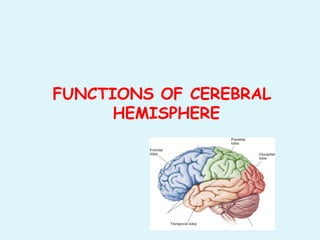
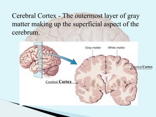
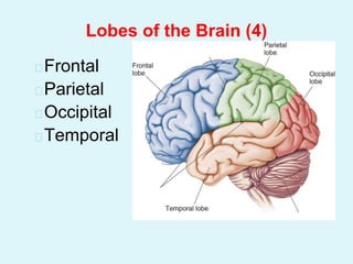
1. The cerebral hemispheres each contain a highly folded cortex and white matter. They contain motor, sensory, and limbic areas.
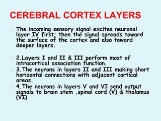
2. The cortex consists of six layers that process sensory signals in different ways and connect to different areas of the brain and body.
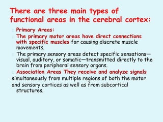
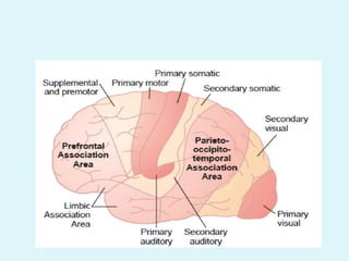
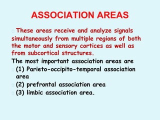
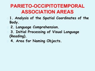
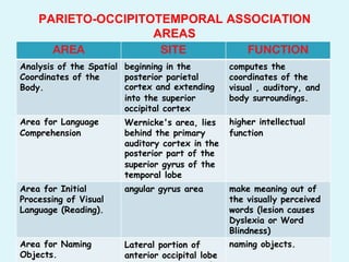
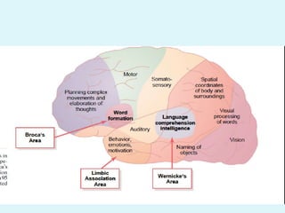
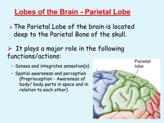
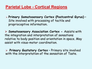
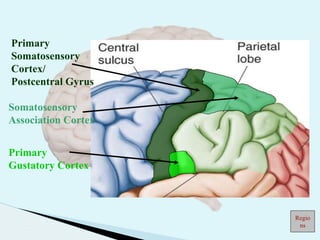
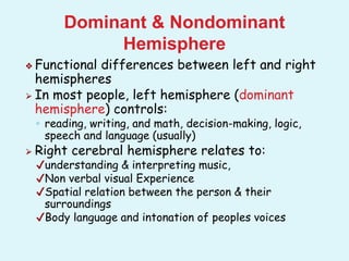
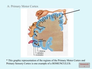
3. There are primary motor and sensory areas that directly connect to muscles and senses, as well as association areas that integrate signals between regions. The main association areas are parietal, prefrontal, and limbic.