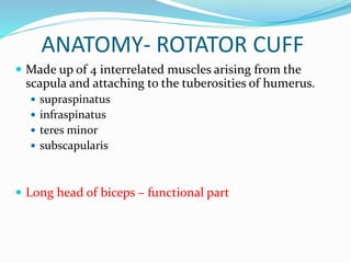
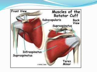
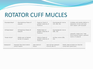
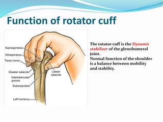
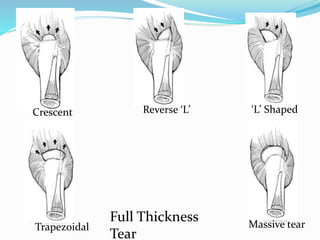
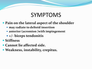
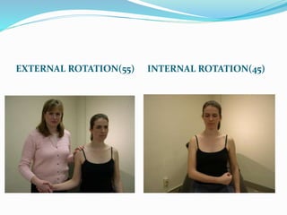
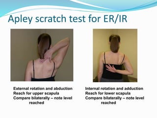
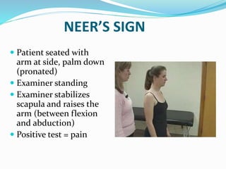
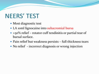
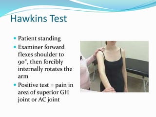
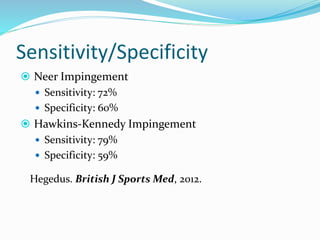
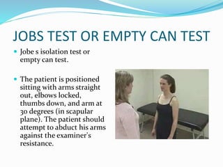
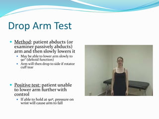
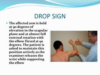
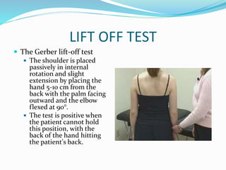
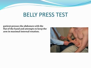
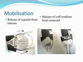
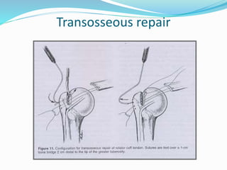
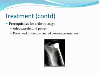
2. INTRODUCTION
In 1834, Smith - first description of a rupture of the
rotator cuff tendon .
Among most common causes of shoulder pain and
instability.
Incidence 5-40% with increasing with advancing age (
>40 years).
Normal senescence process
3.
4. ANATOMY- ROTATOR CUFF
Made up of 4 interrelated muscles arising from the
scapula and attaching to the tuberosities of humerus.
supraspinatus
infraspinatus
teres minor
subscapularis
Long head of biceps – functional part
8. Function of rotator cuff
The rotator cuff is the Dynamic
stabilizer of the glenohumeral
joint.
Normal function of the shoulder
is a balance between mobility
and stability.
9. Function of rotator cuff
Stabilisers of shoulder mainly anterior and posterior
cuff providing fixed fulcrum for concentric rotation of
the humeral head.
Neutralises shearing forces of deltoid in early
abduction.
Initiation of abduction.
Rotation of shoulder.
10. ETIOLOGY
IMPINGEMENT( MC )
TRAUMA
ATTRITION - AGING
ISCHEMIC TENDON
LACK OF NUTRITION TO JOINT
IATROGENIC
11. Different shapes of acromia (Biglianni et al) -anterior
slope
Type 1 - Flat ( 3 % of cuff tears)
Type 2 - Curved (24 % of cuff tears)
Type 3 - Hooked ( 73 % of cuff tears)
12. PATHOLOGY
Torn Rotator Cuff
Can not Counterbalance the upward
pull of the deltoid on the humerus
Not able to Hold the head of the
humerus secure in the glenoid
AHD <6mm
13. Leads to abutement of humeral
head against acromion
Acetabulization: Concave deformity of
under surface of Acromion
14. Narrowing & Arthritis
of Gleno-Humeral Joint
Last stage of Cuff tear
arthropathy with collapse of
humerus head
16. CLASSIFICATION
1. DURATION – ACUTE OR CHRONIC
2. DEGREE OF TEAR- PARTIAL OR FULL THICKNESS
TEAR.
3. ETIOLOGY- TRAUMATIC OR DEGENERATIVE.
4. COFIELD – BASED ON SIZE OF TEAR
SIZE OF TEAR DEGREE
<1 cm SMALL
1- 3 cms MEDIUM
3-5cms LARGE
>5 cms MASSIVE
23. 24
Focused History Questions
Mechanism of Injury
Helps predict injured structure
Example: Fall directly onto anterior/superior
shoulderAC joint injury (shoulder separation)
Example: Arm forcefully abducted and externally
rotated subluxation or anterior dislocation
Example: If chronic pain, note activity that triggers
pain, such as the cocking phase of throwing or the
pull-through phase of swimming
24. 25
Focused History Questions
Mechanism of Injury, continued
Can determine radiological needs
Likelihood of specific conditions varies
Setting (work, recreation, sports, traumatic,
atraumatic)
Age of the patient*
25. 26
Focused History Questions
Location of pain*
Anterior
Lateral
Superior
Posterior
Radiation of pain
Rotator cuff problems often include pain
radiating to upper arm
If pain starts in neck and radiates to shoulder,
consider cervical spine disease
34. 35
Palpation of AC Joint
Patient's arm at his/her
side
Note swelling, pain, and
gapping.
35. 36
Palpation of Bicipital Groove
Patient sitting,
beginning with the arm
straight
Patient actively flexes
biceps muscle while
examiner provides
supination and ER
Examiner palpates the
bicipital groove for pain
36. 37
Range of Motion (ROM)
Evaluate active ROM
If movement limited by pain, weakness, or tightness,
assist passively
Lack of full ROM with active and passive exam is
found in adhesive capsulitis and arthropathy
Evaluate bilaterally for comparison
40. Apley scratch test for ER/IR
Internal rotation and adduction
Reach for lower scapula
Compare bilaterally – note level
reached
External rotation and abduction
Reach for upper scapula
Compare bilaterally – note level
reached
41.
42.
43. NEER’S SIGN
Patient seated with
arm at side, palm down
(pronated)
Examiner standing
Examiner stabilizes
scapula and raises the
arm (between flexion
and abduction)
Positive test = pain
44. NEERS’ TEST
Most diagnostic test
LA 10ml lignocaine into subacromial bursa
>50% relief – rotator cuff tendinitis or partial tear of
bursal surface.
Pain relief but weakness persists – full thickness tears
No relief - incorrect diagnosis or wrong injection
45. Hawkins Test
Patient standing
Examiner forward
flexes shoulder to
90°, then forcibly
internally rotates the
arm
Positive test = pain in
area of superior GH
joint or AC joint
48. JOBS TEST OR EMPTY CAN TEST
Jobe s isolation test or
empty can test.
The patient is positioned
sitting with arms straight
out, elbows locked,
thumbs down, and arm at
30 degrees (in scapular
plane). The patient should
attempt to abduct his arms
against the examiner's
resistance.
49. Drop Arm Test
Method: patient abducts (or
examiner passively abducts)
arm and then slowly lowers it
May be able to lower arm slowly to
90° (deltoid function)
Arm will then drop to side if rotator
cuff tear
Positive test: patient unable
to lower arm further with
control
If able to hold at 90º, pressure on
wrist will cause arm to fall
50.
51. DROP SIGN
The affected arm is held
at 90 degrees of
elevation in the scapular
plane and at almost full
external rotation with
the elbow flexed at 90
degrees. The patient is
asked to maintain this
position actively as the
examiner releases the
wrist while supporting
the elbow
52.
53. LIFT OFF TEST
The Gerber lift-off test
The shoulder is placed
passively in internal
rotation and slight
extension by placing the
hand 5-10 cm from the
back with the palm facing
outward and the elbow
flexed at 90°.
The test is positive when
the patient cannot hold
this position, with the
back of the hand hitting
the patient's back.
54. BELLY PRESS TEST
patient presses the abdomen with the
flat of the hand and attempts to keep the
arm in maximal internal rotation.
55. OTHER TESTS
EXTERNAL ROTATION LAG SIGN- SUPRASPINATUS
AND INFRASPINATUS.
EXTERNAL ROTATION STRESS TEST-
INFRASPINATUS AND TERES MINOR.
58. Cochrane Database Review 2013 –
Hanchard, et al.
Physical tests for shoulder impingements and
local lesions of bursa, tendon or labrum that may
accompany impingement.
33 studies involving 4002 shoulders
59. Cochrane Database Review 2013 –
Hanchard, et al.
There is insufficient evidence upon which to base
selection of physical tests for shoulder impingements,
and local lesions of bursa, tendon or labrum that may
accompany impingement, in primary care. The large
body of literature revealed extreme diversity in the
performance and interpretation of tests, which
hinders synthesis of the evidence and/or clinical
applicability.
61. X RAY
AP VIEW
AXILLARY LATERAL VIEW
SUPRASPINATUS OUTLET VIEW
62. X RAY AP VIEW – ER AND IR VIEWS
The internal rotation view is for detecting Hill-Sachs lesions, and external
rotation for the greater tuberosity and proximal humeral physis in skeletally
immature patients. A true anteroposterior radiograph of the glenohumeral
joint is forarticular cartilage of the glenoid and the humeral head.
69. Conservative management
McLaughlin in 1962 advanced reasons to avoid early
repair
25 % of cadavers had torn cuff -most of them were
asymptomatic
50 % of patients would recover comfortably
Results of early and late repair are similar
Repair did not always permit anatomic restoration
Early diagnosis is difficult
NATURAL HISTORY IS UNPREDICTABLE
70. Review of literature indicates that success rate of
nonoperative treatment ranges from 33% to 92%
Bartolozzi et al (Clin orthop, 1994) reported 66-
75% good or excellent results (mean follow up 20
months). Unfavorable prognostic factors were
Tear> 1 cm2
Symptoms > 1yr
Significant functional impairment
73. Orthotherapy
Term used by Michael Wirth (OCNA 1997)
Interactive exchange between patient and orthopedic
surgeon directed at creating exercise regimen that
gradually improves motion and strength in shoulder girdle.
Three phases:
Phase 1- restore full, painless range of motion. Codman pendulum
exercise followed by passive movements in all direction
Phase 2- designed to strengthened remaining muscles of rotator
cuff, deltoid & scapular muscles
Phase 3- gradual reinstitution of normal activities including work,
hobby and sport.
75. Operative treatment
Patient selection:
Samilson & Binder :
Patient physiologically younger than 60 yrs
Clinically or arthrographically demonstrable full
thickness cuff tear.
Failure to improve on nonoperative management for
minimum of 6 weeks
Need to use shoulder in overhead elevation
Full passive range of motion
Ability & willingness to cooperate
76. Poor prognostic factors
Old age group (physiological age >60 years)
Long history
No history of trauma
Smoker
Multiple steroid injection
Diffuse osteopenia
Grade 3 or less of external rotation
Upward migration of humeral head.
77. Procedures
Repair of tear
open or arthroscopic
Tendon to tendon or tendon to bone
Arthroscopic debridement,SAD and acromioplasty
with mini-open repair.
78. Technique of open repair
Approach- 5 to 7 cm incision extending from lateral
aspect of ant third of acromion to lateral tip of
coracoid
79. Rotator cuff repair:
Assess the nature of tear
Mobilisation –
Release of adhesion
Release of coracohumeral ligament
Interval slide
Subscapularis tendon transfer
Repair – tendon to tendon or tendon to
bone(McLaughlin technique)
82. Advantages of open repair
Easy to do
No special equipment required
Allows direct visualization of cuff repair and
acromioplasty
Good long term follow-up
83. Disadvantages
Deltoid detachment required
False positive studies (arthrogram 2%, MRI 10%) will
lead to unnecessary open exploration
Unrepairable tear will be opened.
Significant intraarticular pathology will be missed
84. Arthroscopic repair of rotator cuff
Advantages :
Lesser morbidity
Ability to identify and treat other pathology
Truly outpatient
Allows to address small undetected tears
Patient acceptance
Disadvantages :
Technically difficult
Implant cost-needs anchor
Increased OR time
High failure rate during learning curve
85. Arthroscopic assisted mini open repair
Lateral portal is
expanded
Useful for small &
moderate shape tears
Results comparable to
open repair
86. Post operative plan.
Arthroscopic
Immd active and passive ROM
Avoid active abduction >60 degree for 3-4 wks
Then electrical stimulation, resisting exercises for 3-4
mths
High demand activities within 4-6 mths
Open
Proceed slowly (deltoid detached)
Avoid active flexion or abduction for 4 wks
Requires 1-2 additional months
87. Partial thickness tear
Surgical options:
Debridement alone
Debridement with arthroscopic subacromial
decompression
Open repair with acromioplasty
Arthroscopic repair
Arthroscopic subacromial decompression with mini
open repair
93. COMPLICATIONS
PROGRESSION OF LESION
ROTATOR CUFF ARTHROPATHY
LONG HEAD OF BICEPS TENDON RUPTURE
ANTEROPOSTERIOR INSTABILITY
94. Cuff tear arthropathy
Radiograph:
Superior translation of
head of humerus
Loss of articular cartilage
Direct articulation of head
with coracoacromial arch
“acetabularization” of
upper glenoid
95. Treatment
Intractable pain unresponsive to conservative
treatment is the strongest indication for surgery
Options :
Shoulder arthrodesis
Hemi replacement arthroplasty
Total shoulder replacement
97. Conclusion
Diagnosis is usually by good history and examination
Non operative management remains the standard
initial care
Surgery in selective active individuals
Arthroscopy - early mobilization and decreased
morbidity
Treatment according to patients functional needs