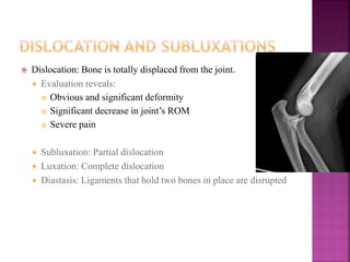
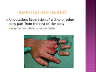
 Trauma:
 In greek means “A Wound”
 Physical injury due to transfer of kinetic energy.
 E=MV2 /2
 Fracture:
 Break in structural continuity of bone.
 Subluxation
 Partial loss of congruity between articular surfaces
 Dislocation:
 Total loss of congruity between articular surfaces
 rapid transport to severely injured patient to trauma centre for
definitive care
 Intial treatment has signifcantly higher chance for survival during
this period
 A standardised protocol for evaluation and treatment of victims of
trauma.
 Developed by James k.Styner, an orthopedic surgeon piloting a
light aircraft, crashed his plane into a field in Nebraska.He was not
satisfied with lack of protocol for such patients.
 ATLS is divided into
 1)Primary survey.
2)Secondary survey.
3)Tertiary survey.
 Airway + (cervical spine immobilisation)
 Breathing+ (high flow 02 )
 Circulation
 Disability/Neurological Assessment
 Exposure/ Environment Control.
 Rapid assessment of ABC’s and addressing life threatening
problems.
-Establishing airway and ventilation ,palcing chest tubes and
controlling active hemorrhage.
 Place large bore IV’s and begin fluid replacement for patients in
shock
 Once hemodynamic stability is obtained,trauma series x rays need
to be taken.
-X ray pelvis(AP view),Chest(AP view), lateral C spine.
 Assessing entire patient for other non life threatening injuires.
 Orthopedic assessment of skeleton
-Splint fractures
-Reduce dislocations
-Evaluate distal pulses and peripheral neurological deficits
 Obtain X-rays and if necessary CT scans of affected areas once
the patient stable enough to shift.
 Periodic vitals monitoring is a must especially while shifting the
patients for investigations like X-rays.
 Look for signs of deterioration and act swiftly in the event of
impending disaster.
 History-note the mechanism of injury.
 Palpation-note swellings and laceration.
 Painful range of movements.
 crepitus- a grating sensation that occurs when two bone ends rub
with each other.
 Abnormal mobility i,e Tibia bends at the middle of its shaft.
 Check pulses,Sensory Exam and motor examination if possible.
 Shortening.
 Abnormal posturing.
 Guarding .
 Exposed bone.
 Dislocation: Bone is totally displaced from the joint.
 Evaluation reveals:
 Obvious and significant deformity
 Significant decrease in joint’s ROM
 Severe pain
 Subluxation: Partial dislocation
 Luxation: Complete dislocation
 Diastasis: Ligaments that hold two bones in place are disrupted
 Amputation: Separation of a limb or other
body part from the rest of the body
 May be complete or incomplete
 Assess for lacerations that communicate with fracture.
-Closed fracture=intact skin over the fracture.
-Open/Compound fracture= laceration communicating
with the fracture.
closed compound
 Hemorrhage control in patients with pelvic fracture
with shock.
----Closed pelvic volume.
 Hemorrhage control of open fractures.
----Direct pressure.
 Restore Pulse by realigning fractures and dislocations.
 4 basic steps:-
-Irrigation and debridement of open wounds.
-Reduction of dislocations.
-Splinting of fractures.
-Looking for signs of Compartment syndromes
 Patients may be classified based on injury:
 Life- or limb-threatening injury or condition
 Life-threatening injuries, simple musculoskeletal
trauma
 Life- or limb-threatening musculoskeletal trauma
 Isolated, non–life- or non–limb-threatening injury
 Priorities should include:
 Identifying the injuries
 Preventing further harm or damage
 Supporting the injured area
 Administering pain medication if necessary
•Bending
•Torsion
•Axial loading
-Tension
-Compression
 Mechanism of injury-traumatic/pathological/stress.
 Anatomical site-bone and location in bone.
 Fracture geometry/type.
 Displacement
-Three planes opf angulation.
-Translation/Shortening
-Rotation.
 Articular involvement
 Soft tissue injury(closed/open)
 Say what it is-Xrays are needed to be taken in
- two planes/two joints/two views.
 Regional location
-Epiphysis/metaphysis/diaphysis.
-Intra-articular/Extra-articular.
 Fracture geometry type-Transverse/oblique/spiral.
 Condition-comminuted/segmental/butterfly.
 Fractures
 Control external bleeding.
 Prevent infection.
 Manage internal bleeding.
 Immobilize.
 Sprains
 Immobilize.
 Chill.
 Elevate.
 Splint.
 Reduce weight bearing.
 Manage pain.
 Decreases pain
 Reduces risk of further damage
 Controls bleeding
 Splinting Principles
1. Make sure the injured area can be seen.
2. Assess and record distal motor and sensory functions.
3. Cover all wounds with a sterile dressing.
4. Do not move the patient before splinting.
5. Fractures—immobilize bone ends and joints.
1. Dislocations—splint entire length of bone.
2. Pad the splint well.
3. Support the injury and minimize movement.
4. Splint knees straight, elbows at right angle.
5. Discontinue traction if patient reports pain
 Rigid splint
 Inflexible device attached to a limb
 Must be padded and long enough
 Use two providers to apply.
 Sling and swathe
 Slings are useful to stabilize upper extremities.
 Swathes add more stabilization for:
 Injuries to clavicle
 Anterior dislocations of the shoulder
 Pneumatic splints
 Stabilize fractures to the lower leg or forearm
 Advantages:
 Slow bleeding
 Minimize swelling
 Pneumatic antishock garment (PASG)
 Used for injuries to lower extremities or pelvis.
 Be sure to check with medical control.
 Document injuries before applying.
 Do not remove in the field.
 Plaster of paris
 Can be moulded with palms
 Easy to apply.
 Cheap.
 Highly susceptible
to damage especially
when wet
 Pop needs to be immersed in
water before application
-Equation
2 (CaSO4·½ H2O) + 3 H2O → 2 (CaSO4.2H2O) + Heat
 Temporary method when operative fixation is
not available for a while
 Can be of two types
 Fracutres associated with severe soft tissue
injuries.
 Fractures associated with neurovascular
damage.
 Severely comminuted unstable fractures.
 Unstable pelvic fractures.
 Infected fractures.
complications :
 Pin tract infection.
 Delayed union.
 Fracttures that need operative fixation.
 Inherently unstable fractures prone to
redisplacement.
 Pathological fractures.
 Polytrauma.
Advantages :
 Shorter hospital stay
 Reduces incidence of malunion and non union
 Enables individual to ambulate earlier.
 Pin and wire fixation
 Screw fixation
 Plate and screw fixation
 Intra medullary fixation
 Stainless steel,titanium,cobalt based
implant.
 Complications:
-Non union
-Implant failure
-Infection
-Refracure
 Devascularization: Loss of blood flow to a body part,
occurring when blood vessels are damaged following a
musculoskeletal injury
 Neurovascular injuries occur when the skeletal system
is compromised.
 Assessment and management
 Assess and reassess pulses.
 Control bleeding.
 Maintain adequate intravascular volume.
 Condition that occurs when pressure is too high
within fascia
 Causes include:
 Overly tight bandages, splints, casts, or PASG
 Fracture, dislocation, crush injury, vascular injury, soft-
tissue injury, bleeding disorder
 Fluid leakage or edema
 Assessment
 The first sign is searing or burning pain out of proportion to
the injury.
 Neurologic symptoms include:
 Paresthesias
 Paralysis of involved muscles
 Pulselessness is a late sign.
 Management
 Elevate the extremity to heart level.
 Apply cold packs.
 Open or loosen constrictive clothing or splint.
 Administer high-flow oxygen and isotonic
crystalloid solution.
 Result of prolonged compressive force that impairs
muscle metabolism and circulation
 When force is released, contents of the muscles
enters the systemic vasculature, resulting in:
 Decreased blood pH
 Hyperkalemia
 Renal dysfunctionBefore releasing the compressing
force:
 Assess the ABCs.
 Administer supplemental oxygen and isotonic
crystalloid solution.
 Establish cardiac monitoring.
 If ECG shows signs of hyperkalemia:
 Administer calcium to stabilize the
myocardium.
 Administer sodium bicarbonate to promote the
intracellular shift of potassium.
 Plan for debridement and damage control
surgery.
 Includes deep vein thrombosis (DVT) and pulmonary
embolism
 Signs and symptoms of DVT include:
 Disproportionate swelling of an extremity
 Discomfort in extremity that worsens with use
 Warmth and erythema of the extremity
 Signs and symptoms of pulmonary embolism include:
 Sudden dyspnea
 Pleuritic chest pain
 Tachypnea
 Right-side heart failure
 Cardiac arrest
•Signs and symptoms of fat
embolism include:
•Tachycardia
•Pulmonary congestion
•Petechiae
•Change in mental status
•Organ dysfunction
•Treatment includes:
•Maintaining an airway
•Supplying adequate oxygen
•Maintaining intravascular
volume
•Providing rapid transport
Orthopedic trauma care

Orthopedic trauma care

  • 2.
     Trauma:  Ingreek means “A Wound”  Physical injury due to transfer of kinetic energy.  E=MV2 /2  Fracture:  Break in structural continuity of bone.  Subluxation  Partial loss of congruity between articular surfaces  Dislocation:  Total loss of congruity between articular surfaces
  • 3.
     rapid transportto severely injured patient to trauma centre for definitive care  Intial treatment has signifcantly higher chance for survival during this period
  • 6.
     A standardisedprotocol for evaluation and treatment of victims of trauma.  Developed by James k.Styner, an orthopedic surgeon piloting a light aircraft, crashed his plane into a field in Nebraska.He was not satisfied with lack of protocol for such patients.  ATLS is divided into  1)Primary survey. 2)Secondary survey. 3)Tertiary survey.
  • 7.
     Airway +(cervical spine immobilisation)  Breathing+ (high flow 02 )  Circulation  Disability/Neurological Assessment  Exposure/ Environment Control.
  • 8.
     Rapid assessmentof ABC’s and addressing life threatening problems. -Establishing airway and ventilation ,palcing chest tubes and controlling active hemorrhage.  Place large bore IV’s and begin fluid replacement for patients in shock  Once hemodynamic stability is obtained,trauma series x rays need to be taken. -X ray pelvis(AP view),Chest(AP view), lateral C spine.
  • 9.
     Assessing entirepatient for other non life threatening injuires.  Orthopedic assessment of skeleton -Splint fractures -Reduce dislocations -Evaluate distal pulses and peripheral neurological deficits  Obtain X-rays and if necessary CT scans of affected areas once the patient stable enough to shift.  Periodic vitals monitoring is a must especially while shifting the patients for investigations like X-rays.  Look for signs of deterioration and act swiftly in the event of impending disaster.
  • 10.
     History-note themechanism of injury.  Palpation-note swellings and laceration.  Painful range of movements.  crepitus- a grating sensation that occurs when two bone ends rub with each other.  Abnormal mobility i,e Tibia bends at the middle of its shaft.  Check pulses,Sensory Exam and motor examination if possible.  Shortening.  Abnormal posturing.  Guarding .  Exposed bone.
  • 11.
     Dislocation: Boneis totally displaced from the joint.  Evaluation reveals:  Obvious and significant deformity  Significant decrease in joint’s ROM  Severe pain  Subluxation: Partial dislocation  Luxation: Complete dislocation  Diastasis: Ligaments that hold two bones in place are disrupted
  • 12.
     Amputation: Separationof a limb or other body part from the rest of the body  May be complete or incomplete
  • 13.
     Assess forlacerations that communicate with fracture. -Closed fracture=intact skin over the fracture. -Open/Compound fracture= laceration communicating with the fracture. closed compound
  • 14.
     Hemorrhage controlin patients with pelvic fracture with shock. ----Closed pelvic volume.  Hemorrhage control of open fractures. ----Direct pressure.  Restore Pulse by realigning fractures and dislocations.  4 basic steps:- -Irrigation and debridement of open wounds. -Reduction of dislocations. -Splinting of fractures. -Looking for signs of Compartment syndromes
  • 15.
     Patients maybe classified based on injury:  Life- or limb-threatening injury or condition  Life-threatening injuries, simple musculoskeletal trauma  Life- or limb-threatening musculoskeletal trauma  Isolated, non–life- or non–limb-threatening injury  Priorities should include:  Identifying the injuries  Preventing further harm or damage  Supporting the injured area  Administering pain medication if necessary
  • 16.
  • 17.
     Mechanism ofinjury-traumatic/pathological/stress.  Anatomical site-bone and location in bone.  Fracture geometry/type.  Displacement -Three planes opf angulation. -Translation/Shortening -Rotation.  Articular involvement  Soft tissue injury(closed/open)
  • 19.
     Say whatit is-Xrays are needed to be taken in - two planes/two joints/two views.  Regional location -Epiphysis/metaphysis/diaphysis. -Intra-articular/Extra-articular.  Fracture geometry type-Transverse/oblique/spiral.  Condition-comminuted/segmental/butterfly.
  • 21.
     Fractures  Controlexternal bleeding.  Prevent infection.  Manage internal bleeding.  Immobilize.  Sprains  Immobilize.  Chill.  Elevate.  Splint.  Reduce weight bearing.  Manage pain.
  • 22.
     Decreases pain Reduces risk of further damage  Controls bleeding  Splinting Principles 1. Make sure the injured area can be seen. 2. Assess and record distal motor and sensory functions. 3. Cover all wounds with a sterile dressing. 4. Do not move the patient before splinting. 5. Fractures—immobilize bone ends and joints.
  • 23.
    1. Dislocations—splint entirelength of bone. 2. Pad the splint well. 3. Support the injury and minimize movement. 4. Splint knees straight, elbows at right angle. 5. Discontinue traction if patient reports pain
  • 24.
     Rigid splint Inflexible device attached to a limb  Must be padded and long enough  Use two providers to apply.  Sling and swathe  Slings are useful to stabilize upper extremities.  Swathes add more stabilization for:  Injuries to clavicle  Anterior dislocations of the shoulder
  • 25.
     Pneumatic splints Stabilize fractures to the lower leg or forearm  Advantages:  Slow bleeding  Minimize swelling  Pneumatic antishock garment (PASG)  Used for injuries to lower extremities or pelvis.  Be sure to check with medical control.  Document injuries before applying.  Do not remove in the field.
  • 26.
     Plaster ofparis  Can be moulded with palms  Easy to apply.  Cheap.  Highly susceptible to damage especially when wet  Pop needs to be immersed in water before application -Equation 2 (CaSO4·½ H2O) + 3 H2O → 2 (CaSO4.2H2O) + Heat
  • 27.
     Temporary methodwhen operative fixation is not available for a while  Can be of two types
  • 29.
     Fracutres associatedwith severe soft tissue injuries.  Fractures associated with neurovascular damage.  Severely comminuted unstable fractures.  Unstable pelvic fractures.  Infected fractures. complications :  Pin tract infection.  Delayed union.
  • 30.
     Fracttures thatneed operative fixation.  Inherently unstable fractures prone to redisplacement.  Pathological fractures.  Polytrauma. Advantages :  Shorter hospital stay  Reduces incidence of malunion and non union  Enables individual to ambulate earlier.
  • 31.
     Pin andwire fixation  Screw fixation  Plate and screw fixation  Intra medullary fixation
  • 32.
     Stainless steel,titanium,cobaltbased implant.  Complications: -Non union -Implant failure -Infection -Refracure
  • 33.
     Devascularization: Lossof blood flow to a body part, occurring when blood vessels are damaged following a musculoskeletal injury  Neurovascular injuries occur when the skeletal system is compromised.  Assessment and management  Assess and reassess pulses.  Control bleeding.  Maintain adequate intravascular volume.
  • 34.
     Condition thatoccurs when pressure is too high within fascia  Causes include:  Overly tight bandages, splints, casts, or PASG  Fracture, dislocation, crush injury, vascular injury, soft- tissue injury, bleeding disorder  Fluid leakage or edema  Assessment  The first sign is searing or burning pain out of proportion to the injury.  Neurologic symptoms include:  Paresthesias  Paralysis of involved muscles  Pulselessness is a late sign.
  • 35.
     Management  Elevatethe extremity to heart level.  Apply cold packs.  Open or loosen constrictive clothing or splint.  Administer high-flow oxygen and isotonic crystalloid solution.
  • 36.
     Result ofprolonged compressive force that impairs muscle metabolism and circulation  When force is released, contents of the muscles enters the systemic vasculature, resulting in:  Decreased blood pH  Hyperkalemia  Renal dysfunctionBefore releasing the compressing force:  Assess the ABCs.  Administer supplemental oxygen and isotonic crystalloid solution.  Establish cardiac monitoring.
  • 37.
     If ECGshows signs of hyperkalemia:  Administer calcium to stabilize the myocardium.  Administer sodium bicarbonate to promote the intracellular shift of potassium.  Plan for debridement and damage control surgery.
  • 38.
     Includes deepvein thrombosis (DVT) and pulmonary embolism  Signs and symptoms of DVT include:  Disproportionate swelling of an extremity  Discomfort in extremity that worsens with use  Warmth and erythema of the extremity  Signs and symptoms of pulmonary embolism include:  Sudden dyspnea  Pleuritic chest pain  Tachypnea  Right-side heart failure  Cardiac arrest
  • 39.
    •Signs and symptomsof fat embolism include: •Tachycardia •Pulmonary congestion •Petechiae •Change in mental status •Organ dysfunction •Treatment includes: •Maintaining an airway •Supplying adequate oxygen •Maintaining intravascular volume •Providing rapid transport