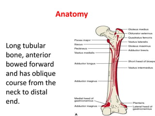
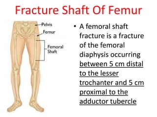
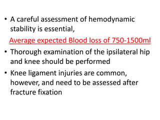
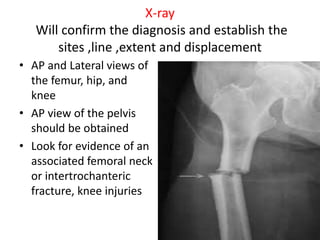
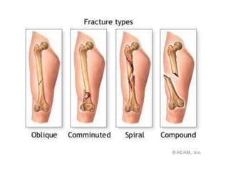
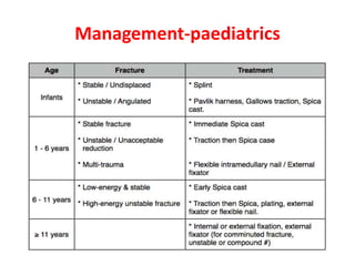
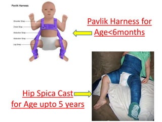
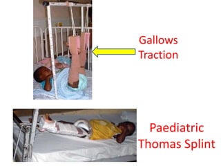
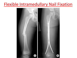
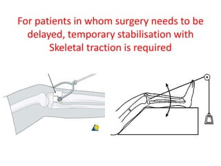
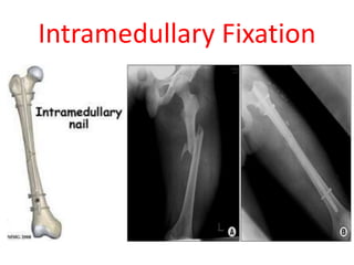
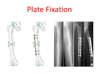
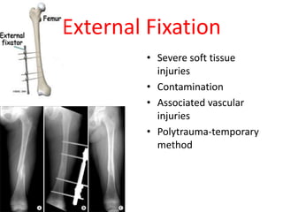
Femoral Shaft Fractures
Dr. Ajay Alex
CMC, Ludhiana
Anatomy
Long tubular
bone, anterior
bowed forward
and has oblique
course from the
neck to distal
end.
Compartments of thigh
Muscle of the thigh are arranged in three
Compartments
1-anterior compartment of thigh
Contains the sartorius and the four large
quadriceps
2-medial compartment of thigh
(gracilis ,pectineus, adductor longus, adductor
brevis, adductor magnus, and obturator
externus)
3-posterior compartment of thigh
contain three large muscle termed the
‘”hamstring”
Femur is surrounded by massive musculature
,which provide the blood supply to femur
Fracture Shaft Of Femur
• A femoral shaft
fracture is a fracture
of the femoral
diaphysis occurring
between 5 cm distal
to the lesser
trochanter and 5 cm
proximal to the
adductor tubercle
Epidemiology
-Common injury due to major violent
trauma
-1 femur fracture/ 10,000 people
more common in people < 25 yr or >65 yr
-Motor vehicle, motorcycle, auto-
pedestrian, fall from height, and gunshot
wound
Mechanism Of Injury
• In Young Adults, almost always the result
of high-energy trauma,
– Motor vehicle accident
– Gunshot injury, or
– Fall from a height
• Pathologic fractures, especially in the
elderly, commonly occur following a trivial
fall
• Stress fractures occur mainly in military
recruits or runners
Symptoms
Diffuse pain or ache, and tenderness and
swelling in the thigh or groin.
Bleeding and bruising in the thigh
(uncommon).
Weakness and inability to bear weight on the
injured leg.
Paleness and deformity
Clinical Evaluation
• A full trauma survey is indicated (ABC)
• The patient is
–Non ambulatory with pain
–Variable gross deformity of thigh
–Swelling,
–Shortening of the affected extremity.
• A careful neurovascular examination is
essential
• A careful assessment of hemodynamic
stability is essential,
Average expected Blood loss of 750-1500ml
• Thorough examination of the ipsilateral hip
and knee should be performed
• Knee ligament injuries are common,
however, and need to be assessed after
fracture fixation
Associated Injuries
• Ipsilateral femur neck, intertroch, distal femur
#s
• Patella, tibia, acetabular, pelvic ring #s
• Soft tissue injuries of knee
• Thoracic & abdominal injuries(5-15%)
X-ray
Will confirm the diagnosis and establish the
sites ,line ,extent and displacement
• AP and Lateral views of
the femur, hip, and
knee
• AP view of the pelvis
should be obtained
• Look for evidence of an
associated femoral neck
or intertrochanteric
fracture, knee injuries
Winquist and Hansen , 1984
* Type 0 - No commination
*Type 1 - Insignificant butterfly
fragment with transverse or short
oblique fracture,
*Type 2 - Large butterfly of less than
50% of the bony width, > 50% of
cortical contact
*Type 3 - Larger butterfly leaving less
than 50% of the cortex in contact
*Type 4 - Segmental commination
AO classification
Management-paediatrics
Pavlik Harness for
Age<6months
Hip Spica Cast
for Age upto 5 years
Gallows
Traction
Paediatric
Thomas Splint
Flexible Intramedullary Nail Fixation
Adults-Non operative
• Traction- Skeletal, thomas splint
• Cast braces- uncommonly used
For patients in whom surgery needs to be
delayed, temporary stabilisation with
Skeletal traction is required
Operative Fixation Options
• Intramedullary Fixation
• Open Reduction and Plate Fixation
• External Fixation
(For Open/ Infected Fractures)
Intramedullary Fixation
Plate Fixation
Indications for plating
• Patients wit extremely narrow medullary
canal
• Fractures around or adjacent previous
malunion
• Fractures extending proximally or distally into
the pertrochanteric or metaphyseal region
• Ipsilateral neck fractures
External Fixation
• Severe soft tissue
injuries
• Contamination
• Associated vascular
injuries
• Polytrauma-temporary
method
COMPLICATION
1-GENERAL
Blood loss, shock ,fat embolism, and acut
respiratory distress are common in high-
energy injuries
COMPLICATION
2-Vascular injury
3-Nerve injury-iatrogenic(femoral, sciatic,
pudendal, peroneal)
4-Thromboembolism
5-Compartment syndrome(1-2%)
6-Infection
7-Delayed union and non-union
8-Joint stiffness, knee & hip pain
9-Heterotrophic ossification

Femur shaft fractures

  • 1.
    Femoral Shaft Fractures Dr.Ajay Alex CMC, Ludhiana
  • 2.
    Anatomy Long tubular bone, anterior bowedforward and has oblique course from the neck to distal end.
  • 3.
    Compartments of thigh Muscleof the thigh are arranged in three Compartments 1-anterior compartment of thigh Contains the sartorius and the four large quadriceps 2-medial compartment of thigh (gracilis ,pectineus, adductor longus, adductor brevis, adductor magnus, and obturator externus)
  • 4.
    3-posterior compartment ofthigh contain three large muscle termed the ‘”hamstring” Femur is surrounded by massive musculature ,which provide the blood supply to femur
  • 7.
    Fracture Shaft OfFemur • A femoral shaft fracture is a fracture of the femoral diaphysis occurring between 5 cm distal to the lesser trochanter and 5 cm proximal to the adductor tubercle
  • 8.
    Epidemiology -Common injury dueto major violent trauma -1 femur fracture/ 10,000 people more common in people < 25 yr or >65 yr -Motor vehicle, motorcycle, auto- pedestrian, fall from height, and gunshot wound
  • 9.
    Mechanism Of Injury •In Young Adults, almost always the result of high-energy trauma, – Motor vehicle accident – Gunshot injury, or – Fall from a height • Pathologic fractures, especially in the elderly, commonly occur following a trivial fall • Stress fractures occur mainly in military recruits or runners
  • 10.
    Symptoms Diffuse pain orache, and tenderness and swelling in the thigh or groin. Bleeding and bruising in the thigh (uncommon). Weakness and inability to bear weight on the injured leg. Paleness and deformity
  • 11.
    Clinical Evaluation • Afull trauma survey is indicated (ABC) • The patient is –Non ambulatory with pain –Variable gross deformity of thigh –Swelling, –Shortening of the affected extremity. • A careful neurovascular examination is essential
  • 12.
    • A carefulassessment of hemodynamic stability is essential, Average expected Blood loss of 750-1500ml • Thorough examination of the ipsilateral hip and knee should be performed • Knee ligament injuries are common, however, and need to be assessed after fracture fixation
  • 13.
    Associated Injuries • Ipsilateralfemur neck, intertroch, distal femur #s • Patella, tibia, acetabular, pelvic ring #s • Soft tissue injuries of knee • Thoracic & abdominal injuries(5-15%)
  • 14.
    X-ray Will confirm thediagnosis and establish the sites ,line ,extent and displacement • AP and Lateral views of the femur, hip, and knee • AP view of the pelvis should be obtained • Look for evidence of an associated femoral neck or intertrochanteric fracture, knee injuries
  • 18.
    Winquist and Hansen, 1984 * Type 0 - No commination *Type 1 - Insignificant butterfly fragment with transverse or short oblique fracture, *Type 2 - Large butterfly of less than 50% of the bony width, > 50% of cortical contact *Type 3 - Larger butterfly leaving less than 50% of the cortex in contact *Type 4 - Segmental commination
  • 20.
  • 21.
  • 22.
    Pavlik Harness for Age<6months HipSpica Cast for Age upto 5 years
  • 23.
  • 24.
  • 25.
    Adults-Non operative • Traction-Skeletal, thomas splint • Cast braces- uncommonly used
  • 26.
    For patients inwhom surgery needs to be delayed, temporary stabilisation with Skeletal traction is required
  • 27.
    Operative Fixation Options •Intramedullary Fixation • Open Reduction and Plate Fixation • External Fixation (For Open/ Infected Fractures)
  • 28.
  • 29.
  • 30.
    Indications for plating •Patients wit extremely narrow medullary canal • Fractures around or adjacent previous malunion • Fractures extending proximally or distally into the pertrochanteric or metaphyseal region • Ipsilateral neck fractures
  • 31.
    External Fixation • Severesoft tissue injuries • Contamination • Associated vascular injuries • Polytrauma-temporary method
  • 32.
    COMPLICATION 1-GENERAL Blood loss, shock,fat embolism, and acut respiratory distress are common in high- energy injuries
  • 33.
    COMPLICATION 2-Vascular injury 3-Nerve injury-iatrogenic(femoral,sciatic, pudendal, peroneal) 4-Thromboembolism 5-Compartment syndrome(1-2%) 6-Infection 7-Delayed union and non-union 8-Joint stiffness, knee & hip pain 9-Heterotrophic ossification