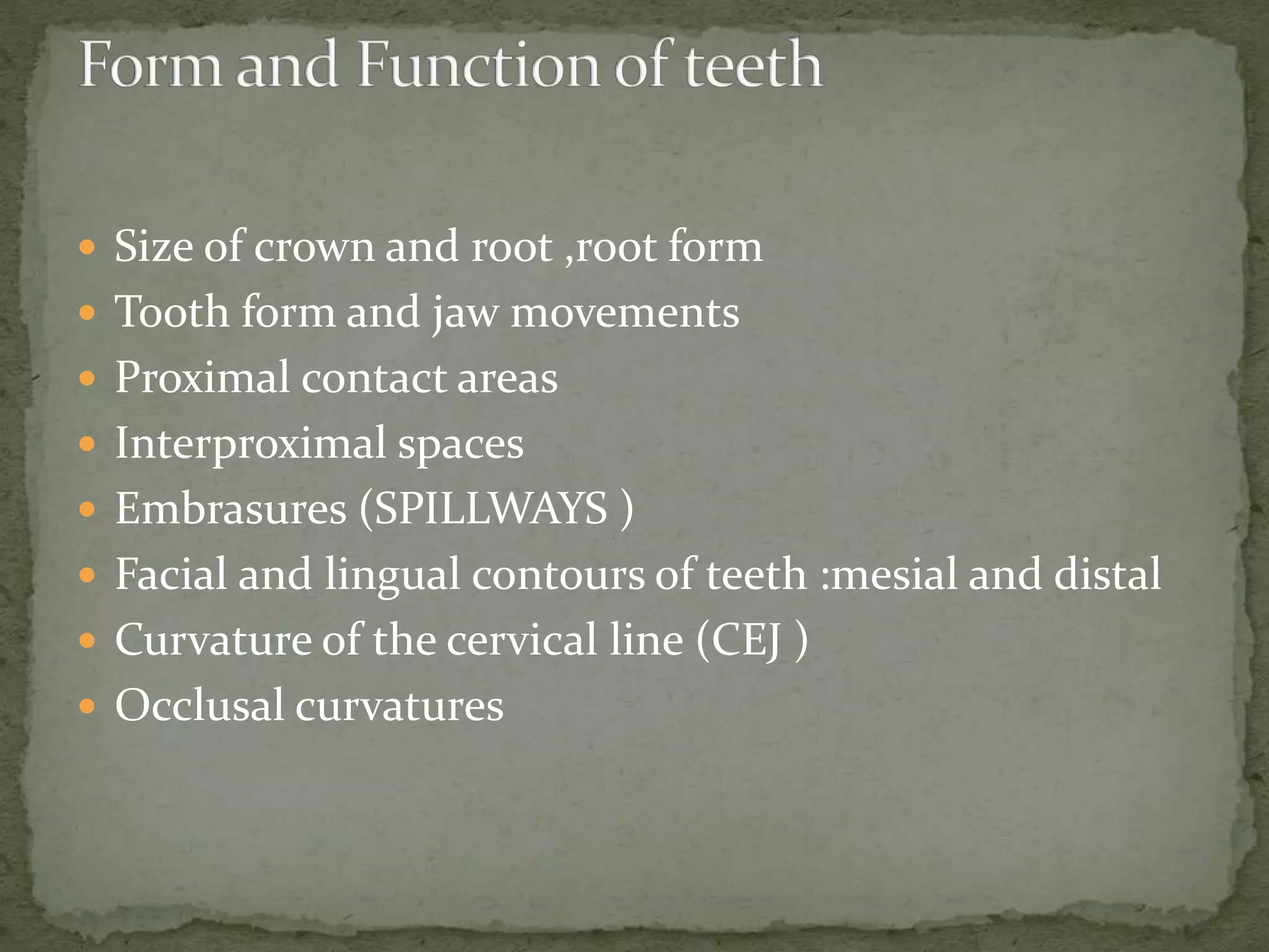
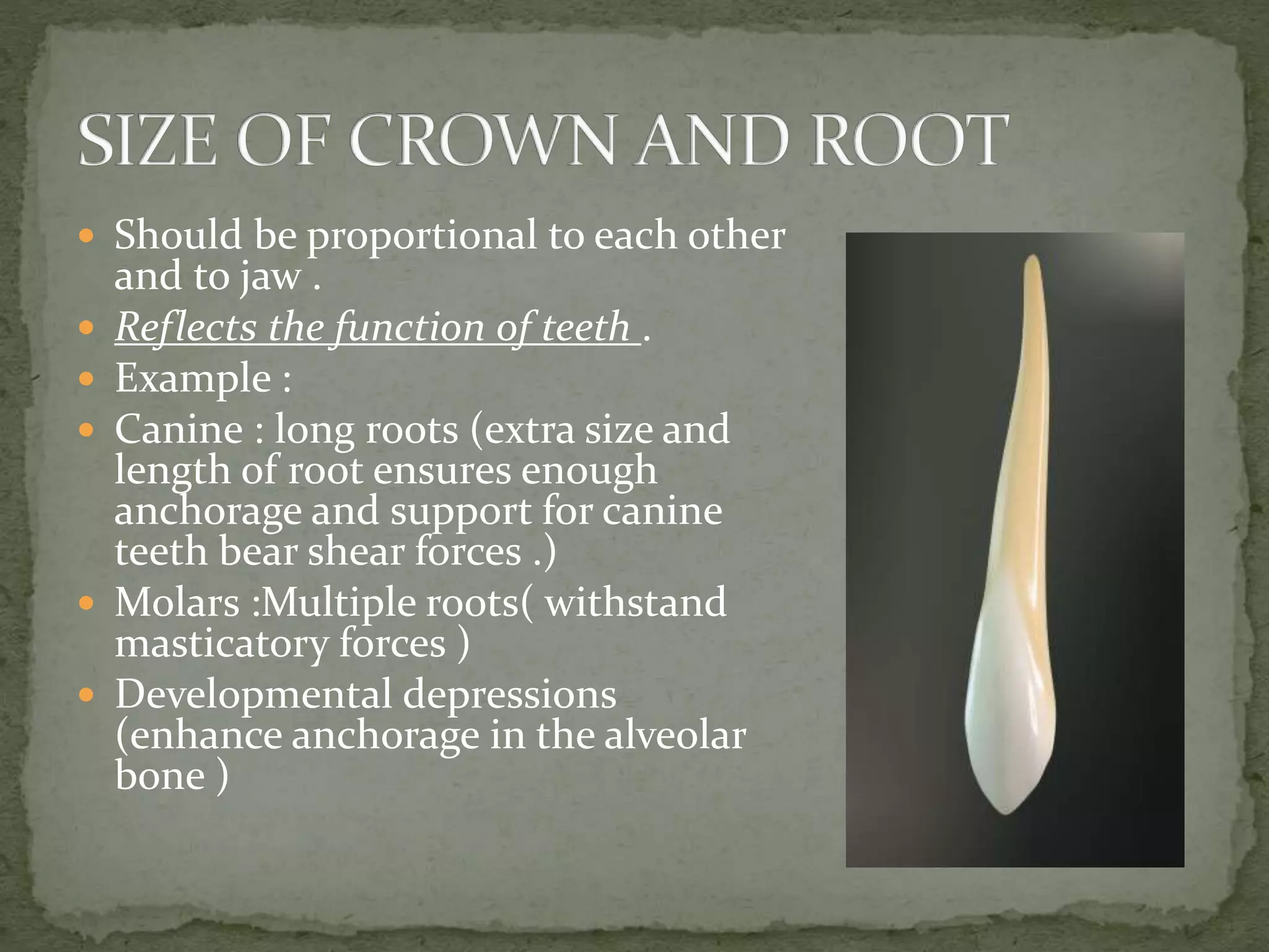
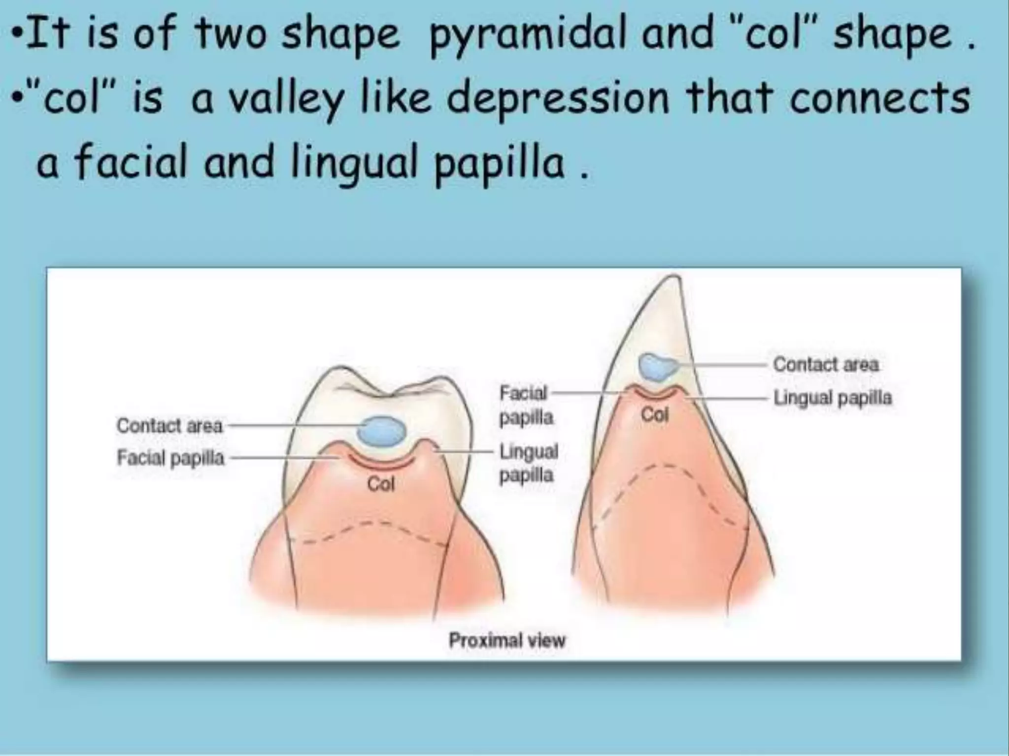
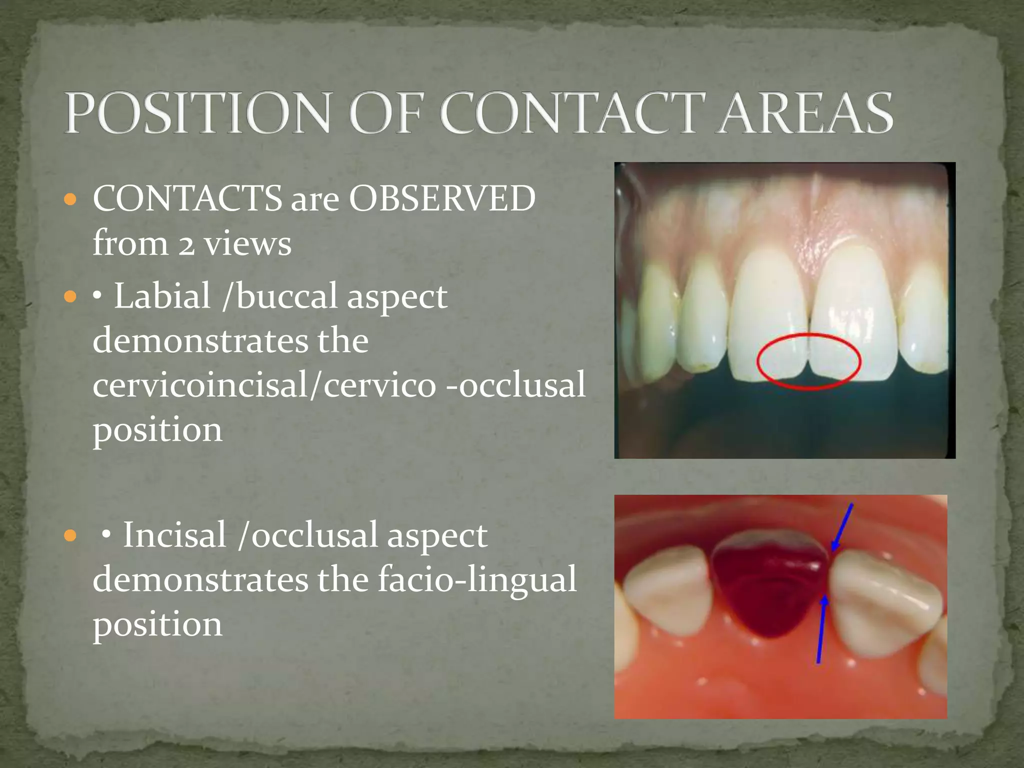
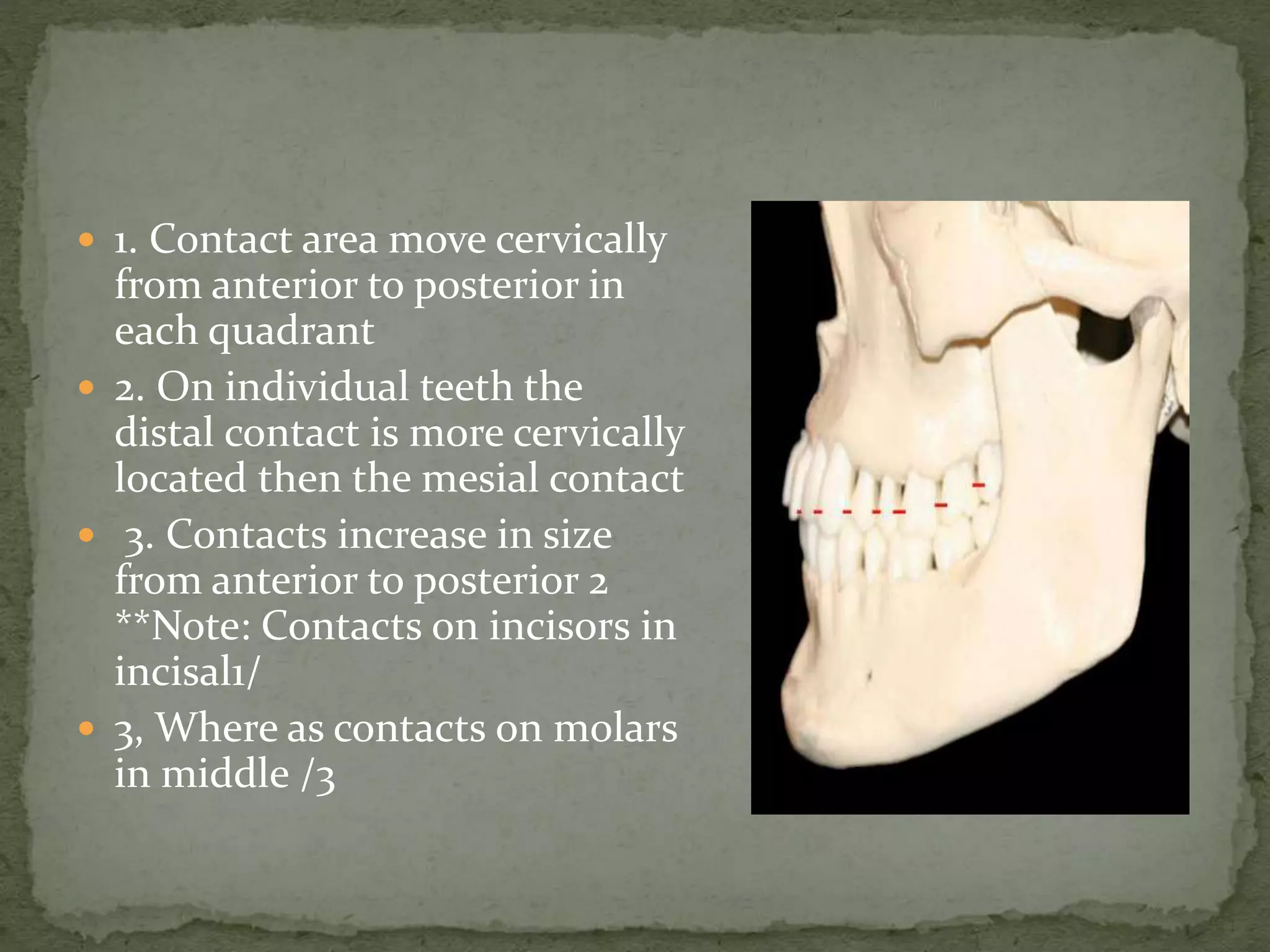
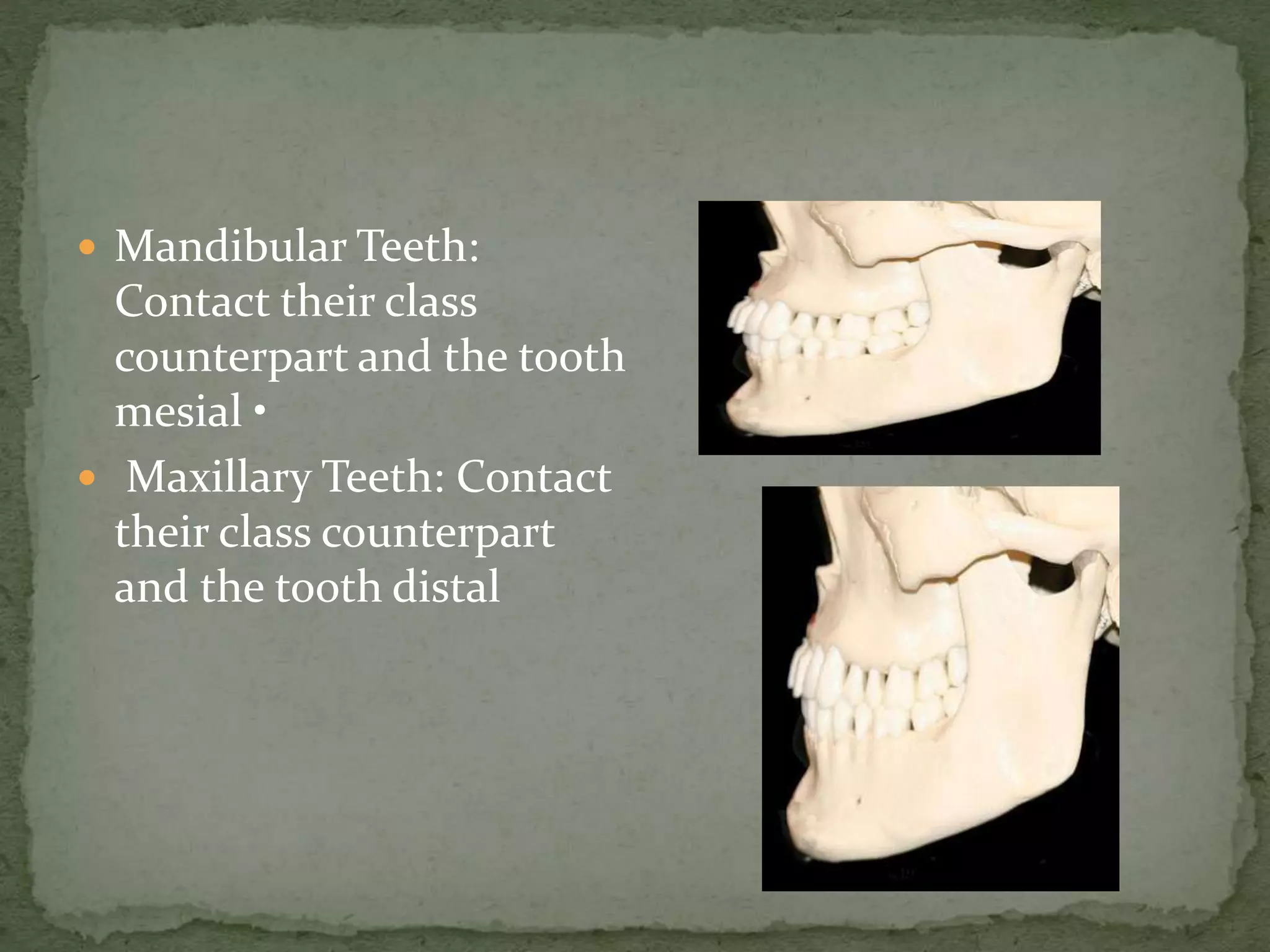
DR MONIKA NEGI
Oral Pathology Microbiology and
Forensic Odontology
Form follows Function
Explains the inter –
relation of the shape of
some part and its
function .
 Size of crown and root ,root form
 Tooth form and jaw movements
 Proximal contact areas
 Interproximal spaces
 Embrasures (SPILLWAYS )
 Facial and lingual contours of teeth :mesial and distal
 Curvature of the cervical line (CEJ )
 Occlusal curvatures
 Should be proportional to each other
and to jaw .
 Reflects the function of teeth .
 Example :
 Canine : long roots (extra size and
length of root ensures enough
anchorage and support for canine
teeth bear shear forces .)
 Molars :Multiple roots( withstand
masticatory forces )
 Developmental depressions
(enhance anchorage in the alveolar
bone )
 Tooth form have direct influence on the jaw movements
.
 Example :
 In animals teeth are conical ,have simple opening and
closing type of jaw movements without lateral
movements .
 Because of interlocking conical form of teeth ,TMJ
morphology ,equal sized jaw and lack of muscles to
carry out lateral movemnts .
 In humans ,jaws are not of same size
 Maxilla overlaps mandibular arch labially and bucally
in horizontal plane .
 TMJ is specialized in humans as the occlusal anatomy
of teeth is complex .
 INCREASING COMPLEXITY OF JAW MOVEMENT IS
ASSOCIATED WITH COMPLEXITY OF OCCLUSAL
ANATOMY OF TEETH .
 From labial and lingual –Teeth are narrower at cervix
mesiodistally than they are towards the occlusal surface .
 Triangular or pyramidal shaped space between the
approximating teeth just cervical to contact area is created .
 BASE –Alveolar process between adjacent teeth
 Triangle are formed by proximal surface of teeth and the
apex of the triangle is at the contact area of the two teeth .
 ACCOMMODATE AND PROTECT INTERPROXIMAL
SPACES .
 Gingival tissue that fills the interproximal space is
called the gingival papilla/ interdental papilla .
 Base=alveolar bone
 • Sides=prox. surfaces of teeth
 • Apex=proximal contact area
 • These triangular shaped areas are
normally filled with gingival
tissues
 Gingiva covers alveolar process of jaw bones
(ATTACHED GINGIVA ) ,extends around the neck of
tooth to form gingival crevice (free/marginal gingiva )
and fills the interdental space .(gingival/interdental
papilla )
 Mucogingival line marks the junction between
attached gingiva and alveolar mucosa .
 The part of interdental gingival tissue that lies below
the contact area and extends faciolingually is called col
 Col is non keratinized and vulnerable to trauma
during mastication and invasion by bacteria .
 Tight contacts and proper interproximal spaces
between adjacent teeth help to protect the col and
interproximal gingival tissue .
 BUCCALLY /LINGUALLY – Roots of teeth taper from
cervix to apices creating enough space between the
roots of adjacent teeth .
 Allows sufficient alveolar bone between one tooth to
another ,so that the teeth are securely anchored in the
jaws .
 Proximal Contact Areas • All
teeth contact adjacent teeth at a
proximal contact area except: –
distal of the last tooth In the
arch – diastemas – pathology.
 CONTACT AREA- it is formed
because of wear of one proximal
surface against another during
physiologic tooth movement.
 MAXILLARY AND
MANDIBULAR CENTRAL
INCISORS ARE THE ONLY
TEETH HAVING MESIAL
SURFACE FACING EACH
OTHER .
 – Ensures food will not be packed between teeth
causing inflammation to the supporting tissues
thereby causing gingivitis & periodontitis
 – It helps stabilize the dental arches by the combined
anchorage of all the teeth in either arch in positive
contact with each other (exception - third molars.)
 – Each tooth is individually supported by these
contact areas.
 CONTACTS are OBSERVED
from 2 views
 • Labial /buccal aspect
demonstrates the
cervicoincisal/cervico -occlusal
position
 • Incisal /occlusal aspect
demonstrates the facio-lingual
position
 • Anterior teeth have
contacts that are generally
centered faciolingually
when view from the incisal
 • Posterior teeth have
contacts that are generally
located buccal to the center
of teeth when viewed from
the occlusal
 HEIGHT OF CROWN DECREASES FROM INCISOR –
MOLAR
 MORE CERVICALLY POSITIONED WHEN MOVING
AWAY FROM THE MIDLINE .
 DISTAL CONTACT AREA IS MORE CERVICALLY
PLACED THAN MESIAL .
 EXCEPT – mand 1ST PREMOLAR (mesial more
cervically )
 Mand CI ( both at same )
 1. Contact area move cervically
from anterior to posterior in
each quadrant
 2. On individual teeth the
distal contact is more cervically
located then the mesial contact
 3. Contacts increase in size
from anterior to posterior 2
**Note: Contacts on incisors in
incisal1/
 3, Where as contacts on molars
in middle /3
 Mandibular Teeth:
Contact their class
counterpart and the tooth
mesial •
 Maxillary Teeth: Contact
their class counterpart
and the tooth distal
 The curvature formed
by two adjacent teeth in
an arch form a spillway
space that is called an
embrasure
 Note the Symmetry of
the embrasure of the
central incisors
 Embrasures are named
relative to the contact
area
 – Facial or lingual
views show the
occlusal/ incisal
embrasures and
cervical/ gingival
embrasures
 – Occlusal views show
the buccal/ labial and
lingual embrasures
 Facial or lingual aspects:
 – The incisal / occlusal
embrasures increase in
size from anterior to
posterior
 – The cervical embrasure
decreases in size from
anterior to posterior
 – The incisal embrasure
of the maxillary
lateral/canine =greatest
or longest
 – The incisal embrasure
of the maxillary
canine/premolar= widest
angle
 Embrasures
 • Mandibular posterior area
occlusal view
 • Arrows denote the facial and
lingual embrasure areas
 • The lingual embrasures are
larger than the facial embrasures,
because of lingual convergence of
the crown.
 • Serves as a spillway for food material during
mastication
 • Prevents food from being forced through the contact
area and injuring the periodontal tissues.
 • Also, Enhances the self-cleansing process of the
tooth by exposing the surfaces to oral fluids,
mechanical cleansing, and friction from the tongue,
lips, and cheeks
 It is a line encircling a
tooth In more or less
horizontal plane,
representing its greatest
circumference.
 The facial and lingual
contours:
 • Protect the gingiva by
the deflection of foods
and help prevent trauma
Buccal Lingual
 Buccal Lingual The
height of contour on
both the facial and
lingual surfaces of
anterior teeth is located
in the cervical third.
 The height of contour of the buccal
surfaces of posterior teeth is located
in the gingival third
 • The height of contour of the lingual
surface of posterior teeth is located
in the middle or occlusal third
 • The average curvature is around
0.5mm, with mandibular posterior
having a lingual curvature of 1 mm. •
Mandibular anteriors having
curvature of less than 0.5mm.
 The CEJ is a stable line whose
curvature is related to the
widths and lengths of the
crown and the location of the
contact areas interproximally
The extent of the curvature
depends on
 • Height of the contact area
above the crown cervix
 • Diameter of the crown
labiolingually.
 The CEJ is curved toward the
apical on the facial and lingual
surfaces of teeth
 • The CEJ is curved toward the
incisal or occlusal surfaces on
the proximal surfaces of teeth
 • The depth of the curvature is
greater on the mesial than on
the distal aspect
 • The depth of curvature on the
CEJ on all surfaces is greater on
anterior teeth and decreases
toward the posterior
 CURVE OF SPEE
 CURVE OF WILSON
 CURVE OF MONSON
 OCCLUSAL CURVATURES
Curve of Spee
 Curvature which begins at
the tip of canines & follows
buccal cusp tips of premolars
& molars posteriorly, when
viewed from their facial aspect
 Curve of Wilson
 The medio-lateral curvature of
the occlusal plane of posterior
teeth.
 Two dimensional & approax.
right angles to that of the Curve
of Spee.
 Complement paths of condyles
during movements of mandible.
 The crowns of mandibular
posterior teeth must incline to
lingual, while crowns of maxillary
posterior teeth must incline toward
buccal
 This curve becomes deeper
posteriorly, so that molars
inclination is greater than that of
premolars
 Because of this curve &
associated tooth inclinations,
buccal cusps of mandibular
molars & lingual cusps of
maxillary molars usually appear
to be longer
 It is a curve obtained by
extending the curve of spee
and curve of wilson to all
cusps and incisal edges .
Form and function of orofacial complex

Form and function of orofacial complex

  • 1.
    DR MONIKA NEGI OralPathology Microbiology and Forensic Odontology
  • 2.
    Form follows Function Explainsthe inter – relation of the shape of some part and its function .
  • 3.
     Size ofcrown and root ,root form  Tooth form and jaw movements  Proximal contact areas  Interproximal spaces  Embrasures (SPILLWAYS )  Facial and lingual contours of teeth :mesial and distal  Curvature of the cervical line (CEJ )  Occlusal curvatures
  • 4.
     Should beproportional to each other and to jaw .  Reflects the function of teeth .  Example :  Canine : long roots (extra size and length of root ensures enough anchorage and support for canine teeth bear shear forces .)  Molars :Multiple roots( withstand masticatory forces )  Developmental depressions (enhance anchorage in the alveolar bone )
  • 5.
     Tooth formhave direct influence on the jaw movements .  Example :  In animals teeth are conical ,have simple opening and closing type of jaw movements without lateral movements .  Because of interlocking conical form of teeth ,TMJ morphology ,equal sized jaw and lack of muscles to carry out lateral movemnts .
  • 6.
     In humans,jaws are not of same size  Maxilla overlaps mandibular arch labially and bucally in horizontal plane .  TMJ is specialized in humans as the occlusal anatomy of teeth is complex .  INCREASING COMPLEXITY OF JAW MOVEMENT IS ASSOCIATED WITH COMPLEXITY OF OCCLUSAL ANATOMY OF TEETH .
  • 8.
     From labialand lingual –Teeth are narrower at cervix mesiodistally than they are towards the occlusal surface .  Triangular or pyramidal shaped space between the approximating teeth just cervical to contact area is created .  BASE –Alveolar process between adjacent teeth  Triangle are formed by proximal surface of teeth and the apex of the triangle is at the contact area of the two teeth .  ACCOMMODATE AND PROTECT INTERPROXIMAL SPACES .  Gingival tissue that fills the interproximal space is called the gingival papilla/ interdental papilla .
  • 9.
     Base=alveolar bone • Sides=prox. surfaces of teeth  • Apex=proximal contact area  • These triangular shaped areas are normally filled with gingival tissues
  • 10.
     Gingiva coversalveolar process of jaw bones (ATTACHED GINGIVA ) ,extends around the neck of tooth to form gingival crevice (free/marginal gingiva ) and fills the interdental space .(gingival/interdental papilla )  Mucogingival line marks the junction between attached gingiva and alveolar mucosa .  The part of interdental gingival tissue that lies below the contact area and extends faciolingually is called col
  • 11.
     Col isnon keratinized and vulnerable to trauma during mastication and invasion by bacteria .  Tight contacts and proper interproximal spaces between adjacent teeth help to protect the col and interproximal gingival tissue .  BUCCALLY /LINGUALLY – Roots of teeth taper from cervix to apices creating enough space between the roots of adjacent teeth .  Allows sufficient alveolar bone between one tooth to another ,so that the teeth are securely anchored in the jaws .
  • 13.
     Proximal ContactAreas • All teeth contact adjacent teeth at a proximal contact area except: – distal of the last tooth In the arch – diastemas – pathology.  CONTACT AREA- it is formed because of wear of one proximal surface against another during physiologic tooth movement.  MAXILLARY AND MANDIBULAR CENTRAL INCISORS ARE THE ONLY TEETH HAVING MESIAL SURFACE FACING EACH OTHER .
  • 14.
     – Ensuresfood will not be packed between teeth causing inflammation to the supporting tissues thereby causing gingivitis & periodontitis  – It helps stabilize the dental arches by the combined anchorage of all the teeth in either arch in positive contact with each other (exception - third molars.)  – Each tooth is individually supported by these contact areas.
  • 15.
     CONTACTS areOBSERVED from 2 views  • Labial /buccal aspect demonstrates the cervicoincisal/cervico -occlusal position  • Incisal /occlusal aspect demonstrates the facio-lingual position
  • 16.
     • Anteriorteeth have contacts that are generally centered faciolingually when view from the incisal  • Posterior teeth have contacts that are generally located buccal to the center of teeth when viewed from the occlusal
  • 17.
     HEIGHT OFCROWN DECREASES FROM INCISOR – MOLAR  MORE CERVICALLY POSITIONED WHEN MOVING AWAY FROM THE MIDLINE .  DISTAL CONTACT AREA IS MORE CERVICALLY PLACED THAN MESIAL .  EXCEPT – mand 1ST PREMOLAR (mesial more cervically )  Mand CI ( both at same )
  • 20.
     1. Contactarea move cervically from anterior to posterior in each quadrant  2. On individual teeth the distal contact is more cervically located then the mesial contact  3. Contacts increase in size from anterior to posterior 2 **Note: Contacts on incisors in incisal1/  3, Where as contacts on molars in middle /3
  • 21.
     Mandibular Teeth: Contacttheir class counterpart and the tooth mesial •  Maxillary Teeth: Contact their class counterpart and the tooth distal
  • 24.
     The curvatureformed by two adjacent teeth in an arch form a spillway space that is called an embrasure  Note the Symmetry of the embrasure of the central incisors
  • 25.
     Embrasures arenamed relative to the contact area  – Facial or lingual views show the occlusal/ incisal embrasures and cervical/ gingival embrasures  – Occlusal views show the buccal/ labial and lingual embrasures
  • 26.
     Facial orlingual aspects:  – The incisal / occlusal embrasures increase in size from anterior to posterior  – The cervical embrasure decreases in size from anterior to posterior  – The incisal embrasure of the maxillary lateral/canine =greatest or longest  – The incisal embrasure of the maxillary canine/premolar= widest angle
  • 27.
     Embrasures  •Mandibular posterior area occlusal view  • Arrows denote the facial and lingual embrasure areas  • The lingual embrasures are larger than the facial embrasures, because of lingual convergence of the crown.
  • 28.
     • Servesas a spillway for food material during mastication  • Prevents food from being forced through the contact area and injuring the periodontal tissues.  • Also, Enhances the self-cleansing process of the tooth by exposing the surfaces to oral fluids, mechanical cleansing, and friction from the tongue, lips, and cheeks
  • 29.
     It isa line encircling a tooth In more or less horizontal plane, representing its greatest circumference.  The facial and lingual contours:  • Protect the gingiva by the deflection of foods and help prevent trauma Buccal Lingual
  • 30.
     Buccal LingualThe height of contour on both the facial and lingual surfaces of anterior teeth is located in the cervical third.
  • 31.
     The heightof contour of the buccal surfaces of posterior teeth is located in the gingival third  • The height of contour of the lingual surface of posterior teeth is located in the middle or occlusal third  • The average curvature is around 0.5mm, with mandibular posterior having a lingual curvature of 1 mm. • Mandibular anteriors having curvature of less than 0.5mm.
  • 34.
     The CEJis a stable line whose curvature is related to the widths and lengths of the crown and the location of the contact areas interproximally The extent of the curvature depends on  • Height of the contact area above the crown cervix  • Diameter of the crown labiolingually.
  • 35.
     The CEJis curved toward the apical on the facial and lingual surfaces of teeth  • The CEJ is curved toward the incisal or occlusal surfaces on the proximal surfaces of teeth  • The depth of the curvature is greater on the mesial than on the distal aspect  • The depth of curvature on the CEJ on all surfaces is greater on anterior teeth and decreases toward the posterior
  • 36.
     CURVE OFSPEE  CURVE OF WILSON  CURVE OF MONSON
  • 38.
     OCCLUSAL CURVATURES Curveof Spee  Curvature which begins at the tip of canines & follows buccal cusp tips of premolars & molars posteriorly, when viewed from their facial aspect
  • 39.
     Curve ofWilson  The medio-lateral curvature of the occlusal plane of posterior teeth.  Two dimensional & approax. right angles to that of the Curve of Spee.  Complement paths of condyles during movements of mandible.
  • 40.
     The crownsof mandibular posterior teeth must incline to lingual, while crowns of maxillary posterior teeth must incline toward buccal  This curve becomes deeper posteriorly, so that molars inclination is greater than that of premolars
  • 41.
     Because ofthis curve & associated tooth inclinations, buccal cusps of mandibular molars & lingual cusps of maxillary molars usually appear to be longer
  • 42.
     It isa curve obtained by extending the curve of spee and curve of wilson to all cusps and incisal edges .