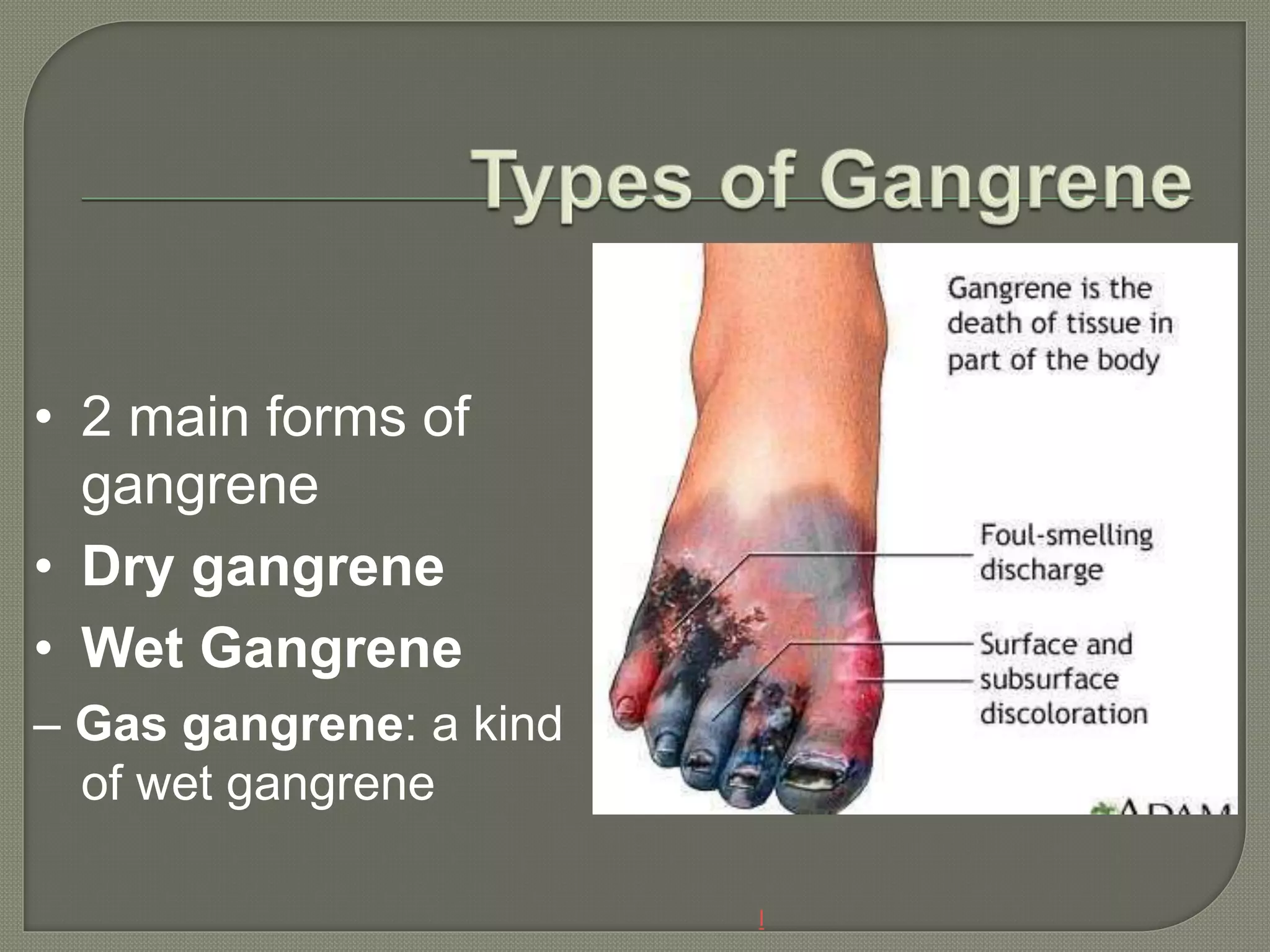
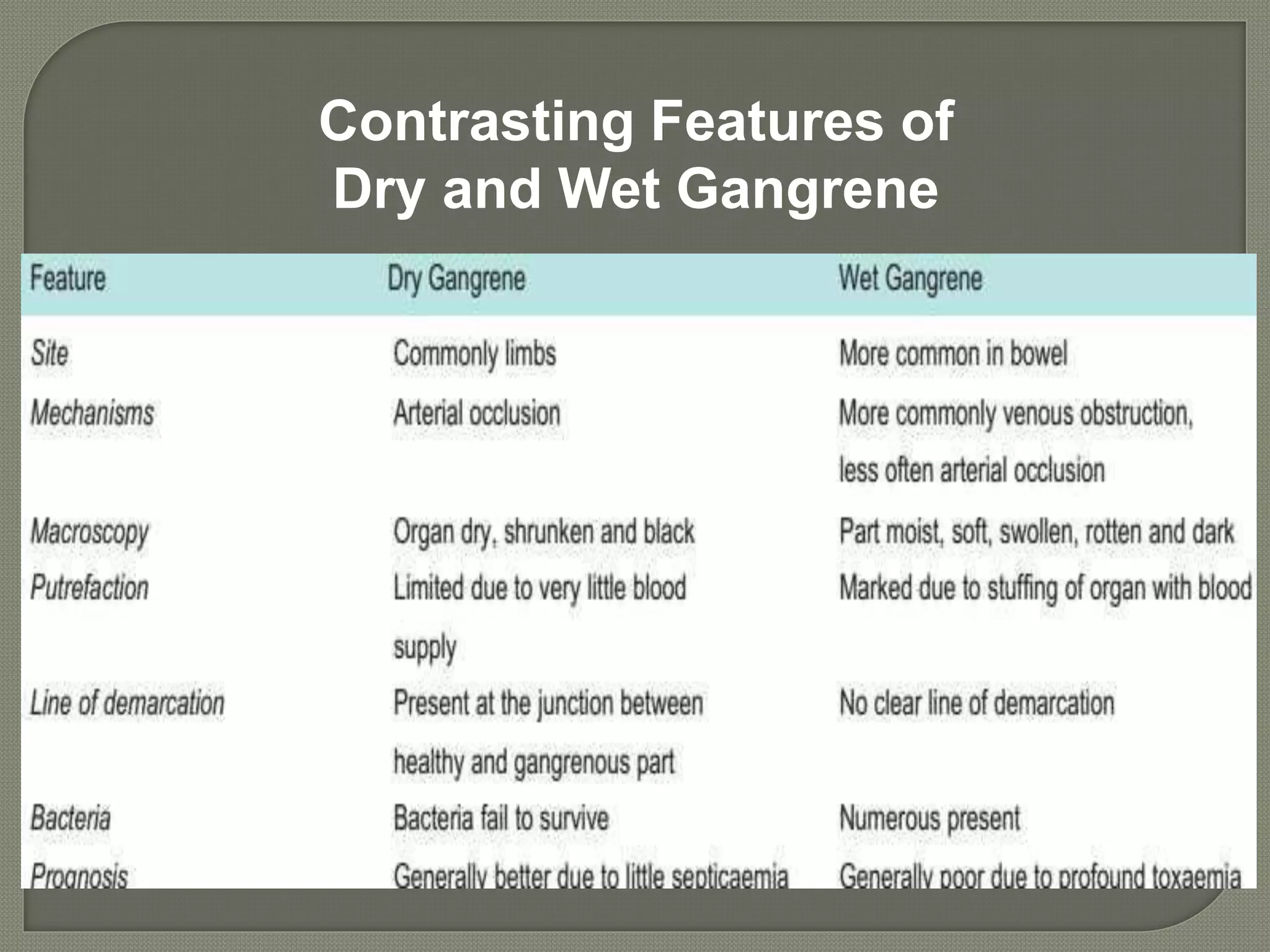
DR.MONIKA NEGI
MDS
ORAL AND
MAXILLOFACIAL
PATHOLOGY
• A form of necrosis of tissue with superadded putrefaction.
• Here the necrosis undergoes liquefaction by the action of
putrefactive bacteria.
• It may be caused either ischemic or inflammatory
• Coagulative Necrosis due to ischaemia
– gangrene of limb
• Gangrenous or necrotising inflammation: primarily
inflammation by virulent bacteria resulting in massive
tissue necrosis.
– Gangrenous appendicitis,
– Gangrenous stomatitis (noma, cancrum oris)
l
• 2 main forms of
gangrene
• Dry gangrene
• Wet Gangrene
– Gas gangrene: a kind
of wet gangrene
• Begins in the distal part of a limb due to ischaemia.
• The gangrene spreads slowly upwards until it reaches a point
where the blood supply is adequate to keep the tissue viable.
• A line of separation is formed at this point between the
gangrenous part and the viable part.
– Toes and feet of an old patient due to arteriosclerosis.
– Thromboangiitis obliterans (Buerger’s disease),
– Raynaud’s disease,
– Trauma
Dry
Gangrene
Dry gangrene
www.facebook.com/notesdental
Grossly
– the affected part is dry, shrunken and dark black, resembling the foot
of a mummy.
– It is black due to liberation of haemoglobin from haemolysed red
blood cells which is acted upon by
hydrogen disulfide (H2S) produced by bacteria resulting
in formation of black iron sulfide.
– The line of separation usually brings about complete separation with
eventual falling off of the gangrenous tissue if it is not removed
surgically
Histologically
– Necrosis with smudging of the tissue.
– The line of separation consists of inflammatory granulation tissue
• Naturally moist tissues and organs such as the mouth, bowel,
lung, cervix,
• develops rapidly due to blockage of venous, and less
commonly, arterial blood flow from thrombosis or embolism.
• The affected part is stuffed with blood which favours the rapid
growth of putrefactive bacteria.
• The toxic products formed by bacteria leads to septicaemia,
and finally death.
Wet Gangrene
• Diabetic foot
– high sugar content in the
necrosed tissue which
favours growth of
bacteria.
• Bed sores
– bed-ridden patient due to
pressure on sites like the
sacrum, buttocks and
heels
• Grossly,
– the affected part is soft, swollen, putrid, rotten and dark.
– The classic example is gangrene of bowel, commonly
due to strangulated hernia
Contrasting Features of
Dry and Wet Gangrene
• Special form of wet gangrene caused by gas-forming
clostridia (gram-positive anaerobic bacteria).
• Bacteria gain entry into the tissues through open
contaminated wounds,eg. road traffic accident
• Results in myonecrosis i.e. necrosis of muscles.
• It produce various toxins which produce necrosis and
oedema locally
• Grossly
– the affected area is swollen, oedematous, painful and crepitant
due to accumulation of gas bubbles within the tissues.
– Subsequently, the affected tissue becomes dark black and foul
smelling.
• Microscopically
– the muscle fibres undergo coagulative necrosis with liquefaction
– Large number of gram-positive bacilli can be identified.
Emergency surgery with excision of
allmuscle and necrotic tissue
Drug of choice – penicillin
Hyperbaric Oxygen therapy
Amputation, if necessary to save the leg
Gangrene

Gangrene

  • 1.
  • 2.
    • A formof necrosis of tissue with superadded putrefaction. • Here the necrosis undergoes liquefaction by the action of putrefactive bacteria. • It may be caused either ischemic or inflammatory • Coagulative Necrosis due to ischaemia – gangrene of limb • Gangrenous or necrotising inflammation: primarily inflammation by virulent bacteria resulting in massive tissue necrosis. – Gangrenous appendicitis, – Gangrenous stomatitis (noma, cancrum oris)
  • 3.
    l • 2 mainforms of gangrene • Dry gangrene • Wet Gangrene – Gas gangrene: a kind of wet gangrene
  • 4.
    • Begins inthe distal part of a limb due to ischaemia. • The gangrene spreads slowly upwards until it reaches a point where the blood supply is adequate to keep the tissue viable. • A line of separation is formed at this point between the gangrenous part and the viable part. – Toes and feet of an old patient due to arteriosclerosis. – Thromboangiitis obliterans (Buerger’s disease), – Raynaud’s disease, – Trauma
  • 5.
  • 6.
    Grossly – the affectedpart is dry, shrunken and dark black, resembling the foot of a mummy. – It is black due to liberation of haemoglobin from haemolysed red blood cells which is acted upon by hydrogen disulfide (H2S) produced by bacteria resulting in formation of black iron sulfide. – The line of separation usually brings about complete separation with eventual falling off of the gangrenous tissue if it is not removed surgically Histologically – Necrosis with smudging of the tissue. – The line of separation consists of inflammatory granulation tissue
  • 7.
    • Naturally moisttissues and organs such as the mouth, bowel, lung, cervix, • develops rapidly due to blockage of venous, and less commonly, arterial blood flow from thrombosis or embolism. • The affected part is stuffed with blood which favours the rapid growth of putrefactive bacteria. • The toxic products formed by bacteria leads to septicaemia, and finally death.
  • 8.
    Wet Gangrene • Diabeticfoot – high sugar content in the necrosed tissue which favours growth of bacteria. • Bed sores – bed-ridden patient due to pressure on sites like the sacrum, buttocks and heels
  • 9.
    • Grossly, – theaffected part is soft, swollen, putrid, rotten and dark. – The classic example is gangrene of bowel, commonly due to strangulated hernia
  • 10.
  • 11.
    • Special formof wet gangrene caused by gas-forming clostridia (gram-positive anaerobic bacteria). • Bacteria gain entry into the tissues through open contaminated wounds,eg. road traffic accident • Results in myonecrosis i.e. necrosis of muscles. • It produce various toxins which produce necrosis and oedema locally
  • 12.
    • Grossly – theaffected area is swollen, oedematous, painful and crepitant due to accumulation of gas bubbles within the tissues. – Subsequently, the affected tissue becomes dark black and foul smelling. • Microscopically – the muscle fibres undergo coagulative necrosis with liquefaction – Large number of gram-positive bacilli can be identified.
  • 13.
    Emergency surgery withexcision of allmuscle and necrotic tissue Drug of choice – penicillin Hyperbaric Oxygen therapy Amputation, if necessary to save the leg