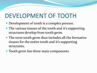
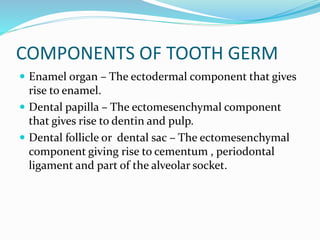
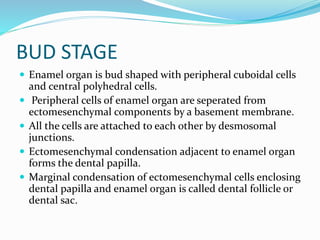
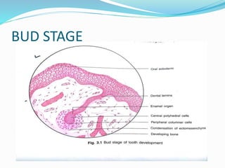
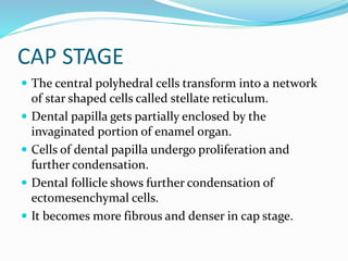
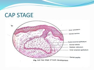
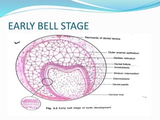
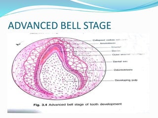
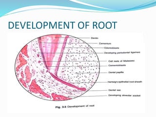
By Dr SURYAGOPAN.P
DEVELOPMENT OF TOOTH
 Development of tooth is a complex process.
 The various tissues of the tooth and it’s supporting
structures develop from tooth germ.
 The term tooth germ thus includes all the formative
tissues for the entire tooth and it’s supporting
structures.
 Tooth germ has three main components
COMPONENTS OF TOOTH GERM
 Enamel organ – The ectodermal component that gives
rise to enamel.
 Dental papilla – The ectomesenchymal component
that gives rise to dentin and pulp.
 Dental follicle or dental sac – The ectomesenchymal
component giving rise to cementum , periodontal
ligament and part of the alveolar socket.
STAGES OF TOOTH DEVELOPMENT
 BUD STAGE
 CAP STAGE
 BELL STAGE- EARLY BELL STAGE
- LATE OR ADVANCED BELL STAGE
BUD STAGE
 Enamel organ is bud shaped with peripheral cuboidal cells
and central polyhedral cells.
 Peripheral cells of enamel organ are seperated from
ectomesenchymal components by a basement membrane.
 All the cells are attached to each other by desmosomal
junctions.
 Ectomesenchymal condensation adjacent to enamel organ
forms the dental papilla.
 Marginal condensation of ectomesenchymal cells enclosing
dental papilla and enamel organ is called dental follicle or
dental sac.
BUD STAGE
CAP STAGE
 Enamel organ increases in size and attain the shape of
a cap by invagination of the deep portion of the bud.
 Cell undergo change in shape so that three seperate
cell groups can be identified.
 Cells lining the convexity or the periphery of the cap
are cuboidal in shape and are called outer enamel
epithelium.
 The cells lining the concave or invaginated portion
change to columnar cells named as inner enamel
epithelium.
CAP STAGE
 The central polyhedral cells transform into a network
of star shaped cells called stellate reticulum.
 Dental papilla gets partially enclosed by the
invaginated portion of enamel organ.
 Cells of dental papilla undergo proliferation and
further condensation.
 Dental follicle shows further condensation of
ectomesenchymal cells.
 It becomes more fibrous and denser in cap stage.
CAP STAGE
EARLY BELL STAGE
EARLY BELL STAGE
 Enamel organ enlarges further and invagination
deepens changing the shape to that of a bell in a
longitudinal section.
 In bell stage 4 different layers of cells are seen in the
enamel organ.
 Cells lining the invaginated portion , the inner enamel
epithelium is composed of single layer of tall columnar
cells that differentiate to ameloblasts(enamel forming
cells)
 During bell stage a new layer appears which is called
stratum intermedium.
EARLY BELL STAGE
 Stratum intermedium is located between inner enamel
epithelium and stellate reticulum and is composed of
2 to 3 layers of squamous cells.
 Stellate reticulum expands further in early bell stage.
 Cells of outer enamel epithelium lining the periphery
of enamel organ flattens to low cuboidal cells.
 At the cervical region of enamel organ outer enamel
epithelium loops inward to join with inner enamel
epithelium. This is called cervical loop.
EARLY BELL STAGE
 During early bell stage enamel organ loses its
connection to oral ectoderm due to degeneration of
dental lamina.
 Remnants of the dental lamina are called cell rests of
serres.
 Successional lamina develops at this stage which is the
primordium for the permenent successor.
EARLY BELL STAGE
 Dental papilla is fully enclosed in the invaginated
portion of the enamel organ in this stage.
 Peripheral cells of dental papilla differentiate into
odontoblasts (dentin forming cells) under the
organizing influence of inner enamel epithelial cells.
 Dental follicle become more fibrous with 3 layers i.e.
Inner cellular, outer fibrous layer and middle loose
connective tissue.
ADVANCED BELL STAGE
ADVANCED BELL STAGE
 Differentiating feature between early and advanced
bell stage is formation of hard tissues.
 Enamel organ shows 4 different layers, inner enamel
epithelium, stratum intermedium, stellate reticulum
and outer enamel epithelium.
 Histological difference from early bell stage are
- Hard tissue formation
- Collapsed stellate reticulum and folding of outer
enamel epithelium bringing capillaries of the dental
follicle nearer to the ameloblasts.
ADVANCED BELL STAGE
 Ameloblasts are brought closer to the dental follicle
which now becomes their source of nutrition.
 Dental papilla shows differentiated odontoblast at the
periphery.
 Dental follicle is distinct enclosing enamel organ and
dental papilla.
DEVELOPMENT OF ROOT
DEVELOPMENT OF ROOT
 Root development occurs in advanced bell stage after
the enamel and dentin formation reaches the cervical
region of tooth.
 The cervical loop which is composed of outer and
inner enamel epithelium, proliferates to form
Hertwig’s epithelial root sheath which determines the
size , shape and number of roots to be formed.
 Inner cells of HERS exert an organizing influence on
dental papilla cells to differentiate in to odontoblasts
that deposit radicular dentin.
DEVELOPMENT OF ROOT
 Once the root dentin is formed Hertwig’s epithelial root
sheath degenerate allowing the dental follicle cells to come
in contact with the dentin.
 The cells of dental follicle differentiate to form
cementoblasts and lays down cementum over the root
dentin.
 As the cementum formation progresses the rest of the
dental follicle becomes more fibrous and develop into
periodontal ligament.
 The remnants of Hertwig’s epithelial root sheath remain in
peridontal ligament and are called ‘cell rests of malassez.’
...........................................................................................................

Development of tooth

  • 1.
  • 2.
    DEVELOPMENT OF TOOTH Development of tooth is a complex process.  The various tissues of the tooth and it’s supporting structures develop from tooth germ.  The term tooth germ thus includes all the formative tissues for the entire tooth and it’s supporting structures.  Tooth germ has three main components
  • 3.
    COMPONENTS OF TOOTHGERM  Enamel organ – The ectodermal component that gives rise to enamel.  Dental papilla – The ectomesenchymal component that gives rise to dentin and pulp.  Dental follicle or dental sac – The ectomesenchymal component giving rise to cementum , periodontal ligament and part of the alveolar socket.
  • 4.
    STAGES OF TOOTHDEVELOPMENT  BUD STAGE  CAP STAGE  BELL STAGE- EARLY BELL STAGE - LATE OR ADVANCED BELL STAGE
  • 5.
    BUD STAGE  Enamelorgan is bud shaped with peripheral cuboidal cells and central polyhedral cells.  Peripheral cells of enamel organ are seperated from ectomesenchymal components by a basement membrane.  All the cells are attached to each other by desmosomal junctions.  Ectomesenchymal condensation adjacent to enamel organ forms the dental papilla.  Marginal condensation of ectomesenchymal cells enclosing dental papilla and enamel organ is called dental follicle or dental sac.
  • 6.
  • 7.
    CAP STAGE  Enamelorgan increases in size and attain the shape of a cap by invagination of the deep portion of the bud.  Cell undergo change in shape so that three seperate cell groups can be identified.  Cells lining the convexity or the periphery of the cap are cuboidal in shape and are called outer enamel epithelium.  The cells lining the concave or invaginated portion change to columnar cells named as inner enamel epithelium.
  • 8.
    CAP STAGE  Thecentral polyhedral cells transform into a network of star shaped cells called stellate reticulum.  Dental papilla gets partially enclosed by the invaginated portion of enamel organ.  Cells of dental papilla undergo proliferation and further condensation.  Dental follicle shows further condensation of ectomesenchymal cells.  It becomes more fibrous and denser in cap stage.
  • 9.
  • 10.
  • 11.
    EARLY BELL STAGE Enamel organ enlarges further and invagination deepens changing the shape to that of a bell in a longitudinal section.  In bell stage 4 different layers of cells are seen in the enamel organ.  Cells lining the invaginated portion , the inner enamel epithelium is composed of single layer of tall columnar cells that differentiate to ameloblasts(enamel forming cells)  During bell stage a new layer appears which is called stratum intermedium.
  • 12.
    EARLY BELL STAGE Stratum intermedium is located between inner enamel epithelium and stellate reticulum and is composed of 2 to 3 layers of squamous cells.  Stellate reticulum expands further in early bell stage.  Cells of outer enamel epithelium lining the periphery of enamel organ flattens to low cuboidal cells.  At the cervical region of enamel organ outer enamel epithelium loops inward to join with inner enamel epithelium. This is called cervical loop.
  • 13.
    EARLY BELL STAGE During early bell stage enamel organ loses its connection to oral ectoderm due to degeneration of dental lamina.  Remnants of the dental lamina are called cell rests of serres.  Successional lamina develops at this stage which is the primordium for the permenent successor.
  • 14.
    EARLY BELL STAGE Dental papilla is fully enclosed in the invaginated portion of the enamel organ in this stage.  Peripheral cells of dental papilla differentiate into odontoblasts (dentin forming cells) under the organizing influence of inner enamel epithelial cells.  Dental follicle become more fibrous with 3 layers i.e. Inner cellular, outer fibrous layer and middle loose connective tissue.
  • 15.
  • 16.
    ADVANCED BELL STAGE Differentiating feature between early and advanced bell stage is formation of hard tissues.  Enamel organ shows 4 different layers, inner enamel epithelium, stratum intermedium, stellate reticulum and outer enamel epithelium.  Histological difference from early bell stage are - Hard tissue formation - Collapsed stellate reticulum and folding of outer enamel epithelium bringing capillaries of the dental follicle nearer to the ameloblasts.
  • 17.
    ADVANCED BELL STAGE Ameloblasts are brought closer to the dental follicle which now becomes their source of nutrition.  Dental papilla shows differentiated odontoblast at the periphery.  Dental follicle is distinct enclosing enamel organ and dental papilla.
  • 18.
  • 19.
    DEVELOPMENT OF ROOT Root development occurs in advanced bell stage after the enamel and dentin formation reaches the cervical region of tooth.  The cervical loop which is composed of outer and inner enamel epithelium, proliferates to form Hertwig’s epithelial root sheath which determines the size , shape and number of roots to be formed.  Inner cells of HERS exert an organizing influence on dental papilla cells to differentiate in to odontoblasts that deposit radicular dentin.
  • 20.
    DEVELOPMENT OF ROOT Once the root dentin is formed Hertwig’s epithelial root sheath degenerate allowing the dental follicle cells to come in contact with the dentin.  The cells of dental follicle differentiate to form cementoblasts and lays down cementum over the root dentin.  As the cementum formation progresses the rest of the dental follicle becomes more fibrous and develop into periodontal ligament.  The remnants of Hertwig’s epithelial root sheath remain in peridontal ligament and are called ‘cell rests of malassez.’
  • 21.