SlideShare a Scribd company logo
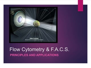
Flow Cytometry & F.A.C.S.
PRINCIPLES AND APPLICATIONS
What is Flow Cytometry?
‘Flow Cytometry’ as the name suggests is a technique for cell
counting and measurement of different properties of the cell
(‘cyto’= cell; ‘metry’=count/measurement).
It is a laser based technology that measures and analyses
different physical and chemical properties of the cells/particles
flowing in a stream of fluid through a beam of light.
A Flow Cytometer
Place your
sample here
Historical Perspective: Evolution of Flow
Cytometry
17th
Century
1934
1947 to
1949
1953 19651879 1968
1970s
onwards…
Development of
light microscope
by
Leeuwenhoek.
Principles of Droplet
formation by Lord
Rayleigh.
Counting of RBCs
by Moldavan by
forcing a
suspension of cells
through capillary
tube.
Development of
Coulter Principle by
Wallace Coulter and
counting of RBCs
using the first Coulter
Counter.
Optical counting of
RBCs by Crosland-
Taylor by use of
laminar flow
principles
Development of
electrostatic
inkjet droplet
deflection by
Richard Sweet
Application of
Sweet’s principle
and Coulter principle
to develop the first
cell sorter by M.
Fulwyler
Development of
fluorescence
based cell sorter
by Wolfgang
Gohde
Development of
FACS and other
advances.
Principles of working of Flow
Cytometer
Coulter
Principle
Principle
s of
Laminar
Flow
Electro
statics
Optics &
Light
Scatteri
ng
Flow
Cytome
try
Components of a Flow Cytometer
A flow cytometer is made up of three main systems: fluidics, optics, and
electronics.
 The fluidics system transports particles in a stream to the laser beam for
interrogation.
 The optics system consists of lasers to illuminate the particles in the
sample stream and optical filters to direct the resulting light signals to the
appropriate detectors.
 The electronics system converts the detected light signals into electronic
signals that can be processed by the computer. For some instruments
equipped with a sorting feature, the electronics system is also capable of
initiating sorting decisions to charge and deflect particles.
Working of a Flow Cytometer
 In the flow cytometer, particles are carried to the laser intercept in a fluid
stream. Any suspended particle or cell from 0.2–150 micrometers in size is
suitable for analysis.
 The portion of the fluid stream where particles are located is called the sample
core.
 When particles pass through the laser intercept, they scatter laser light. Any
fluorescent molecules present on the particle fluoresce.
 The scattered and fluorescent light is collected by appropriately positioned
lenses.
 A combination of beam splitters and filters steers the scattered and fluorescent
light to the appropriate detectors.
 The detectors produce electronic signals proportional to the optical signals
striking them.
Applications of Flow
Cytometry
Flow cytometry is the sine qua non (without which, nothing)of the modern
researcher’s toolbox.
Flow cytometry measures multiple characteristics of individual particles
flowing in single file in a stream of fluid. Light scattering at different angles can
distinguish differences in size and internal complexity, whereas light emitted
from fluorescently labeled antibodies can identify a wide array of cell surface
and cytoplasmic antigens. This approach makes flow cytometry a powerful
tool for detailed analysis of complex populations in a short period of time.
Applications
Immunophenotyping
Cell subsets are measured by labeling
population-specific proteins with a fluorescent
tag on the cell surface. In clinical labs,
immunophenotyping is useful in diagnosing
hematological malignancies such as
lymphomas and leukemia.
Cell Sorting
The cell sorter is a specialized flow
cytometer with the ability to physically
isolate cells of interest into separate
collection tubes. The sorter uses
sophisticated electronics and fluidics to
identify and "kick" the cells of interest out of
the fluidic stream into a test tube.
DNA Content Analysis
The measurement of cellular DNA content by
flow cytometry uses fluorescent dyes, such as
propidium iodide, that intercalate into the DNA
helical structure. The fluorescent signal is directly
proportional to the amount of DNA in the nucleus
and can identify gross gains or losses in DNA.
Cell Cycle Analysis
Flow cytometry can analyze replication
states using fluorescent dyes to measure
the four distinct phases of the cell cycle.
Along with determining cell cycle replication
states, the assay can measure cell
aneuploidy associated with chromosomal
abnormalities.
Apoptosis
The two distinct types of cell death,
apoptosis and necrosis, can be
distinguished by flow cytometry on the basis
of differences in morphological, biochemical
and molecular changes occurring in the
dying cells.
Cell Proliferation Assays
The flow cytometer can measure proliferation by
labeling resting cells with a cell membrane
fluorescent dye, carboxyfluorescein succinimidyl
ester (CFSE). When the cells are activated, they
begin to proliferate and undergo mitosis. As the
cells divide, half of the original dye is passed on
to each daughter cell. By measuring the
reduction of the fluorescence signal, researchers
can calculate cellular activation and proliferation.
Fluorescence Activated Cell
Sorting (FACS)
 Consider a group of lymphocytes from a mouse that have been stained with
green fluorescent antibodies specific for CD4 (e.g., fluorescein isothiocyanate,
or FITC anti-CD4) and red fluorescent antibodies specific for CD8 (e.g.,
phycoerythrin, or PE anti-CD8).
 Both the labeled cells generate SSC and FSC as they pass through the laser
beam creating voltage pulses that are recorded by the computer.
 However, each labeled cell will also emit light of specific wavelength as a
result of the fluorescent label. For instance, CD4 cells will emit green
fluorescent light of wavelength 525-530 nm while CD8 cells emit orange light
of wavelength 560 nm. These fluorescent signals pass through the
Photomultiplier tubes and generate voltage pulses.
 The software integrates all the information for a particular cell allowing
characterization of individual cells.
Clinical Applications: DNA
Content Analysis
 Investigators are currently using techniques of DNA flow cytometry to measure ploidy status
(DNA content) and proliferative potential (S phase fraction) in a wide variety of solid tumors.
These measurements have shown relevance for diagnosis, prognosis, and treatment for
patients with cancer.
 The measurement of cellular DNA content by flow cytometry uses fluorescent dyes, such as
propidium iodide, that intercalate into the DNA helical structure. The fluorescent signal is
directly proportional to the amount of DNA in the nucleus and can identify gross gains or losses
in DNA.
 Abnormal DNA content, also known as “DNA content aneuploidy”, can be determined in a tumor
cell population. DNA aneuploidy generally is associated with malignancy; however, certain
benign conditions may appear aneuploid.
 Cell Cycle Analysis: This technique is based on the premise that cells in G0 or G1 phases of
the cell cycle possess a normal diploid chromosomal, and hence DNA content (2n) whereas
cells in G2 and just prior to mitosis (M) contain exactly twice this amount (4n). As DNA is
synthesized during S-phase, cells are found with a DNA content ranging between 2n and 4n. A
histogram plot of DNA content against cell numbers gives the classical DNA profile for a
proliferating cell culture.
Flow Cytometry and Ecology
 Assessments of diversity, abundance, and activity of water column
microorganisms are fundamental to studies in aquatic microbiology.
 Currently, most applications of flow cytometry to environmental samples make
use of various morphological and physiological characteristics of the cells (e.g.,
size and pigment content of photosynthetic organisms).
 These criteria generally are not sufficient for identification at the genus or
species level. Staining with DNA-specific fluorochromes offers information
about numbers of bacterial cells but not about their identity.
 The combined use of dyes that bind preferentially to G- C or A. T base pairs
has been used to distinguish organisms of different G+C content
Flow Cytometry and Cancer
Research
 The prognosis of patients with cancer is largely determined by the specific histological diagnosis,
tumor mass stage, and host performance status.
 Quantitative cytology in the form of flow cytometry has greatly advanced the objective elucidation of
tumor cell heterogeneity by using probes that discriminate tumor and normal cells and assess
differentiate as well as proliferative tumor cell properties.
 Both DNA content analysis and FACS can be utilised in cancer research.
 Abnormal nuclear DMA content is a conclusive marker of malignancy and is found with increasing
frequency in leukemia (23% among 793 patients), in lymphoma (53% among 360 patients), and in
myeloma (76% among 177 patients), as well as in solid tumors (75% among 3611 patients), for an
overall incidence of 67% in 4941 patients.
 Flow cytometric immunophenotyping (FCI) aids in the differentiation of chronic lymphocytic leukemia
(CLL) from mantle cell lymphoma (MCL); however, overlapping phenotypes may occur. CD11c
expression has been reported in up to 90% of CLL cases but has rarely been reported in MCL.
Whether CD11c can be used to exclude MCL has not been directly addressed. FCI reports were
reviewed for 90 MCL cases (44 patients) and 355 CLL/small lymphocytic lymphoma (SLL) cases (158
patients).
References
 http://flowcytometry.berkeley.edu/pdfs/Basic%20Flow%20Cytometry.pdf
 http://www.azom.com/article.aspx?ArticleID=6020
 https://en.wikipedia.org/wiki/Flow_cytometry
Thank You.

More Related Content

What's hot

Flow cytometry
Flow cytometry Flow cytometry
Flow cytometry
RatnakumariP
 
Fluorescence- Activated Cell Sorter (FACS)
Fluorescence- Activated Cell Sorter (FACS)Fluorescence- Activated Cell Sorter (FACS)
Fluorescence- Activated Cell Sorter (FACS)
Nidhi Parikh
 
Flow cytometry
Flow cytometryFlow cytometry
Flow cytometry
Pradeep Singh Narwat
 
Flow cytometry
Flow cytometryFlow cytometry
Flow cytometry
Sreenivasa Reddy Thalla
 
Flow Cytometry - basics, principles and applications
Flow Cytometry - basics, principles and applicationsFlow Cytometry - basics, principles and applications
Flow Cytometry - basics, principles and applications
Ankit Raiyani
 
Principle and applications of flow cytometry
Principle and applications of flow cytometryPrinciple and applications of flow cytometry
Principle and applications of flow cytometry
Dinesh Gangoda
 
Kumc introduction to flow cytometry
Kumc introduction to flow cytometryKumc introduction to flow cytometry
Kumc introduction to flow cytometry
Richard Hastings
 
Flow Cytometry technique
Flow Cytometry technique Flow Cytometry technique
Flow Cytometry technique
Md Fayezur Rahaman
 
Cytokine signaling pathway
Cytokine signaling pathwayCytokine signaling pathway
Cytokine signaling pathway
Juliet Abisha
 
Single Cell Isolation
Single Cell IsolationSingle Cell Isolation
Single Cell Isolation
QIAGEN
 
Presentation on flow cytometry1
Presentation on flow cytometry1Presentation on flow cytometry1
Presentation on flow cytometry1
Nagendra sharma
 
3D tumor spheroid models for in vitro therapeutic screening: a systematic app...
3D tumor spheroid models for in vitro therapeutic screening: a systematic app...3D tumor spheroid models for in vitro therapeutic screening: a systematic app...
3D tumor spheroid models for in vitro therapeutic screening: a systematic app...
Arun kumar
 
Adaptive Immune Response to Viral Infections
Adaptive Immune Response to Viral InfectionsAdaptive Immune Response to Viral Infections
Adaptive Immune Response to Viral Infections
Mousumi Bora
 
Induced Pluripotent Stem Cells (iPSCs)
Induced Pluripotent Stem Cells (iPSCs)Induced Pluripotent Stem Cells (iPSCs)
Induced Pluripotent Stem Cells (iPSCs)
Garry D. Lasaga
 
Flow cytometry: Principles and Applications
Flow cytometry: Principles and ApplicationsFlow cytometry: Principles and Applications
Flow cytometry: Principles and Applications
Juhi Arora
 
Application of Fluorescence Activated-Cell Sorting (FACS) in separation of di...
Application of Fluorescence Activated-Cell Sorting (FACS) in separation of di...Application of Fluorescence Activated-Cell Sorting (FACS) in separation of di...
Application of Fluorescence Activated-Cell Sorting (FACS) in separation of di...
Goh Mei Ying
 
Tumor suppressor gene
Tumor suppressor geneTumor suppressor gene
Tumor suppressor gene
Amani Alismail
 
Flow cytometry and fluorescence activated cell sorting (FACS)
Flow cytometry and fluorescence activated cell sorting (FACS)Flow cytometry and fluorescence activated cell sorting (FACS)
Flow cytometry and fluorescence activated cell sorting (FACS)
Abu Sufiyan Chhipa
 
FlowCytometry Basics
FlowCytometry BasicsFlowCytometry Basics
FlowCytometry BasicsAnna Öberg
 
basic concept of molecular pathology
basic concept of molecular pathologybasic concept of molecular pathology
basic concept of molecular pathology
University Of Wuerzburg,Germany
 

What's hot (20)

Flow cytometry
Flow cytometry Flow cytometry
Flow cytometry
 
Fluorescence- Activated Cell Sorter (FACS)
Fluorescence- Activated Cell Sorter (FACS)Fluorescence- Activated Cell Sorter (FACS)
Fluorescence- Activated Cell Sorter (FACS)
 
Flow cytometry
Flow cytometryFlow cytometry
Flow cytometry
 
Flow cytometry
Flow cytometryFlow cytometry
Flow cytometry
 
Flow Cytometry - basics, principles and applications
Flow Cytometry - basics, principles and applicationsFlow Cytometry - basics, principles and applications
Flow Cytometry - basics, principles and applications
 
Principle and applications of flow cytometry
Principle and applications of flow cytometryPrinciple and applications of flow cytometry
Principle and applications of flow cytometry
 
Kumc introduction to flow cytometry
Kumc introduction to flow cytometryKumc introduction to flow cytometry
Kumc introduction to flow cytometry
 
Flow Cytometry technique
Flow Cytometry technique Flow Cytometry technique
Flow Cytometry technique
 
Cytokine signaling pathway
Cytokine signaling pathwayCytokine signaling pathway
Cytokine signaling pathway
 
Single Cell Isolation
Single Cell IsolationSingle Cell Isolation
Single Cell Isolation
 
Presentation on flow cytometry1
Presentation on flow cytometry1Presentation on flow cytometry1
Presentation on flow cytometry1
 
3D tumor spheroid models for in vitro therapeutic screening: a systematic app...
3D tumor spheroid models for in vitro therapeutic screening: a systematic app...3D tumor spheroid models for in vitro therapeutic screening: a systematic app...
3D tumor spheroid models for in vitro therapeutic screening: a systematic app...
 
Adaptive Immune Response to Viral Infections
Adaptive Immune Response to Viral InfectionsAdaptive Immune Response to Viral Infections
Adaptive Immune Response to Viral Infections
 
Induced Pluripotent Stem Cells (iPSCs)
Induced Pluripotent Stem Cells (iPSCs)Induced Pluripotent Stem Cells (iPSCs)
Induced Pluripotent Stem Cells (iPSCs)
 
Flow cytometry: Principles and Applications
Flow cytometry: Principles and ApplicationsFlow cytometry: Principles and Applications
Flow cytometry: Principles and Applications
 
Application of Fluorescence Activated-Cell Sorting (FACS) in separation of di...
Application of Fluorescence Activated-Cell Sorting (FACS) in separation of di...Application of Fluorescence Activated-Cell Sorting (FACS) in separation of di...
Application of Fluorescence Activated-Cell Sorting (FACS) in separation of di...
 
Tumor suppressor gene
Tumor suppressor geneTumor suppressor gene
Tumor suppressor gene
 
Flow cytometry and fluorescence activated cell sorting (FACS)
Flow cytometry and fluorescence activated cell sorting (FACS)Flow cytometry and fluorescence activated cell sorting (FACS)
Flow cytometry and fluorescence activated cell sorting (FACS)
 
FlowCytometry Basics
FlowCytometry BasicsFlowCytometry Basics
FlowCytometry Basics
 
basic concept of molecular pathology
basic concept of molecular pathologybasic concept of molecular pathology
basic concept of molecular pathology
 

Viewers also liked

Flow cytometry ready
Flow cytometry readyFlow cytometry ready
Flow cytometry readyYra Yunus
 
Flow cytometry
Flow cytometryFlow cytometry
Flow cytometry
bilal musharaf
 
Featured Presentation FIRN Kona 2009
Featured Presentation FIRN Kona 2009Featured Presentation FIRN Kona 2009
Featured Presentation FIRN Kona 2009
TOKBLS
 
Facs DiVa training Garvan MLC Flow Cytometry Facility
Facs DiVa training Garvan MLC Flow Cytometry FacilityFacs DiVa training Garvan MLC Flow Cytometry Facility
Facs DiVa training Garvan MLC Flow Cytometry Facility
Robert (Rob) Salomon
 
Flow cytometry
Flow cytometryFlow cytometry
Flow cytometry
Anushi Jain
 
Cytokines by Dr. Komal Lohi, LTMMC, Sion
Cytokines by Dr. Komal Lohi, LTMMC, SionCytokines by Dr. Komal Lohi, LTMMC, Sion
Cytokines by Dr. Komal Lohi, LTMMC, Sion
Dr. Komal Lohi
 
Analysis of Plant Genomes Using Flow Cytometry
Analysis of Plant Genomes Using Flow CytometryAnalysis of Plant Genomes Using Flow Cytometry
Analysis of Plant Genomes Using Flow Cytometry
International Institute of Tropical Agriculture
 
ELISA Vs ELISPOT - Principle, Procedure, Advantages
ELISA Vs ELISPOT - Principle, Procedure, AdvantagesELISA Vs ELISPOT - Principle, Procedure, Advantages
ELISA Vs ELISPOT - Principle, Procedure, Advantages
ajithnandanam
 
Cytokines
CytokinesCytokines
Cytokines
RESHMASOMAN3
 
Acute leukemias 1-csbrp
Acute leukemias 1-csbrpAcute leukemias 1-csbrp
Acute leukemias 1-csbrp
Prasad CSBR
 
Introduction to Flow Cytometry
Introduction to Flow CytometryIntroduction to Flow Cytometry
Introduction to Flow Cytometry
Robert (Rob) Salomon
 
How to Become a Thought Leader in Your Niche
How to Become a Thought Leader in Your NicheHow to Become a Thought Leader in Your Niche
How to Become a Thought Leader in Your Niche
Leslie Samuel
 

Viewers also liked (13)

Flow cytometry ready
Flow cytometry readyFlow cytometry ready
Flow cytometry ready
 
Flow cytometry
Flow cytometryFlow cytometry
Flow cytometry
 
Featured Presentation FIRN Kona 2009
Featured Presentation FIRN Kona 2009Featured Presentation FIRN Kona 2009
Featured Presentation FIRN Kona 2009
 
Facs DiVa training Garvan MLC Flow Cytometry Facility
Facs DiVa training Garvan MLC Flow Cytometry FacilityFacs DiVa training Garvan MLC Flow Cytometry Facility
Facs DiVa training Garvan MLC Flow Cytometry Facility
 
Flow cytometry
Flow cytometryFlow cytometry
Flow cytometry
 
Cytokines by Dr. Komal Lohi, LTMMC, Sion
Cytokines by Dr. Komal Lohi, LTMMC, SionCytokines by Dr. Komal Lohi, LTMMC, Sion
Cytokines by Dr. Komal Lohi, LTMMC, Sion
 
Analysis of Plant Genomes Using Flow Cytometry
Analysis of Plant Genomes Using Flow CytometryAnalysis of Plant Genomes Using Flow Cytometry
Analysis of Plant Genomes Using Flow Cytometry
 
ELISA Vs ELISPOT - Principle, Procedure, Advantages
ELISA Vs ELISPOT - Principle, Procedure, AdvantagesELISA Vs ELISPOT - Principle, Procedure, Advantages
ELISA Vs ELISPOT - Principle, Procedure, Advantages
 
Antigen antibody
Antigen antibodyAntigen antibody
Antigen antibody
 
Cytokines
CytokinesCytokines
Cytokines
 
Acute leukemias 1-csbrp
Acute leukemias 1-csbrpAcute leukemias 1-csbrp
Acute leukemias 1-csbrp
 
Introduction to Flow Cytometry
Introduction to Flow CytometryIntroduction to Flow Cytometry
Introduction to Flow Cytometry
 
How to Become a Thought Leader in Your Niche
How to Become a Thought Leader in Your NicheHow to Become a Thought Leader in Your Niche
How to Become a Thought Leader in Your Niche
 

Similar to Rohan gupta 2015 b1ab651p facs

Applications of Flow Cytometry | Cell Analysis
Applications of Flow Cytometry | Cell AnalysisApplications of Flow Cytometry | Cell Analysis
Applications of Flow Cytometry | Cell Analysis
University of The Punjab
 
Flow cytometry
Flow cytometryFlow cytometry
Flow cytometry
SaniyaTabassum3
 
Flowcytometry 1
Flowcytometry 1Flowcytometry 1
Flowcytometry 1
SUNIL KUMAR PEDDANA
 
Laboratory diagnosis of neoplasm
Laboratory diagnosis of neoplasmLaboratory diagnosis of neoplasm
Laboratory diagnosis of neoplasm
Sunita Patil
 
Flow Cytometry: Guide to Understanding Applications and Benefits | The Lifesc...
Flow Cytometry: Guide to Understanding Applications and Benefits | The Lifesc...Flow Cytometry: Guide to Understanding Applications and Benefits | The Lifesc...
Flow Cytometry: Guide to Understanding Applications and Benefits | The Lifesc...
The Lifesciences Magazine
 
Identification of Rare and Novel Alleles in FFPE Tumor Samples | ESHG 2015 Po...
Identification of Rare and Novel Alleles in FFPE Tumor Samples | ESHG 2015 Po...Identification of Rare and Novel Alleles in FFPE Tumor Samples | ESHG 2015 Po...
Identification of Rare and Novel Alleles in FFPE Tumor Samples | ESHG 2015 Po...
Thermo Fisher Scientific
 
Analytical chemistry 2013 qian liu
Analytical chemistry 2013 qian liuAnalytical chemistry 2013 qian liu
Analytical chemistry 2013 qian liu
Qian Liu, phD
 
Liquid Biopsy Cancer Prevention and Monitoring
Liquid Biopsy Cancer Prevention and MonitoringLiquid Biopsy Cancer Prevention and Monitoring
Liquid Biopsy Cancer Prevention and Monitoring
David Tjahjono,MD,MBA(UK)
 
Medcrave Group - Microfluidic technologies
Medcrave Group - Microfluidic technologiesMedcrave Group - Microfluidic technologies
Medcrave Group - Microfluidic technologies
MedCrave
 
Immunoflowcytometry, the basics and applications.pptx
Immunoflowcytometry, the basics and applications.pptxImmunoflowcytometry, the basics and applications.pptx
Immunoflowcytometry, the basics and applications.pptx
AmirRaziq1
 
Qualitative Flow Cytometric Analysis of Malaysian
Qualitative Flow Cytometric Analysis of MalaysianQualitative Flow Cytometric Analysis of Malaysian
Qualitative Flow Cytometric Analysis of MalaysianMohadese Hashem Boroojerdi
 
Nano Flow Cytometer by NanoFCM Inc.
Nano Flow Cytometer by NanoFCM Inc.Nano Flow Cytometer by NanoFCM Inc.
Nano Flow Cytometer by NanoFCM Inc.
AninditaGuha8
 
Flowcytometry
FlowcytometryFlowcytometry
Flowcytometry
Badheeb
 
Flowcytometry
FlowcytometryFlowcytometry
Flowcytometry
eman youssif
 
Asok dikshit early detection of cancer cells
Asok dikshit early detection of cancer cellsAsok dikshit early detection of cancer cells
Asok dikshit early detection of cancer cellssatyendraurinfo
 
Second Review Research in quantifying lymphocytes.pptx
Second Review Research in quantifying lymphocytes.pptxSecond Review Research in quantifying lymphocytes.pptx
Second Review Research in quantifying lymphocytes.pptx
KerenEvangelineI
 
Pathology-Today-2016-Q2
Pathology-Today-2016-Q2Pathology-Today-2016-Q2
Pathology-Today-2016-Q2Gary Weiland
 
flow cytometry by aksharaditya shukla.
flow cytometry by aksharaditya shukla.flow cytometry by aksharaditya shukla.
flow cytometry by aksharaditya shukla.
Aksharaditya Shukla
 
Use of flow cytometric immunophenotyping by teresa lotodo
Use of flow cytometric immunophenotyping by teresa lotodoUse of flow cytometric immunophenotyping by teresa lotodo
Use of flow cytometric immunophenotyping by teresa lotodo
Kesho Conference
 

Similar to Rohan gupta 2015 b1ab651p facs (20)

Applications of Flow Cytometry | Cell Analysis
Applications of Flow Cytometry | Cell AnalysisApplications of Flow Cytometry | Cell Analysis
Applications of Flow Cytometry | Cell Analysis
 
Flow cytometry
Flow cytometryFlow cytometry
Flow cytometry
 
Flowcytometry 1
Flowcytometry 1Flowcytometry 1
Flowcytometry 1
 
Laboratory diagnosis of neoplasm
Laboratory diagnosis of neoplasmLaboratory diagnosis of neoplasm
Laboratory diagnosis of neoplasm
 
Journal of Surgical Oncology - Barnabas
Journal of Surgical Oncology - BarnabasJournal of Surgical Oncology - Barnabas
Journal of Surgical Oncology - Barnabas
 
Flow Cytometry: Guide to Understanding Applications and Benefits | The Lifesc...
Flow Cytometry: Guide to Understanding Applications and Benefits | The Lifesc...Flow Cytometry: Guide to Understanding Applications and Benefits | The Lifesc...
Flow Cytometry: Guide to Understanding Applications and Benefits | The Lifesc...
 
Identification of Rare and Novel Alleles in FFPE Tumor Samples | ESHG 2015 Po...
Identification of Rare and Novel Alleles in FFPE Tumor Samples | ESHG 2015 Po...Identification of Rare and Novel Alleles in FFPE Tumor Samples | ESHG 2015 Po...
Identification of Rare and Novel Alleles in FFPE Tumor Samples | ESHG 2015 Po...
 
Analytical chemistry 2013 qian liu
Analytical chemistry 2013 qian liuAnalytical chemistry 2013 qian liu
Analytical chemistry 2013 qian liu
 
Liquid Biopsy Cancer Prevention and Monitoring
Liquid Biopsy Cancer Prevention and MonitoringLiquid Biopsy Cancer Prevention and Monitoring
Liquid Biopsy Cancer Prevention and Monitoring
 
Medcrave Group - Microfluidic technologies
Medcrave Group - Microfluidic technologiesMedcrave Group - Microfluidic technologies
Medcrave Group - Microfluidic technologies
 
Immunoflowcytometry, the basics and applications.pptx
Immunoflowcytometry, the basics and applications.pptxImmunoflowcytometry, the basics and applications.pptx
Immunoflowcytometry, the basics and applications.pptx
 
Qualitative Flow Cytometric Analysis of Malaysian
Qualitative Flow Cytometric Analysis of MalaysianQualitative Flow Cytometric Analysis of Malaysian
Qualitative Flow Cytometric Analysis of Malaysian
 
Nano Flow Cytometer by NanoFCM Inc.
Nano Flow Cytometer by NanoFCM Inc.Nano Flow Cytometer by NanoFCM Inc.
Nano Flow Cytometer by NanoFCM Inc.
 
Flowcytometry
FlowcytometryFlowcytometry
Flowcytometry
 
Flowcytometry
FlowcytometryFlowcytometry
Flowcytometry
 
Asok dikshit early detection of cancer cells
Asok dikshit early detection of cancer cellsAsok dikshit early detection of cancer cells
Asok dikshit early detection of cancer cells
 
Second Review Research in quantifying lymphocytes.pptx
Second Review Research in quantifying lymphocytes.pptxSecond Review Research in quantifying lymphocytes.pptx
Second Review Research in quantifying lymphocytes.pptx
 
Pathology-Today-2016-Q2
Pathology-Today-2016-Q2Pathology-Today-2016-Q2
Pathology-Today-2016-Q2
 
flow cytometry by aksharaditya shukla.
flow cytometry by aksharaditya shukla.flow cytometry by aksharaditya shukla.
flow cytometry by aksharaditya shukla.
 
Use of flow cytometric immunophenotyping by teresa lotodo
Use of flow cytometric immunophenotyping by teresa lotodoUse of flow cytometric immunophenotyping by teresa lotodo
Use of flow cytometric immunophenotyping by teresa lotodo
 

Recently uploaded

Richard's entangled aventures in wonderland
Richard's entangled aventures in wonderlandRichard's entangled aventures in wonderland
Richard's entangled aventures in wonderland
Richard Gill
 
platelets- lifespan -Clot retraction-disorders.pptx
platelets- lifespan -Clot retraction-disorders.pptxplatelets- lifespan -Clot retraction-disorders.pptx
platelets- lifespan -Clot retraction-disorders.pptx
muralinath2
 
platelets_clotting_biogenesis.clot retractionpptx
platelets_clotting_biogenesis.clot retractionpptxplatelets_clotting_biogenesis.clot retractionpptx
platelets_clotting_biogenesis.clot retractionpptx
muralinath2
 
Lab report on liquid viscosity of glycerin
Lab report on liquid viscosity of glycerinLab report on liquid viscosity of glycerin
Lab report on liquid viscosity of glycerin
ossaicprecious19
 
4. An Overview of Sugarcane White Leaf Disease in Vietnam.pdf
4. An Overview of Sugarcane White Leaf Disease in Vietnam.pdf4. An Overview of Sugarcane White Leaf Disease in Vietnam.pdf
4. An Overview of Sugarcane White Leaf Disease in Vietnam.pdf
ssuserbfdca9
 
Nucleic Acid-its structural and functional complexity.
Nucleic Acid-its structural and functional complexity.Nucleic Acid-its structural and functional complexity.
Nucleic Acid-its structural and functional complexity.
Nistarini College, Purulia (W.B) India
 
Observation of Io’s Resurfacing via Plume Deposition Using Ground-based Adapt...
Observation of Io’s Resurfacing via Plume Deposition Using Ground-based Adapt...Observation of Io’s Resurfacing via Plume Deposition Using Ground-based Adapt...
Observation of Io’s Resurfacing via Plume Deposition Using Ground-based Adapt...
Sérgio Sacani
 
general properties of oerganologametal.ppt
general properties of oerganologametal.pptgeneral properties of oerganologametal.ppt
general properties of oerganologametal.ppt
IqrimaNabilatulhusni
 
ESR_factors_affect-clinic significance-Pathysiology.pptx
ESR_factors_affect-clinic significance-Pathysiology.pptxESR_factors_affect-clinic significance-Pathysiology.pptx
ESR_factors_affect-clinic significance-Pathysiology.pptx
muralinath2
 
Leaf Initiation, Growth and Differentiation.pdf
Leaf Initiation, Growth and Differentiation.pdfLeaf Initiation, Growth and Differentiation.pdf
Leaf Initiation, Growth and Differentiation.pdf
RenuJangid3
 
Body fluids_tonicity_dehydration_hypovolemia_hypervolemia.pptx
Body fluids_tonicity_dehydration_hypovolemia_hypervolemia.pptxBody fluids_tonicity_dehydration_hypovolemia_hypervolemia.pptx
Body fluids_tonicity_dehydration_hypovolemia_hypervolemia.pptx
muralinath2
 
In silico drugs analogue design: novobiocin analogues.pptx
In silico drugs analogue design: novobiocin analogues.pptxIn silico drugs analogue design: novobiocin analogues.pptx
In silico drugs analogue design: novobiocin analogues.pptx
AlaminAfendy1
 
role of pramana in research.pptx in science
role of pramana in research.pptx in sciencerole of pramana in research.pptx in science
role of pramana in research.pptx in science
sonaliswain16
 
THE IMPORTANCE OF MARTIAN ATMOSPHERE SAMPLE RETURN.
THE IMPORTANCE OF MARTIAN ATMOSPHERE SAMPLE RETURN.THE IMPORTANCE OF MARTIAN ATMOSPHERE SAMPLE RETURN.
THE IMPORTANCE OF MARTIAN ATMOSPHERE SAMPLE RETURN.
Sérgio Sacani
 
Citrus Greening Disease and its Management
Citrus Greening Disease and its ManagementCitrus Greening Disease and its Management
Citrus Greening Disease and its Management
subedisuryaofficial
 
GBSN - Biochemistry (Unit 5) Chemistry of Lipids
GBSN - Biochemistry (Unit 5) Chemistry of LipidsGBSN - Biochemistry (Unit 5) Chemistry of Lipids
GBSN - Biochemistry (Unit 5) Chemistry of Lipids
Areesha Ahmad
 
Astronomy Update- Curiosity’s exploration of Mars _ Local Briefs _ leadertele...
Astronomy Update- Curiosity’s exploration of Mars _ Local Briefs _ leadertele...Astronomy Update- Curiosity’s exploration of Mars _ Local Briefs _ leadertele...
Astronomy Update- Curiosity’s exploration of Mars _ Local Briefs _ leadertele...
NathanBaughman3
 
erythropoiesis-I_mechanism& clinical significance.pptx
erythropoiesis-I_mechanism& clinical significance.pptxerythropoiesis-I_mechanism& clinical significance.pptx
erythropoiesis-I_mechanism& clinical significance.pptx
muralinath2
 
Structures and textures of metamorphic rocks
Structures and textures of metamorphic rocksStructures and textures of metamorphic rocks
Structures and textures of metamorphic rocks
kumarmathi863
 
Hemoglobin metabolism_pathophysiology.pptx
Hemoglobin metabolism_pathophysiology.pptxHemoglobin metabolism_pathophysiology.pptx
Hemoglobin metabolism_pathophysiology.pptx
muralinath2
 

Recently uploaded (20)

Richard's entangled aventures in wonderland
Richard's entangled aventures in wonderlandRichard's entangled aventures in wonderland
Richard's entangled aventures in wonderland
 
platelets- lifespan -Clot retraction-disorders.pptx
platelets- lifespan -Clot retraction-disorders.pptxplatelets- lifespan -Clot retraction-disorders.pptx
platelets- lifespan -Clot retraction-disorders.pptx
 
platelets_clotting_biogenesis.clot retractionpptx
platelets_clotting_biogenesis.clot retractionpptxplatelets_clotting_biogenesis.clot retractionpptx
platelets_clotting_biogenesis.clot retractionpptx
 
Lab report on liquid viscosity of glycerin
Lab report on liquid viscosity of glycerinLab report on liquid viscosity of glycerin
Lab report on liquid viscosity of glycerin
 
4. An Overview of Sugarcane White Leaf Disease in Vietnam.pdf
4. An Overview of Sugarcane White Leaf Disease in Vietnam.pdf4. An Overview of Sugarcane White Leaf Disease in Vietnam.pdf
4. An Overview of Sugarcane White Leaf Disease in Vietnam.pdf
 
Nucleic Acid-its structural and functional complexity.
Nucleic Acid-its structural and functional complexity.Nucleic Acid-its structural and functional complexity.
Nucleic Acid-its structural and functional complexity.
 
Observation of Io’s Resurfacing via Plume Deposition Using Ground-based Adapt...
Observation of Io’s Resurfacing via Plume Deposition Using Ground-based Adapt...Observation of Io’s Resurfacing via Plume Deposition Using Ground-based Adapt...
Observation of Io’s Resurfacing via Plume Deposition Using Ground-based Adapt...
 
general properties of oerganologametal.ppt
general properties of oerganologametal.pptgeneral properties of oerganologametal.ppt
general properties of oerganologametal.ppt
 
ESR_factors_affect-clinic significance-Pathysiology.pptx
ESR_factors_affect-clinic significance-Pathysiology.pptxESR_factors_affect-clinic significance-Pathysiology.pptx
ESR_factors_affect-clinic significance-Pathysiology.pptx
 
Leaf Initiation, Growth and Differentiation.pdf
Leaf Initiation, Growth and Differentiation.pdfLeaf Initiation, Growth and Differentiation.pdf
Leaf Initiation, Growth and Differentiation.pdf
 
Body fluids_tonicity_dehydration_hypovolemia_hypervolemia.pptx
Body fluids_tonicity_dehydration_hypovolemia_hypervolemia.pptxBody fluids_tonicity_dehydration_hypovolemia_hypervolemia.pptx
Body fluids_tonicity_dehydration_hypovolemia_hypervolemia.pptx
 
In silico drugs analogue design: novobiocin analogues.pptx
In silico drugs analogue design: novobiocin analogues.pptxIn silico drugs analogue design: novobiocin analogues.pptx
In silico drugs analogue design: novobiocin analogues.pptx
 
role of pramana in research.pptx in science
role of pramana in research.pptx in sciencerole of pramana in research.pptx in science
role of pramana in research.pptx in science
 
THE IMPORTANCE OF MARTIAN ATMOSPHERE SAMPLE RETURN.
THE IMPORTANCE OF MARTIAN ATMOSPHERE SAMPLE RETURN.THE IMPORTANCE OF MARTIAN ATMOSPHERE SAMPLE RETURN.
THE IMPORTANCE OF MARTIAN ATMOSPHERE SAMPLE RETURN.
 
Citrus Greening Disease and its Management
Citrus Greening Disease and its ManagementCitrus Greening Disease and its Management
Citrus Greening Disease and its Management
 
GBSN - Biochemistry (Unit 5) Chemistry of Lipids
GBSN - Biochemistry (Unit 5) Chemistry of LipidsGBSN - Biochemistry (Unit 5) Chemistry of Lipids
GBSN - Biochemistry (Unit 5) Chemistry of Lipids
 
Astronomy Update- Curiosity’s exploration of Mars _ Local Briefs _ leadertele...
Astronomy Update- Curiosity’s exploration of Mars _ Local Briefs _ leadertele...Astronomy Update- Curiosity’s exploration of Mars _ Local Briefs _ leadertele...
Astronomy Update- Curiosity’s exploration of Mars _ Local Briefs _ leadertele...
 
erythropoiesis-I_mechanism& clinical significance.pptx
erythropoiesis-I_mechanism& clinical significance.pptxerythropoiesis-I_mechanism& clinical significance.pptx
erythropoiesis-I_mechanism& clinical significance.pptx
 
Structures and textures of metamorphic rocks
Structures and textures of metamorphic rocksStructures and textures of metamorphic rocks
Structures and textures of metamorphic rocks
 
Hemoglobin metabolism_pathophysiology.pptx
Hemoglobin metabolism_pathophysiology.pptxHemoglobin metabolism_pathophysiology.pptx
Hemoglobin metabolism_pathophysiology.pptx
 

Rohan gupta 2015 b1ab651p facs

  • 1. Flow Cytometry & F.A.C.S. PRINCIPLES AND APPLICATIONS
  • 2. What is Flow Cytometry? ‘Flow Cytometry’ as the name suggests is a technique for cell counting and measurement of different properties of the cell (‘cyto’= cell; ‘metry’=count/measurement). It is a laser based technology that measures and analyses different physical and chemical properties of the cells/particles flowing in a stream of fluid through a beam of light.
  • 3. A Flow Cytometer Place your sample here
  • 4. Historical Perspective: Evolution of Flow Cytometry 17th Century 1934 1947 to 1949 1953 19651879 1968 1970s onwards… Development of light microscope by Leeuwenhoek. Principles of Droplet formation by Lord Rayleigh. Counting of RBCs by Moldavan by forcing a suspension of cells through capillary tube. Development of Coulter Principle by Wallace Coulter and counting of RBCs using the first Coulter Counter. Optical counting of RBCs by Crosland- Taylor by use of laminar flow principles Development of electrostatic inkjet droplet deflection by Richard Sweet Application of Sweet’s principle and Coulter principle to develop the first cell sorter by M. Fulwyler Development of fluorescence based cell sorter by Wolfgang Gohde Development of FACS and other advances.
  • 5. Principles of working of Flow Cytometer Coulter Principle Principle s of Laminar Flow Electro statics Optics & Light Scatteri ng Flow Cytome try
  • 6. Components of a Flow Cytometer A flow cytometer is made up of three main systems: fluidics, optics, and electronics.  The fluidics system transports particles in a stream to the laser beam for interrogation.  The optics system consists of lasers to illuminate the particles in the sample stream and optical filters to direct the resulting light signals to the appropriate detectors.  The electronics system converts the detected light signals into electronic signals that can be processed by the computer. For some instruments equipped with a sorting feature, the electronics system is also capable of initiating sorting decisions to charge and deflect particles.
  • 7. Working of a Flow Cytometer  In the flow cytometer, particles are carried to the laser intercept in a fluid stream. Any suspended particle or cell from 0.2–150 micrometers in size is suitable for analysis.  The portion of the fluid stream where particles are located is called the sample core.  When particles pass through the laser intercept, they scatter laser light. Any fluorescent molecules present on the particle fluoresce.  The scattered and fluorescent light is collected by appropriately positioned lenses.  A combination of beam splitters and filters steers the scattered and fluorescent light to the appropriate detectors.  The detectors produce electronic signals proportional to the optical signals striking them.
  • 8. Applications of Flow Cytometry Flow cytometry is the sine qua non (without which, nothing)of the modern researcher’s toolbox. Flow cytometry measures multiple characteristics of individual particles flowing in single file in a stream of fluid. Light scattering at different angles can distinguish differences in size and internal complexity, whereas light emitted from fluorescently labeled antibodies can identify a wide array of cell surface and cytoplasmic antigens. This approach makes flow cytometry a powerful tool for detailed analysis of complex populations in a short period of time.
  • 9. Applications Immunophenotyping Cell subsets are measured by labeling population-specific proteins with a fluorescent tag on the cell surface. In clinical labs, immunophenotyping is useful in diagnosing hematological malignancies such as lymphomas and leukemia. Cell Sorting The cell sorter is a specialized flow cytometer with the ability to physically isolate cells of interest into separate collection tubes. The sorter uses sophisticated electronics and fluidics to identify and "kick" the cells of interest out of the fluidic stream into a test tube. DNA Content Analysis The measurement of cellular DNA content by flow cytometry uses fluorescent dyes, such as propidium iodide, that intercalate into the DNA helical structure. The fluorescent signal is directly proportional to the amount of DNA in the nucleus and can identify gross gains or losses in DNA. Cell Cycle Analysis Flow cytometry can analyze replication states using fluorescent dyes to measure the four distinct phases of the cell cycle. Along with determining cell cycle replication states, the assay can measure cell aneuploidy associated with chromosomal abnormalities. Apoptosis The two distinct types of cell death, apoptosis and necrosis, can be distinguished by flow cytometry on the basis of differences in morphological, biochemical and molecular changes occurring in the dying cells. Cell Proliferation Assays The flow cytometer can measure proliferation by labeling resting cells with a cell membrane fluorescent dye, carboxyfluorescein succinimidyl ester (CFSE). When the cells are activated, they begin to proliferate and undergo mitosis. As the cells divide, half of the original dye is passed on to each daughter cell. By measuring the reduction of the fluorescence signal, researchers can calculate cellular activation and proliferation.
  • 10. Fluorescence Activated Cell Sorting (FACS)  Consider a group of lymphocytes from a mouse that have been stained with green fluorescent antibodies specific for CD4 (e.g., fluorescein isothiocyanate, or FITC anti-CD4) and red fluorescent antibodies specific for CD8 (e.g., phycoerythrin, or PE anti-CD8).  Both the labeled cells generate SSC and FSC as they pass through the laser beam creating voltage pulses that are recorded by the computer.  However, each labeled cell will also emit light of specific wavelength as a result of the fluorescent label. For instance, CD4 cells will emit green fluorescent light of wavelength 525-530 nm while CD8 cells emit orange light of wavelength 560 nm. These fluorescent signals pass through the Photomultiplier tubes and generate voltage pulses.  The software integrates all the information for a particular cell allowing characterization of individual cells.
  • 11.
  • 12. Clinical Applications: DNA Content Analysis  Investigators are currently using techniques of DNA flow cytometry to measure ploidy status (DNA content) and proliferative potential (S phase fraction) in a wide variety of solid tumors. These measurements have shown relevance for diagnosis, prognosis, and treatment for patients with cancer.  The measurement of cellular DNA content by flow cytometry uses fluorescent dyes, such as propidium iodide, that intercalate into the DNA helical structure. The fluorescent signal is directly proportional to the amount of DNA in the nucleus and can identify gross gains or losses in DNA.  Abnormal DNA content, also known as “DNA content aneuploidy”, can be determined in a tumor cell population. DNA aneuploidy generally is associated with malignancy; however, certain benign conditions may appear aneuploid.  Cell Cycle Analysis: This technique is based on the premise that cells in G0 or G1 phases of the cell cycle possess a normal diploid chromosomal, and hence DNA content (2n) whereas cells in G2 and just prior to mitosis (M) contain exactly twice this amount (4n). As DNA is synthesized during S-phase, cells are found with a DNA content ranging between 2n and 4n. A histogram plot of DNA content against cell numbers gives the classical DNA profile for a proliferating cell culture.
  • 13. Flow Cytometry and Ecology  Assessments of diversity, abundance, and activity of water column microorganisms are fundamental to studies in aquatic microbiology.  Currently, most applications of flow cytometry to environmental samples make use of various morphological and physiological characteristics of the cells (e.g., size and pigment content of photosynthetic organisms).  These criteria generally are not sufficient for identification at the genus or species level. Staining with DNA-specific fluorochromes offers information about numbers of bacterial cells but not about their identity.  The combined use of dyes that bind preferentially to G- C or A. T base pairs has been used to distinguish organisms of different G+C content
  • 14. Flow Cytometry and Cancer Research  The prognosis of patients with cancer is largely determined by the specific histological diagnosis, tumor mass stage, and host performance status.  Quantitative cytology in the form of flow cytometry has greatly advanced the objective elucidation of tumor cell heterogeneity by using probes that discriminate tumor and normal cells and assess differentiate as well as proliferative tumor cell properties.  Both DNA content analysis and FACS can be utilised in cancer research.  Abnormal nuclear DMA content is a conclusive marker of malignancy and is found with increasing frequency in leukemia (23% among 793 patients), in lymphoma (53% among 360 patients), and in myeloma (76% among 177 patients), as well as in solid tumors (75% among 3611 patients), for an overall incidence of 67% in 4941 patients.  Flow cytometric immunophenotyping (FCI) aids in the differentiation of chronic lymphocytic leukemia (CLL) from mantle cell lymphoma (MCL); however, overlapping phenotypes may occur. CD11c expression has been reported in up to 90% of CLL cases but has rarely been reported in MCL. Whether CD11c can be used to exclude MCL has not been directly addressed. FCI reports were reviewed for 90 MCL cases (44 patients) and 355 CLL/small lymphocytic lymphoma (SLL) cases (158 patients).