VIP Service Call Girls Sindhi Colony 📳 7877925207 For 18+ VIP Call Girl At Th...
Mlt neutrophils5
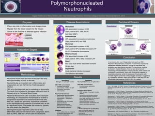
1. Purpose
• Play a key role in inflammation and phagocytosis
• Migrate from the blood vessel into the tissues
• Serve as the first line of defense against infection
Maturation Stages
Methodology
Disease Associations
Results
Peripheral Smears
Conclusion
• In conclusion, the use of diagnostics tools such as; CBC ,
Peripheral Blood Smears and Stains are extremely important to
differentiate between leukemia's, stages of maturation and
qualitative and quantitative disorders. The technician must be
proficient in the identification of normal and abnormal cell
morphology. The identification of toxic granules, Dohle bodies and
cytoplasmic vacuolization, along with neutrophilia suggests a
Quantitative disorder, while the presence of neutropenia with
Chediak Higashi syndrome or May-Hegglin anomaly is suggestive
of a qualitative disorder. Stains such as MPO, SBB and LAP can
differentiate the presence of AML or ALL in a patient.
References
Myeloid
stem
cell
Pluripotent
stem cell
IL-1
IL-3
IL-6
Quantitative
Qualitative
Quantitative
Changes in neutrophil number
CBC WBC concentration
• Increased neutrophils
(neutrophilia), “Shift to the left”
• Decreased neutrophils
(neutropenia)
Peripheral Blood Smear
• Neutrophilia-Toxic granulation,
Dohle bodies, Cytoplasmic
vacuolization→ observed in
infections
• Neutropenia-risk of increase
bacterial infection
Staphylococcus aureus,
Streptococcus viridans and
gram-negative organisms
Urinalysis
• Urine culture; look for presence
of bacteria (neutrophilia and
acquired neutropenia)
Qualitative
Neutrophil Deficiency or dysfunction
CBC WBC Concentration:
• Low-decreased neutrophils
(neutropenia)
Peripheral Blood Smear
• WBC Variable in neutrophil function
defects
• Generally unremarkable besides
with Chediak Higashi syndrome
(very large granules) and Neutrophil
specific granule-deficiency
disorders (bilobed nuclei w/
abnormal nuclear membranes and
absent secondary granules)
Stain
• Nitroblue tetrazolium dye test (NBT)-
used to indirectly detect the
production of superoxide by
neutrophils
• Myeloperoxidase stains
Polymorphonucleated
Neutrophils
Angela Thetford and Tiffany Smither | MLT 1042 Hematology| Professor Domenici| College of Southern Maryland| March 27, 2015
• Neutrophils are the most abundant leukocyte in the body
with approximately 50-70% of the cells.
• Neutrophils play an important role in inflammation and
phagocytosis and are the first line of defense against
infection.
• CBC is the first diagnostic test in evaluating an abnormality.
The result of an increased or decreased neutrophil count is
followed up by an Peripheral Blood Smear.
• Peripheral Blood Smear is the evaluation of the neutrophil
morphology. It is important for the technician to be able to
recognize the appearance of normal and abnormal cells,
their size , shape and granularity. Importance is stressed
on the identification and presence of immature cells, as this
is a significant finding. The greater the immaturity, typically,
the more severe the diagnosis.
• Staining techniques are another useful tool to help
differentiate cells and help narrow down a diagnosis. Stains
such as, MPO, SBB and LAP are used for neutrophils.
1. Margination
2. Diapedesis
3. Chemotaxis
4. Phagocytosis
http://www.ecellulitis.com/wp-content/uploads/2012/07/9160594_s.jpg?b74eb9
Myeloblast
AML associated increased in BM
Stain positive MPO, SBB, NCAE
Last blast t(8;21)
Promyelocyte
APL associated increased promyelocytes
Stain positive MPO and SBB
Myelocyte
CML associated increased in BM
Stain positive MP and SBB, Deceased LAP
t(9;22) Philadelphia chromosome
Metamyelocyte
CNL associated increase in BM
Stain positive MPO, SBB, increased LAP
Band
RA, Gout acute stress associated increase
Wright stain
Segmented
Acute bacterial infection-increased
Viral infection-decreased
• Carr, J., & Rodak, B. (2009). Section 2 Neutrophils; Section 4 Leukocytes. In Clinical
hematology atlas (4th ed., pp. 44-57;135-141;145-162). St. Louis, Mo.: Saunders
Elsevier.
• Harmening, D. (2009). Chapter 1 Morphology of Human Blood and Marrow cells:
Hematopoiesis. In Clinical hematology and fundamentals of hemostasis (5th ed., pp.
16-20; 36-38). Philadelphia, PA: F. A. Davis.
• Harmening, D. (2009). Chapter 5 Cell biology, Disorders of Neutrophils, Infectious
Mononucleosis, and Reactive Lymphocytosis. In Clinical hematology and
fundamentals of hemostasis (5th ed., pp. 306-318). Philadelphia, PA: F. A. Davis.
• Harmening, D. (2009). Chapter 16 Introduction to Leukemia and the Acute
Leukemias. In Clinical hematology and fundamentals of hemostasis (5th ed., pp. 332-
334;338-339;343-347;363). Philadelphia, PA: F. A. Davis.
• Harmening, D. (2009). Chapter 17 Chronic Myeloproliferative Disorders I: Chronic
Myelogenous Leukemia. In Clinical hematology and fundamentals of hemostasis (5th
ed., pp. 372-376; 379). Philadelphia, PA: F. A. Davis.
• Inflammation. (n.d.). Retrieved March 22, 2015, from http://ecellulitis.com
• Rashidi, H., & Nguyen, J. (n.d.). HematologyOutlines - Atlas. Retrieved March 22,
2015, from http://hematologyoutlines.com/atlas.html