SlideShare a Scribd company logo
1 of 20
Immunohistochemistry (IHC)
-SAMSUDEEN.S
Methods of protein detection
• ELISA
• Gel Electrophoresis
• Western blot
• Immunoprecipitation
• Spectrophotometry
• Enzyme assays
• X-ray crystallography
• NMR
• Immunohistochemistry
Immunohistochemistry (IHC)
Immunohistochemistry (IHC) combines
histological, immunological and biochemical
techniques for the identification of specific
tissue components by means of a specific
antigen/antibody reaction tagged with a
visible label.
IHC makes it possible to visualize the
distribution and localization of specific cellular
components within a cell or tissue.
History
• IHC takes its name from the roots "immuno",
refer to antibodies and "histo", meaning
tissue.
• Albert Coons conceptualized and first
implemented the procedure in 1941
Immunohistochemical staining
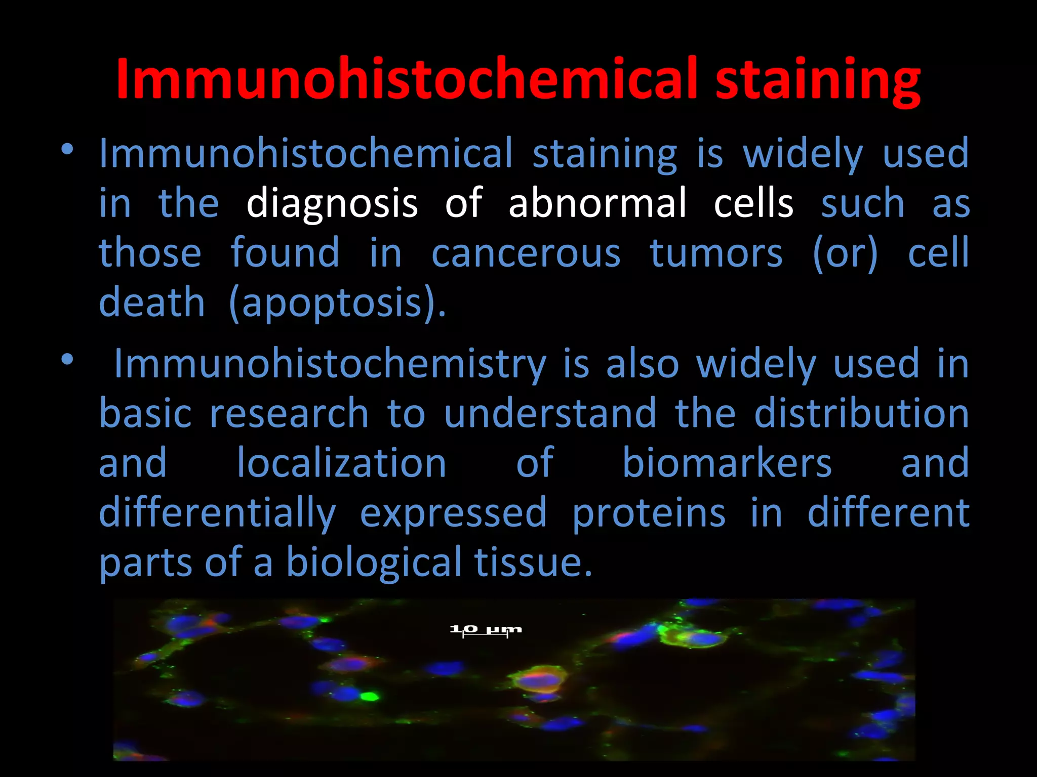
• Immunohistochemical staining is widely used
in the diagnosis of abnormal cells such as
those found in cancerous tumors (or) cell
death (apoptosis).
• Immunohistochemistry is also widely used in
basic research to understand the distribution
and localization of biomarkers and
differentially expressed proteins in different
parts of a biological tissue.
Immunohistochemical
staining
Preparation of the sample
• Preparation of the sample is critical to
maintain cell morphology, tissue architecture
and the antigenicity of target epitopes. This
requires proper tissue collection, fixation and
sectioning. A solution of paraformaldehyde is
often used to fix tissue.
Preparing tissue slices
• The tissue may then be sliced or used whole,
dependent upon the purpose of the experiment or the
tissue itself.
• Before sectioning, the tissue sample may be
embedded in a medium, like paraffin wax or
cryomedia.
• Sections can be sliced on a variety of instruments,
most commonly a microtome, cryostat, or vibratome.
• Specimens are typically sliced at a range of 3 μm-5 μm.
The slices are then mounted on slides, dehydrated
using alcohol washes of increasing concentrations
(e.g., 50%, 75%, 90%, 95%, 100%) and cleared using a
detergent like xylene before being imaged under a
microscope.
Antibody types
• The antibodies used for specific detection can
be
– monoclonal antibody.
– polyclonal antibody.
Monoclonal Antibody
• Monoclonal antibodies are antibodies that
are made by identical immune cells that are
all clones of a unique parent cell.
• Monoclonal antibodies can have monovalent
affinity in that they bind to the same epitope.
Polyclonal Antibody
• Polyclonal antibodies are antibodies that  are 
secreted by different B cell lineages within the 
body. They are a collection of immunoglobulin 
molecules that react against a specific antigen, 
each identifying a different epitope.
Sample labeling
• For  immunohistochemical  detection  strategies, 
antibodies  are  classified  as  primary  or  secondary 
reagents. 
• Primary  antibodies  are  raised  against  an  antigen  of 
interest  and  are  typically  unconjugated  (unlabeled), 
while  secondary  antibodies  are  raised  against 
immunoglobulins of the primary antibody species. 
• The  secondary  antibody  is  usually  conjugated  to  a 
linker  molecule,  such  as  biotin,  that  then  recruits 
reporter molecules, or the secondary antibody itself is 
directly bound to the reporter molecule.
Immunohistochemical reporters
• Reporter  molecules  vary  based  on  the  nature  of  the 
detection method, the most popular being chromogenic and 
fluorescence  detection  mediated  by  an  enzyme  or  a 
fluorophore respectively. 
• With chromogenic reporters, an enzyme label reacts with a 
substrate to yield an intensely colored product that can be 
analyzed with an ordinary light microscope. While the list of 
enzyme  substrates  is  extensive,  alkaline  phosphatase  (AP) 
and horseradish peroxidase (HRP) are the two enzymes used 
most extensively as labels for protein detection.
• An array of chromogenic, fluorogenic and chemiluminescent 
substrates is available for use with either enzyme, including 
DAB  (3,3'-diaminobenzidine)  which  produce  a  brown  or 
purple  staining,  respectively,  wherever  the  enzymes  are 
bound.
Target antigen detection methods
• There are 2 types of methods in targeting 
antigen in Immunohistochemical staining.
• They are : Direct method & Indirect method.
Direct method
•    The direct method is a one-step staining method 
and involves a labeled antibody reacting directly with 
the  antigen  in  tissue  sections.  While  this  technique 
utilizes  only  one  antibody  and  therefore  is  simple 
and rapid, the sensitivity is lower due to little signal 
amplification, in contrast to indirect approaches.
Indirect method
• The indirect method involves an unlabeled primary antibody
(first layer) that binds to the target antigen in the tissue and a
labeled secondary antibody (second layer) that reacts with the
primary antibody. This method is more sensitive than direct
detection strategies because of signal amplification due to the
binding of several secondary antibodies to each primary
antibody if the secondary antibody is conjugated to the
fluorescent (or) enzyme Reporter.
Counterstains
• After immunohistochemical staining of the target
antigen, a second stain is often applied to provide
contrast that helps the primary stain stand out. Many
of these stains show specificity for specific classes of
biomolecules, while others will stain the whole cell.
• Both chromogenic and fluorescent dyes are available
for IHC to provide a vast array of reagents to fit every
experimental design, and include: hematoxylin,
Hoechst stain and DAPI (4',6-diamidino-2-
phenylindole) are commonly used.
• Hoechst can be used to stain living cells as it can pass
the cell membrane, whereas DAPI can't .
Examples of some commonly used markers include:
BrdU: used to identify replicating cells. Used to identify tumors
as well as in neuroscience research.
Cytokeratins: used for identification of carcinomas but may
also be expressed in some sarcomas.
CD15 and CD30 : used for Hodgkin's disease
Alpha fetoprotein: for yolk sac tumors and hepatocellular
carcinoma
CD117 (KIT): for gastrointestinal stromal tumors (GIST) and
mast cell tumors
CD10 (CALLA): for renal cell carcinoma and acute lymphoblastic
leukemia
Prostate specific antigen (PSA): for prostate cancer estrogens
and progesterone receptor (ER & PR) staining are used both diagnostically
(breast and Gynecologic tumors) as well as prognostic in breast cancer and
predictive of response to therapy (estrogen receptor)
CD20 identification of B-cell lymphomas
CD3 identification of T-cell lymphomas
Mapping protein expression
• Immunohistochemistry can also be used for a
more general protein profiling. The Human
Protein Atlas displays a map of protein
expression in normal human organs and tissues
and organs. The combination of
immunohistochemistry and tissue microarrays
provides protein expression patterns in a large
number of different tissue types.
• Immunohistochemistry is also used for protein
profiling in the most common forms of human
cancer.
Immunohistochemistry

More Related Content

What's hot (20)

Immunohistochemistry
ImmunohistochemistryImmunohistochemistry
Immunohistochemistry
 
Enzyme histochemistry
Enzyme histochemistryEnzyme histochemistry
Enzyme histochemistry
 
Immunohistochemistry
Immunohistochemistry Immunohistochemistry
Immunohistochemistry
 
1378968135.74359 immunohistochemistry
1378968135.74359  immunohistochemistry1378968135.74359  immunohistochemistry
1378968135.74359 immunohistochemistry
 
principle of Immunohistochemistry and its use in diagnostics
principle of Immunohistochemistry and its use in diagnosticsprinciple of Immunohistochemistry and its use in diagnostics
principle of Immunohistochemistry and its use in diagnostics
 
immunohistochemistry
immunohistochemistryimmunohistochemistry
immunohistochemistry
 
Special stain in histopathology
Special stain in histopathologySpecial stain in histopathology
Special stain in histopathology
 
Fixation of tissues
Fixation of tissuesFixation of tissues
Fixation of tissues
 
Immunofluorescence
ImmunofluorescenceImmunofluorescence
Immunofluorescence
 
Immunocytochemistry Protocol
Immunocytochemistry ProtocolImmunocytochemistry Protocol
Immunocytochemistry Protocol
 
Cancer immunology
Cancer immunologyCancer immunology
Cancer immunology
 
Immunohistochemistry
ImmunohistochemistryImmunohistochemistry
Immunohistochemistry
 
Immunofluorescence
ImmunofluorescenceImmunofluorescence
Immunofluorescence
 
CYTOLOGY PPT.pptx
CYTOLOGY PPT.pptxCYTOLOGY PPT.pptx
CYTOLOGY PPT.pptx
 
8 kirti staining
8  kirti staining8  kirti staining
8 kirti staining
 
Molecular diagnostic techniques
Molecular diagnostic techniquesMolecular diagnostic techniques
Molecular diagnostic techniques
 
Cryostat and frozen section
Cryostat and frozen sectionCryostat and frozen section
Cryostat and frozen section
 
Cell block
Cell blockCell block
Cell block
 
Flow cytometry
Flow cytometryFlow cytometry
Flow cytometry
 
Counter immunoelectrophoresis
Counter immunoelectrophoresisCounter immunoelectrophoresis
Counter immunoelectrophoresis
 

Similar to Immunohistochemistry

IHC.pptx
IHC.pptxIHC.pptx
IHC.pptxHaail
 
Huzaifa umar (biomedical science and instrumentation presentation)
Huzaifa umar (biomedical science and instrumentation presentation)Huzaifa umar (biomedical science and instrumentation presentation)
Huzaifa umar (biomedical science and instrumentation presentation)Huzaifa Umar
 
Antibody production.pptx
Antibody production.pptxAntibody production.pptx
Antibody production.pptxAlphoncePancras
 
MONOCLONAL ANTIBODY.pdf
MONOCLONAL ANTIBODY.pdfMONOCLONAL ANTIBODY.pdf
MONOCLONAL ANTIBODY.pdfRoop
 
Monoclonal antibody production by hybridoma technology
Monoclonal antibody production by hybridoma technologyMonoclonal antibody production by hybridoma technology
Monoclonal antibody production by hybridoma technologyHasnat Tariq
 
BTEC849 Lecture slides.ppt
BTEC849 Lecture slides.pptBTEC849 Lecture slides.ppt
BTEC849 Lecture slides.pptMuhammad574698
 
Monoclonal Antibodies Hybridoma Technique
Monoclonal Antibodies Hybridoma Technique  Monoclonal Antibodies Hybridoma Technique
Monoclonal Antibodies Hybridoma Technique Iqbal Ahmed Rana
 
monoclonalantibodies.pdf
monoclonalantibodies.pdfmonoclonalantibodies.pdf
monoclonalantibodies.pdfMaryamUmar33
 
Immunoflowcytometry, the basics and applications.pptx
Immunoflowcytometry, the basics and applications.pptxImmunoflowcytometry, the basics and applications.pptx
Immunoflowcytometry, the basics and applications.pptxAmirRaziq1
 
Hybridoma technology and production of monoclonal antibody
Hybridoma technology and production of monoclonal antibodyHybridoma technology and production of monoclonal antibody
Hybridoma technology and production of monoclonal antibodyRajpal Choudhary
 

Similar to Immunohistochemistry (20)

Immunohistochemistry (IHC)
Immunohistochemistry (IHC)Immunohistochemistry (IHC)
Immunohistochemistry (IHC)
 
IHC.pptx
IHC.pptxIHC.pptx
IHC.pptx
 
mono clonals.pptx
mono clonals.pptxmono clonals.pptx
mono clonals.pptx
 
Huzaifa umar (biomedical science and instrumentation presentation)
Huzaifa umar (biomedical science and instrumentation presentation)Huzaifa umar (biomedical science and instrumentation presentation)
Huzaifa umar (biomedical science and instrumentation presentation)
 
Antibody production.pptx
Antibody production.pptxAntibody production.pptx
Antibody production.pptx
 
MONOCLONAL ANTIBODY.pdf
MONOCLONAL ANTIBODY.pdfMONOCLONAL ANTIBODY.pdf
MONOCLONAL ANTIBODY.pdf
 
Monoclonal antibody production by hybridoma technology
Monoclonal antibody production by hybridoma technologyMonoclonal antibody production by hybridoma technology
Monoclonal antibody production by hybridoma technology
 
Immunohistochemistry
ImmunohistochemistryImmunohistochemistry
Immunohistochemistry
 
Monoclonal Antibodies
Monoclonal Antibodies Monoclonal Antibodies
Monoclonal Antibodies
 
BTEC849 Lecture slides.ppt
BTEC849 Lecture slides.pptBTEC849 Lecture slides.ppt
BTEC849 Lecture slides.ppt
 
Hybridoma
HybridomaHybridoma
Hybridoma
 
Monoclonal Antibodies Hybridoma Technique
Monoclonal Antibodies Hybridoma Technique  Monoclonal Antibodies Hybridoma Technique
Monoclonal Antibodies Hybridoma Technique
 
Monoclonal antibody
Monoclonal antibodyMonoclonal antibody
Monoclonal antibody
 
Flow cytometry
Flow cytometryFlow cytometry
Flow cytometry
 
Monoclonal antibodies dr. asm
Monoclonal antibodies dr. asmMonoclonal antibodies dr. asm
Monoclonal antibodies dr. asm
 
Monoclonal antibodies
Monoclonal antibodiesMonoclonal antibodies
Monoclonal antibodies
 
monoclonalantibodies.pdf
monoclonalantibodies.pdfmonoclonalantibodies.pdf
monoclonalantibodies.pdf
 
Immunoflowcytometry, the basics and applications.pptx
Immunoflowcytometry, the basics and applications.pptxImmunoflowcytometry, the basics and applications.pptx
Immunoflowcytometry, the basics and applications.pptx
 
Monoclonal antibody
Monoclonal antibodyMonoclonal antibody
Monoclonal antibody
 
Hybridoma technology and production of monoclonal antibody
Hybridoma technology and production of monoclonal antibodyHybridoma technology and production of monoclonal antibody
Hybridoma technology and production of monoclonal antibody
 

More from SamsuDeen12

Most probable number or multiple tube fermentation technique
Most probable number or multiple tube fermentation techniqueMost probable number or multiple tube fermentation technique
Most probable number or multiple tube fermentation techniqueSamsuDeen12
 
Production of tetracyclin and cephalosporin
Production of tetracyclin and cephalosporinProduction of tetracyclin and cephalosporin
Production of tetracyclin and cephalosporinSamsuDeen12
 
Products of fish
Products of fishProducts of fish
Products of fishSamsuDeen12
 

More from SamsuDeen12 (10)

Most probable number or multiple tube fermentation technique
Most probable number or multiple tube fermentation techniqueMost probable number or multiple tube fermentation technique
Most probable number or multiple tube fermentation technique
 
Herpesviruses
HerpesvirusesHerpesviruses
Herpesviruses
 
cDNA Library
cDNA LibrarycDNA Library
cDNA Library
 
Production of tetracyclin and cephalosporin
Production of tetracyclin and cephalosporinProduction of tetracyclin and cephalosporin
Production of tetracyclin and cephalosporin
 
Products of fish
Products of fishProducts of fish
Products of fish
 
Paramecium
ParameciumParamecium
Paramecium
 
Tuberculosis
TuberculosisTuberculosis
Tuberculosis
 
Crossing over
Crossing overCrossing over
Crossing over
 
Sage
SageSage
Sage
 
Mitosis
MitosisMitosis
Mitosis
 

Recently uploaded

Broad bean, Lima Bean, Jack bean, Ullucus.pptx
Broad bean, Lima Bean, Jack bean, Ullucus.pptxBroad bean, Lima Bean, Jack bean, Ullucus.pptx
Broad bean, Lima Bean, Jack bean, Ullucus.pptxjana861314
 
Isotopic evidence of long-lived volcanism on Io
Isotopic evidence of long-lived volcanism on IoIsotopic evidence of long-lived volcanism on Io
Isotopic evidence of long-lived volcanism on IoSérgio Sacani
 
PossibleEoarcheanRecordsoftheGeomagneticFieldPreservedintheIsuaSupracrustalBe...
PossibleEoarcheanRecordsoftheGeomagneticFieldPreservedintheIsuaSupracrustalBe...PossibleEoarcheanRecordsoftheGeomagneticFieldPreservedintheIsuaSupracrustalBe...
PossibleEoarcheanRecordsoftheGeomagneticFieldPreservedintheIsuaSupracrustalBe...Sérgio Sacani
 
Biological Classification BioHack (3).pdf
Biological Classification BioHack (3).pdfBiological Classification BioHack (3).pdf
Biological Classification BioHack (3).pdfmuntazimhurra
 
Botany 4th semester file By Sumit Kumar yadav.pdf
Botany 4th semester file By Sumit Kumar yadav.pdfBotany 4th semester file By Sumit Kumar yadav.pdf
Botany 4th semester file By Sumit Kumar yadav.pdfSumit Kumar yadav
 
Recombinant DNA technology (Immunological screening)
Recombinant DNA technology (Immunological screening)Recombinant DNA technology (Immunological screening)
Recombinant DNA technology (Immunological screening)PraveenaKalaiselvan1
 
GFP in rDNA Technology (Biotechnology).pptx
GFP in rDNA Technology (Biotechnology).pptxGFP in rDNA Technology (Biotechnology).pptx
GFP in rDNA Technology (Biotechnology).pptxAleenaTreesaSaji
 
Artificial Intelligence In Microbiology by Dr. Prince C P
Artificial Intelligence In Microbiology by Dr. Prince C PArtificial Intelligence In Microbiology by Dr. Prince C P
Artificial Intelligence In Microbiology by Dr. Prince C PPRINCE C P
 
Hubble Asteroid Hunter III. Physical properties of newly found asteroids
Hubble Asteroid Hunter III. Physical properties of newly found asteroidsHubble Asteroid Hunter III. Physical properties of newly found asteroids
Hubble Asteroid Hunter III. Physical properties of newly found asteroidsSérgio Sacani
 
Formation of low mass protostars and their circumstellar disks
Formation of low mass protostars and their circumstellar disksFormation of low mass protostars and their circumstellar disks
Formation of low mass protostars and their circumstellar disksSérgio Sacani
 
Boyles law module in the grade 10 science
Boyles law module in the grade 10 scienceBoyles law module in the grade 10 science
Boyles law module in the grade 10 sciencefloriejanemacaya1
 
Raman spectroscopy.pptx M Pharm, M Sc, Advanced Spectral Analysis
Raman spectroscopy.pptx M Pharm, M Sc, Advanced Spectral AnalysisRaman spectroscopy.pptx M Pharm, M Sc, Advanced Spectral Analysis
Raman spectroscopy.pptx M Pharm, M Sc, Advanced Spectral AnalysisDiwakar Mishra
 
Natural Polymer Based Nanomaterials
Natural Polymer Based NanomaterialsNatural Polymer Based Nanomaterials
Natural Polymer Based NanomaterialsAArockiyaNisha
 
Animal Communication- Auditory and Visual.pptx
Animal Communication- Auditory and Visual.pptxAnimal Communication- Auditory and Visual.pptx
Animal Communication- Auditory and Visual.pptxUmerFayaz5
 
TEST BANK For Radiologic Science for Technologists, 12th Edition by Stewart C...
TEST BANK For Radiologic Science for Technologists, 12th Edition by Stewart C...TEST BANK For Radiologic Science for Technologists, 12th Edition by Stewart C...
TEST BANK For Radiologic Science for Technologists, 12th Edition by Stewart C...ssifa0344
 
Green chemistry and Sustainable development.pptx
Green chemistry  and Sustainable development.pptxGreen chemistry  and Sustainable development.pptx
Green chemistry and Sustainable development.pptxRajatChauhan518211
 
Pests of cotton_Sucking_Pests_Dr.UPR.pdf
Pests of cotton_Sucking_Pests_Dr.UPR.pdfPests of cotton_Sucking_Pests_Dr.UPR.pdf
Pests of cotton_Sucking_Pests_Dr.UPR.pdfPirithiRaju
 
Call Us ≽ 9953322196 ≼ Call Girls In Mukherjee Nagar(Delhi) |
Call Us ≽ 9953322196 ≼ Call Girls In Mukherjee Nagar(Delhi) |Call Us ≽ 9953322196 ≼ Call Girls In Mukherjee Nagar(Delhi) |
Call Us ≽ 9953322196 ≼ Call Girls In Mukherjee Nagar(Delhi) |aasikanpl
 

Recently uploaded (20)

Broad bean, Lima Bean, Jack bean, Ullucus.pptx
Broad bean, Lima Bean, Jack bean, Ullucus.pptxBroad bean, Lima Bean, Jack bean, Ullucus.pptx
Broad bean, Lima Bean, Jack bean, Ullucus.pptx
 
Isotopic evidence of long-lived volcanism on Io
Isotopic evidence of long-lived volcanism on IoIsotopic evidence of long-lived volcanism on Io
Isotopic evidence of long-lived volcanism on Io
 
PossibleEoarcheanRecordsoftheGeomagneticFieldPreservedintheIsuaSupracrustalBe...
PossibleEoarcheanRecordsoftheGeomagneticFieldPreservedintheIsuaSupracrustalBe...PossibleEoarcheanRecordsoftheGeomagneticFieldPreservedintheIsuaSupracrustalBe...
PossibleEoarcheanRecordsoftheGeomagneticFieldPreservedintheIsuaSupracrustalBe...
 
9953056974 Young Call Girls In Mahavir enclave Indian Quality Escort service
9953056974 Young Call Girls In Mahavir enclave Indian Quality Escort service9953056974 Young Call Girls In Mahavir enclave Indian Quality Escort service
9953056974 Young Call Girls In Mahavir enclave Indian Quality Escort service
 
Biological Classification BioHack (3).pdf
Biological Classification BioHack (3).pdfBiological Classification BioHack (3).pdf
Biological Classification BioHack (3).pdf
 
Botany 4th semester file By Sumit Kumar yadav.pdf
Botany 4th semester file By Sumit Kumar yadav.pdfBotany 4th semester file By Sumit Kumar yadav.pdf
Botany 4th semester file By Sumit Kumar yadav.pdf
 
Recombinant DNA technology (Immunological screening)
Recombinant DNA technology (Immunological screening)Recombinant DNA technology (Immunological screening)
Recombinant DNA technology (Immunological screening)
 
GFP in rDNA Technology (Biotechnology).pptx
GFP in rDNA Technology (Biotechnology).pptxGFP in rDNA Technology (Biotechnology).pptx
GFP in rDNA Technology (Biotechnology).pptx
 
Artificial Intelligence In Microbiology by Dr. Prince C P
Artificial Intelligence In Microbiology by Dr. Prince C PArtificial Intelligence In Microbiology by Dr. Prince C P
Artificial Intelligence In Microbiology by Dr. Prince C P
 
Hubble Asteroid Hunter III. Physical properties of newly found asteroids
Hubble Asteroid Hunter III. Physical properties of newly found asteroidsHubble Asteroid Hunter III. Physical properties of newly found asteroids
Hubble Asteroid Hunter III. Physical properties of newly found asteroids
 
Formation of low mass protostars and their circumstellar disks
Formation of low mass protostars and their circumstellar disksFormation of low mass protostars and their circumstellar disks
Formation of low mass protostars and their circumstellar disks
 
Boyles law module in the grade 10 science
Boyles law module in the grade 10 scienceBoyles law module in the grade 10 science
Boyles law module in the grade 10 science
 
Raman spectroscopy.pptx M Pharm, M Sc, Advanced Spectral Analysis
Raman spectroscopy.pptx M Pharm, M Sc, Advanced Spectral AnalysisRaman spectroscopy.pptx M Pharm, M Sc, Advanced Spectral Analysis
Raman spectroscopy.pptx M Pharm, M Sc, Advanced Spectral Analysis
 
Natural Polymer Based Nanomaterials
Natural Polymer Based NanomaterialsNatural Polymer Based Nanomaterials
Natural Polymer Based Nanomaterials
 
Animal Communication- Auditory and Visual.pptx
Animal Communication- Auditory and Visual.pptxAnimal Communication- Auditory and Visual.pptx
Animal Communication- Auditory and Visual.pptx
 
TEST BANK For Radiologic Science for Technologists, 12th Edition by Stewart C...
TEST BANK For Radiologic Science for Technologists, 12th Edition by Stewart C...TEST BANK For Radiologic Science for Technologists, 12th Edition by Stewart C...
TEST BANK For Radiologic Science for Technologists, 12th Edition by Stewart C...
 
Green chemistry and Sustainable development.pptx
Green chemistry  and Sustainable development.pptxGreen chemistry  and Sustainable development.pptx
Green chemistry and Sustainable development.pptx
 
Pests of cotton_Sucking_Pests_Dr.UPR.pdf
Pests of cotton_Sucking_Pests_Dr.UPR.pdfPests of cotton_Sucking_Pests_Dr.UPR.pdf
Pests of cotton_Sucking_Pests_Dr.UPR.pdf
 
Call Us ≽ 9953322196 ≼ Call Girls In Mukherjee Nagar(Delhi) |
Call Us ≽ 9953322196 ≼ Call Girls In Mukherjee Nagar(Delhi) |Call Us ≽ 9953322196 ≼ Call Girls In Mukherjee Nagar(Delhi) |
Call Us ≽ 9953322196 ≼ Call Girls In Mukherjee Nagar(Delhi) |
 
CELL -Structural and Functional unit of life.pdf
CELL -Structural and Functional unit of life.pdfCELL -Structural and Functional unit of life.pdf
CELL -Structural and Functional unit of life.pdf
 

Immunohistochemistry

  • 2. Methods of protein detection • ELISA • Gel Electrophoresis • Western blot • Immunoprecipitation • Spectrophotometry • Enzyme assays • X-ray crystallography • NMR • Immunohistochemistry
  • 3. Immunohistochemistry (IHC) Immunohistochemistry (IHC) combines histological, immunological and biochemical techniques for the identification of specific tissue components by means of a specific antigen/antibody reaction tagged with a visible label. IHC makes it possible to visualize the distribution and localization of specific cellular components within a cell or tissue.
  • 4. History • IHC takes its name from the roots "immuno", refer to antibodies and "histo", meaning tissue. • Albert Coons conceptualized and first implemented the procedure in 1941
  • 5. Immunohistochemical staining • Immunohistochemical staining is widely used in the diagnosis of abnormal cells such as those found in cancerous tumors (or) cell death (apoptosis). • Immunohistochemistry is also widely used in basic research to understand the distribution and localization of biomarkers and differentially expressed proteins in different parts of a biological tissue.
  • 7. Preparation of the sample • Preparation of the sample is critical to maintain cell morphology, tissue architecture and the antigenicity of target epitopes. This requires proper tissue collection, fixation and sectioning. A solution of paraformaldehyde is often used to fix tissue.
  • 8. Preparing tissue slices • The tissue may then be sliced or used whole, dependent upon the purpose of the experiment or the tissue itself. • Before sectioning, the tissue sample may be embedded in a medium, like paraffin wax or cryomedia. • Sections can be sliced on a variety of instruments, most commonly a microtome, cryostat, or vibratome. • Specimens are typically sliced at a range of 3 μm-5 μm. The slices are then mounted on slides, dehydrated using alcohol washes of increasing concentrations (e.g., 50%, 75%, 90%, 95%, 100%) and cleared using a detergent like xylene before being imaged under a microscope.
  • 9. Antibody types • The antibodies used for specific detection can be – monoclonal antibody. – polyclonal antibody.
  • 10. Monoclonal Antibody • Monoclonal antibodies are antibodies that are made by identical immune cells that are all clones of a unique parent cell. • Monoclonal antibodies can have monovalent affinity in that they bind to the same epitope.
  • 11. Polyclonal Antibody • Polyclonal antibodies are antibodies that  are  secreted by different B cell lineages within the  body. They are a collection of immunoglobulin  molecules that react against a specific antigen,  each identifying a different epitope.
  • 12. Sample labeling • For  immunohistochemical  detection  strategies,  antibodies  are  classified  as  primary  or  secondary  reagents.  • Primary  antibodies  are  raised  against  an  antigen  of  interest  and  are  typically  unconjugated  (unlabeled),  while  secondary  antibodies  are  raised  against  immunoglobulins of the primary antibody species.  • The  secondary  antibody  is  usually  conjugated  to  a  linker  molecule,  such  as  biotin,  that  then  recruits  reporter molecules, or the secondary antibody itself is  directly bound to the reporter molecule.
  • 13. Immunohistochemical reporters • Reporter  molecules  vary  based  on  the  nature  of  the  detection method, the most popular being chromogenic and  fluorescence  detection  mediated  by  an  enzyme  or  a  fluorophore respectively.  • With chromogenic reporters, an enzyme label reacts with a  substrate to yield an intensely colored product that can be  analyzed with an ordinary light microscope. While the list of  enzyme  substrates  is  extensive,  alkaline  phosphatase  (AP)  and horseradish peroxidase (HRP) are the two enzymes used  most extensively as labels for protein detection. • An array of chromogenic, fluorogenic and chemiluminescent  substrates is available for use with either enzyme, including  DAB  (3,3'-diaminobenzidine)  which  produce  a  brown  or  purple  staining,  respectively,  wherever  the  enzymes  are  bound.
  • 14. Target antigen detection methods • There are 2 types of methods in targeting  antigen in Immunohistochemical staining. • They are : Direct method & Indirect method.
  • 15. Direct method •    The direct method is a one-step staining method  and involves a labeled antibody reacting directly with  the  antigen  in  tissue  sections.  While  this  technique  utilizes  only  one  antibody  and  therefore  is  simple  and rapid, the sensitivity is lower due to little signal  amplification, in contrast to indirect approaches.
  • 16. Indirect method • The indirect method involves an unlabeled primary antibody (first layer) that binds to the target antigen in the tissue and a labeled secondary antibody (second layer) that reacts with the primary antibody. This method is more sensitive than direct detection strategies because of signal amplification due to the binding of several secondary antibodies to each primary antibody if the secondary antibody is conjugated to the fluorescent (or) enzyme Reporter.
  • 17. Counterstains • After immunohistochemical staining of the target antigen, a second stain is often applied to provide contrast that helps the primary stain stand out. Many of these stains show specificity for specific classes of biomolecules, while others will stain the whole cell. • Both chromogenic and fluorescent dyes are available for IHC to provide a vast array of reagents to fit every experimental design, and include: hematoxylin, Hoechst stain and DAPI (4',6-diamidino-2- phenylindole) are commonly used. • Hoechst can be used to stain living cells as it can pass the cell membrane, whereas DAPI can't .
  • 18. Examples of some commonly used markers include: BrdU: used to identify replicating cells. Used to identify tumors as well as in neuroscience research. Cytokeratins: used for identification of carcinomas but may also be expressed in some sarcomas. CD15 and CD30 : used for Hodgkin's disease Alpha fetoprotein: for yolk sac tumors and hepatocellular carcinoma CD117 (KIT): for gastrointestinal stromal tumors (GIST) and mast cell tumors CD10 (CALLA): for renal cell carcinoma and acute lymphoblastic leukemia Prostate specific antigen (PSA): for prostate cancer estrogens and progesterone receptor (ER & PR) staining are used both diagnostically (breast and Gynecologic tumors) as well as prognostic in breast cancer and predictive of response to therapy (estrogen receptor) CD20 identification of B-cell lymphomas CD3 identification of T-cell lymphomas
  • 19. Mapping protein expression • Immunohistochemistry can also be used for a more general protein profiling. The Human Protein Atlas displays a map of protein expression in normal human organs and tissues and organs. The combination of immunohistochemistry and tissue microarrays provides protein expression patterns in a large number of different tissue types. • Immunohistochemistry is also used for protein profiling in the most common forms of human cancer.