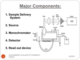
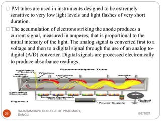
Flame photometry is a technique used to determine the concentration of certain metal ions in a sample by measuring the intensity of light emitted from their atoms when excited in a flame. The sample is nebulized and introduced into a flame, where the metal atoms are excited and emit light of characteristic wavelengths. This emitted light is separated into its component wavelengths by a monochromator and measured with a detector, allowing quantification of metal ion concentrations in the original sample. Potential sources of interference include overlap of emission lines from different elements and effects of high ion concentrations on atom excitation.
Introduction
working principle
fragmentation process
general rules for fragmentation
general modes of fragmentation
metastable ions
isotopic peaks
applications
Unlike a spectrometer (which is any instrument that can measure the
properties of light over a range of wavelengths), a spectrophotometer
measures only the intensity of light as a function of its wavelength.
Interfaces in chromatography [LC-MS, GC-MS, HPTLC, LC, GC]Shikha Popali
THE INTERFACES OF CHROMATOGRAPHY INCLUDES THE CHROMATOGRAPHY CRITEREA WHERE THE DIFFERENT CHROMATOGRAPHY ARE EXPLAINED IN DETAIL WITH PRACTICAL EXAMPLES AND THEIR IMAGES.
Pharmaceuticals: In some pharmaceutical manufacturing processes, minute quantities of a catalyst used in the process (usually a metal) are sometimes present in the final product. By using AAS the amount of catalyst present can be determined.
Atomic absorption spectroscopy (AAS) and atomic emission spectroscopy (AES) is a spectro analytical procedure for the quantitative determination of chemical elements by free atoms in the gaseous state.
Atomic absorption spectroscopy is based on absorption of light by free metallic ions.
In analytical chemistry the technique is used for determining the concentration of a particular element (the analyte) in a sample to be analyzed. AAS can be used to determine over 70 different elements in solution, or directly in solid samples via electrothermal vaporization
Atomic absorption spectrometry (AAS) is an analytical technique that measures the concentrations of elements.
Atomic absorption is so sensitive that it can measure down to parts per billion of a gram (µg dm–3 ) in a sample.
The technique makes use of the wavelengths of light specifically absorbed by an element. They correspond to the energies needed to promote electrons from one energy level to another, higher, energy level.
Atomic absorption spectrometry has many uses in different areas of chemistry.
Clinical analysis : Analysing metals in biological fluids such as blood and urine.
Environmental analysis: Monitoring our environment – eg finding out the levels of various elements in rivers, seawater, drinking water, air, petrol and drinks such as wine, beer and fruit drinks.
The technique makes use of the atomic absorption spectrum of a sample in order to assess the concentration of specific analytes within it. It requires standards with known analyte content to establish the relation between the measured absorbance and the analyte concentration and relies therefore on the [Beer–Lambert law].
The electrons within an atom exist at various energy levels. When the atom is exposed to its own unique wavelength, it can absorb the energy (photons) and electrons move from a ground state to excited states.
The radiant energy absorbed by the electrons is directly related to the transition that occurs during this process.
Furthermore, since the electronic structure of every element is unique, the radiation absorbed represents a unique property of each individual element and it can be measured.
An atomic absorption spectrometer uses these basic principles and applies them in practical quantitative analysis
A typical atomic absorption spectrometer consists of four main components:
Atomization
Light source,
Atomization system,
Monochromator &
Detection system
Atomization can be carried out either by a flame or furnace.
Heat energy is utilized in atomic absorption spectroscopy to convert metallic elements to atomic dissociated vapor.
The temperature should be controlled very carefully for the conversion of atomic vapor.
At too high temperatures, atoms
a brief discussion of AAS, an analytical technique use for heavy metal analysis. Atomic absorption spectroscopy is a quantitative method of analysis of any kind of sample; that is applicable to many metals
AAS can be used to determine over 70 different elements in solution, or directly in solid samples via electro thermal vaporization.
Atomic Absorption Spectroscopy is a very common technique for detecting metals and metalloids in samples.
It is very reliable and simple to use.
It also measures the concentration of metals in the sample.
Atomic Absorption Spectroscopy is an analytical technique that measures the concentration of an element by measuring the amount of light that is absorbed at a characteristic wavelength when it passes through cloud of atoms
As the number of atoms in the light path increases, the amount of light absorbed increases.
Applications: Presence of metals as an impurity or in alloys can be perform.
Level of metals could be detected in tissue samples like Aluminum in blood and Copper in brain tissues.
Due to wear and tear there are different sorts of metals which are given in the lubrication oils which could be determined for the analysis of conditions of machines.
Determination of elements in the agricultural samples.
Water sample analysis (e.g. Ca, Mg, Fe, Si, Al, Ba content).
Food sample analysis.
Analysis of animal feedstuffs (e.g. Mn, Fe, Cu, Cr, Se, Zn).
Analysis of additives in lubricating oils and greases (Ba, Ca, Na, Li, Zn, Mg). analysis of soils.
Clinical sample analysis (blood samples: whole blood, plasma, serum; Ca, Mg, Li, Na, K, Fe).
Analysis of Environmental samples such as- drinking water, ocean water, soil.
Pharmaceutical sample Analysis: Estimation of zinc in insulin preparation, calcium in calcium salt is done by using AAS. Principle: The sample, in solution, is aspirated as a spray into a chamber, where it is mixed with air and fuel.
This mixture passes through baffles, here large drops fall and are drained off. Only fine droplets reach the flame.
Light from the hollow-cathode lamp passes through the sample of ground-state atoms in the flame.
The amount of light absorbed is proportional to the concentration.
The element being determined must be reduced to the elemental state, vaporized, and imposed in the beam of the radiation in the source.
When a ground-state atom absorbs light energy, an excited atom is produced.
The excited atom then returns to the ground state, emitting light of the same energy as it absorbed.
The flame sample thus contains a dynamic population of ground-state and excited atoms, both absorbing and emitting radiant energy. The emitted energy from the flame will go in all directions, and it will be a steady emission.
Because the purpose of the instrument is to measure the amount of light absorbed, the light detector must be able to distinguish between the light beam emitted by the hollow cathode lamp and that emitted by excited atoms in the flame.
A presentation containing the Principle, shematic diagram, omponents of the instrument, working of the instrument, application, advantages and disadvantages of the instrument.
Atomic absorption spectroscopy is a method of elemental analysis. It is particularly useful for determining trace metals in liquids and is most independent of molecular form of the metal in sample.
Slide 1: Title Slide
Extrachromosomal Inheritance
Slide 2: Introduction to Extrachromosomal Inheritance
Definition: Extrachromosomal inheritance refers to the transmission of genetic material that is not found within the nucleus.
Key Components: Involves genes located in mitochondria, chloroplasts, and plasmids.
Slide 3: Mitochondrial Inheritance
Mitochondria: Organelles responsible for energy production.
Mitochondrial DNA (mtDNA): Circular DNA molecule found in mitochondria.
Inheritance Pattern: Maternally inherited, meaning it is passed from mothers to all their offspring.
Diseases: Examples include Leber’s hereditary optic neuropathy (LHON) and mitochondrial myopathy.
Slide 4: Chloroplast Inheritance
Chloroplasts: Organelles responsible for photosynthesis in plants.
Chloroplast DNA (cpDNA): Circular DNA molecule found in chloroplasts.
Inheritance Pattern: Often maternally inherited in most plants, but can vary in some species.
Examples: Variegation in plants, where leaf color patterns are determined by chloroplast DNA.
Slide 5: Plasmid Inheritance
Plasmids: Small, circular DNA molecules found in bacteria and some eukaryotes.
Features: Can carry antibiotic resistance genes and can be transferred between cells through processes like conjugation.
Significance: Important in biotechnology for gene cloning and genetic engineering.
Slide 6: Mechanisms of Extrachromosomal Inheritance
Non-Mendelian Patterns: Do not follow Mendel’s laws of inheritance.
Cytoplasmic Segregation: During cell division, organelles like mitochondria and chloroplasts are randomly distributed to daughter cells.
Heteroplasmy: Presence of more than one type of organellar genome within a cell, leading to variation in expression.
Slide 7: Examples of Extrachromosomal Inheritance
Four O’clock Plant (Mirabilis jalapa): Shows variegated leaves due to different cpDNA in leaf cells.
Petite Mutants in Yeast: Result from mutations in mitochondrial DNA affecting respiration.
Slide 8: Importance of Extrachromosomal Inheritance
Evolution: Provides insight into the evolution of eukaryotic cells.
Medicine: Understanding mitochondrial inheritance helps in diagnosing and treating mitochondrial diseases.
Agriculture: Chloroplast inheritance can be used in plant breeding and genetic modification.
Slide 9: Recent Research and Advances
Gene Editing: Techniques like CRISPR-Cas9 are being used to edit mitochondrial and chloroplast DNA.
Therapies: Development of mitochondrial replacement therapy (MRT) for preventing mitochondrial diseases.
Slide 10: Conclusion
Summary: Extrachromosomal inheritance involves the transmission of genetic material outside the nucleus and plays a crucial role in genetics, medicine, and biotechnology.
Future Directions: Continued research and technological advancements hold promise for new treatments and applications.
Slide 11: Questions and Discussion
Invite Audience: Open the floor for any questions or further discussion on the topic.
Cancer cell metabolism: special Reference to Lactate PathwayAADYARAJPANDEY1
Normal Cell Metabolism:
Cellular respiration describes the series of steps that cells use to break down sugar and other chemicals to get the energy we need to function.
Energy is stored in the bonds of glucose and when glucose is broken down, much of that energy is released.
Cell utilize energy in the form of ATP.
The first step of respiration is called glycolysis. In a series of steps, glycolysis breaks glucose into two smaller molecules - a chemical called pyruvate. A small amount of ATP is formed during this process.
Most healthy cells continue the breakdown in a second process, called the Kreb's cycle. The Kreb's cycle allows cells to “burn” the pyruvates made in glycolysis to get more ATP.
The last step in the breakdown of glucose is called oxidative phosphorylation (Ox-Phos).
It takes place in specialized cell structures called mitochondria. This process produces a large amount of ATP. Importantly, cells need oxygen to complete oxidative phosphorylation.
If a cell completes only glycolysis, only 2 molecules of ATP are made per glucose. However, if the cell completes the entire respiration process (glycolysis - Kreb's - oxidative phosphorylation), about 36 molecules of ATP are created, giving it much more energy to use.
IN CANCER CELL:
Unlike healthy cells that "burn" the entire molecule of sugar to capture a large amount of energy as ATP, cancer cells are wasteful.
Cancer cells only partially break down sugar molecules. They overuse the first step of respiration, glycolysis. They frequently do not complete the second step, oxidative phosphorylation.
This results in only 2 molecules of ATP per each glucose molecule instead of the 36 or so ATPs healthy cells gain. As a result, cancer cells need to use a lot more sugar molecules to get enough energy to survive.
introduction to WARBERG PHENOMENA:
WARBURG EFFECT Usually, cancer cells are highly glycolytic (glucose addiction) and take up more glucose than do normal cells from outside.
Otto Heinrich Warburg (; 8 October 1883 – 1 August 1970) In 1931 was awarded the Nobel Prize in Physiology for his "discovery of the nature and mode of action of the respiratory enzyme.
WARNBURG EFFECT : cancer cells under aerobic (well-oxygenated) conditions to metabolize glucose to lactate (aerobic glycolysis) is known as the Warburg effect. Warburg made the observation that tumor slices consume glucose and secrete lactate at a higher rate than normal tissues.
This pdf is about the Schizophrenia.
For more details visit on YouTube; @SELF-EXPLANATORY;
https://www.youtube.com/channel/UCAiarMZDNhe1A3Rnpr_WkzA/videos
Thanks...!
Richard's entangled aventures in wonderlandRichard Gill
Since the loophole-free Bell experiments of 2020 and the Nobel prizes in physics of 2022, critics of Bell's work have retreated to the fortress of super-determinism. Now, super-determinism is a derogatory word - it just means "determinism". Palmer, Hance and Hossenfelder argue that quantum mechanics and determinism are not incompatible, using a sophisticated mathematical construction based on a subtle thinning of allowed states and measurements in quantum mechanics, such that what is left appears to make Bell's argument fail, without altering the empirical predictions of quantum mechanics. I think however that it is a smoke screen, and the slogan "lost in math" comes to my mind. I will discuss some other recent disproofs of Bell's theorem using the language of causality based on causal graphs. Causal thinking is also central to law and justice. I will mention surprising connections to my work on serial killer nurse cases, in particular the Dutch case of Lucia de Berk and the current UK case of Lucy Letby.
Earliest Galaxies in the JADES Origins Field: Luminosity Function and Cosmic ...Sérgio Sacani
We characterize the earliest galaxy population in the JADES Origins Field (JOF), the deepest
imaging field observed with JWST. We make use of the ancillary Hubble optical images (5 filters
spanning 0.4−0.9µm) and novel JWST images with 14 filters spanning 0.8−5µm, including 7 mediumband filters, and reaching total exposure times of up to 46 hours per filter. We combine all our data
at > 2.3µm to construct an ultradeep image, reaching as deep as ≈ 31.4 AB mag in the stack and
30.3-31.0 AB mag (5σ, r = 0.1” circular aperture) in individual filters. We measure photometric
redshifts and use robust selection criteria to identify a sample of eight galaxy candidates at redshifts
z = 11.5 − 15. These objects show compact half-light radii of R1/2 ∼ 50 − 200pc, stellar masses of
M⋆ ∼ 107−108M⊙, and star-formation rates of SFR ∼ 0.1−1 M⊙ yr−1
. Our search finds no candidates
at 15 < z < 20, placing upper limits at these redshifts. We develop a forward modeling approach to
infer the properties of the evolving luminosity function without binning in redshift or luminosity that
marginalizes over the photometric redshift uncertainty of our candidate galaxies and incorporates the
impact of non-detections. We find a z = 12 luminosity function in good agreement with prior results,
and that the luminosity function normalization and UV luminosity density decline by a factor of ∼ 2.5
from z = 12 to z = 14. We discuss the possible implications of our results in the context of theoretical
models for evolution of the dark matter halo mass function.
3. INTRODUCTION :
“The determination of elemental
composition by its
electromagnetic or mass
spectrum.”
Atomic Spectroscopy is assumed
to be the oldest instrumental
method for the determination of
elements.
These techniques are introduced
in the mid of 19th Century, when
Bunsen and Kirchhoff showed that
the radiation emitted from the
flames depends on the
characteristic element present in
8/2/2021
3
RAJARAMBAPU COLLEGE OF PHARMACY,
SANGLI
5. WHAT IS FLAME PHOTOMETRY ?
8/2/2021
RAJARAMBAPU COLLEGE OF PHARMACY,
SANGLI
5
• Flame photometry (more accurately called Flame Atomic Emission
Spectrometry)is a branch of spectroscopy in which the species
examined in the spectrometer are in the form of atoms.
• A photoelectric flame photometer is an instrument used in
inorganic chemical analysis to determine the concentration
of certain metal ions among them sodium, potassium,
calcium and lithium.
• Flame Photometry is based on measurement of intensity of
the light emitted when a metal is introduced into flame.
– The wavelength of colour tells what the element is
(qualitative)
– The colour's intensity tells us how much of the element
present
(quantitative).
6. 8/2/2021
RAJARAMBAPU COLLEGE OF PHARMACY,
SANGLI
6
• The basic principle upon which Atomic Spectroscopy
works is based on the fact that "Matter absorbs light at
the same wavelength at which it emits light".
• Atoms of elements - subjected to hot flame specific
quantum of thermal energy absorbed by orbital
electrons - become unstable at high energy level -
release energy as photons of particular wavelength -
change back to ground state.
• When a metal salt solution is burned, the metal
provides a colored flame and each metal ion gives a
different colored flame.
• Flame tests, therefore, can be used to test for the
absence or presence of a metal ion.
7. PRINCIPLE :
8/2/2021
RAJARAMBAPU COLLEGE OF PHARMACY,
SANGLI
7
• Liquid sample contaning metal salt solution
is introduced into a flame.
• Solvent is first vaporized, leaving particles
of solid salt which is then vaporised into
gaseous state
• Gaseous molecule dissociate to give
neutral atoms which can be excited (made
unstable) by thermal energy of flame
• The unstable excited atoms emit photons
while returning to lower energy state
• The measurement of emitted photons
forms the basis of flame photometry.
8. 8/2/2021
RAJARAMBAPU COLLEGE OF PHARMACY,
SANGLI
8
• Under constant and controlled conditions, the light intensity of
the characteristic wavelength produced by each of the atoms
is directly proportional to the number of atoms that are
emitting energy, which in turn is directly proportional to the
concentration of the substance of interest in the sample.
• Various metals emit a characteristic colour of light
when heated.
11. Structure of Flame:
8/2/2021
RAJARAMBAPU COLLEGE OF PHARMACY,
SANGLI
11
As seen in the figure, the flame
may be divided into the following
regions or zones.
– Preheating zones
– Primary reaction zone or inner
zone
– Internal zone
– Secondary reaction zone
12. 8/2/2021
RAJARAMBAPU COLLEGE OF PHARMACY,
SANGLI
12
• preheating zone- In this, combustion mixture is heated
to the ignition temperature by thermal conduction from the
primary reaction zone.
• primary reaction zone- This zone is about 0.1 mm thick
at atmospheric pressure
– There is no thermodynamic equilibrium in this zone and the
concentration of ions and free radicals is very high.
– This region is not used for flame photometry.
• interconal zone – It can extend up to considerable
height. The maximum temperature is achieved just
above the tip of the inner zone.
– This zone is used for flame photometry.
• secondary reaction zone - In this zone, the products of
the combustion processes are burnt to stable
molecular species by the surrounding air.
16. 1. Sample Delivery System:
8/2/2021
RAJARAMBAPU COLLEGE OF PHARMACY,
SANGLI
16
There are three components for introducing liquid sample:
• Nebulizer – it breaks up the liquid into small droplets.
– Nebulization the is conversion of a sample to a mist of finely
divided droplets using a jet of compressed gas.
– The flow carries the sample into the atomization region.
– Pneumatic Nebulizers: (most common)
• Aerosol modifier – it removes large droplets from the
stream and allow only smaller droplets than a certain
size to pass
• Flame or Atomizer – it converts the analyte into free atoms.
17. 2. Source:
8/2/2021
RAJARAMBAPU COLLEGE OF PHARMACY,
SANGLI
17
• A Burner used to spray the sample solution into fine
droplets.
• Several burners and fuel+oxidant combinations have been
used to produce analytical flame including: Premixed,
Mecker, Total consumption, Lundergarh, Shielded burner,
and Nitrous oxideacetylene flames
• Pre-mixed Burner:
– widely used because uniformity in flame intensity
– In this energy type of burner , aspirated sample , fuel and
oxidant are thoroughly mixed before reaching the burner
opening.
18. 8/2/2021
RAJARAMBAPU COLLEGE OF PHARMACY,
SANGLI
18
• Total Consumption Burner:
– In this fuel and oxidant are hydrogen and oxygen gases
– Sample solution is aspirated through a capillary by high
pressure of fuel and Oxidant and burnt at the tip of burner
– Entire sample is consumed.
Pre-mixed burner Total Consumption Burner
19. MECKER BURNER:
8/2/2021
RAJARAMBAPU COLLEGE OF PHARMACY,
SANGLI
19
This burner was used earlier and employed natural gas and
oxygen. Produces relatively low temp. and low excitation
energies. This are best used for ALKALI metals only. Now-a-
days it is not used.
20. LUNDERGRAPH BURNER :
8/2/2021
RAJARAMBAPU COLLEGE OF PHARMACY,
SANGLI
20
In this sample and air is mixed in a chamber, this mixed
composition is send to fuel nozzle where it is atomized.
Here the sample reaches
the flame is only about 5%.
21. SHIELDED BURNERS :
8/2/2021
RAJARAMBAPU COLLEGE OF PHARMACY,
SANGLI
21
In this flame was shielded from the ambient atmosphere
by a stream of inert gas. Shielding is done to get better
analytical sensitivity and quieter flame.
22. NITROUS OXIDE ACETYLENE FLAME:
8/2/2021
RAJARAMBAPU COLLEGE OF PHARMACY,
SANGLI
22
These flames were superior to other flames for
effectively producing free atoms. The drawback of it is
the high temperature reduces its usefulness for the
determination of alkali metals as they are easily ionized
and Intense background emission, which makes the
measurement of metal emission very difficult
23. 3. MONOCHROMATOR :
8/2/2021
RAJARAMBAPU COLLEGE OF PHARMACY,
SANGLI
23
1. MIRRORS :
The radiation from the flame is emitted in all the directions in
space. Much of the radiation is lost and loss of signal results.
A mirror is located behind the burner to reflect the radiation
back to the entrance slit of the monochromator. The
reflecting surface of the mirror is front-faced.
24. 8/2/2021
RAJARAMBAPU COLLEGE OF PHARMACY,
SANGLI
24
2. SLITS :
The entrance and exit slits are used before and after the
dispersion elements.
The entrance slit cuts off most if radiation from the
surroundings and allows only the radiation from the
flame and the mirror reflection of flame to enter the
optical system.
The exit slit is placed after the monochromator and
allows only the selected wavelength range to pass
through the detector.
25. 4. DETECTORS :
8/2/2021
RAJARAMBAPU COLLEGE OF PHARMACY,
SANGLI
25
1. PHOTOMULTIPLIER TUBE :
The third major type of light detector is the photomultiplier (PM)
tube, which detects and amplifies radiant energy.
Incident light strikes the coated cathode, emitting electrons. The
electrons are attracted to a series of anodes, known as
dynodes,each having a successively higher positive voltage These
dynodes are of a material that gives off many secondary electrons
when hit by single electrons. Initial electron emission at the
cathode triggers a multiple cascade of electrons within the PM tube
itself. Because of this amplification, the PM tube is 200 times more
sensitive than the phototube.
26. 8/2/2021
RAJARAMBAPU COLLEGE OF PHARMACY,
SANGLI
26
PM tubes are used in instruments designed to be extremely
sensitive to very low light levels and light flashes of very short
duration.
The accumulation of electrons striking the anode produces a
current signal, measured in amperes, that is proportional to the
initial intensity of the light. The analog signal is converted first to a
voltage and then to a digital signal through the use of an analog to-
digital (A/D) converter. Digital signals are processed electronically
to produce absorbance readings.
27. 5. READ OUT DEVICES :
8/2/2021
RAJARAMBAPU COLLEGE OF PHARMACY,
SANGLI
27
In the past nearly all spectrophotometer used ammeters or
galvanometers. Newer digital devices and printers have now
replaced these, and many instruments relay their electrical
output directly to computer circuits where calculations are
performed, allowing direct
reporting of sample concentration.
Microprocessor and recorders.
28. INTERFERENCES :
8/2/2021
RAJARAMBAPU COLLEGE OF PHARMACY,
SANGLI
28
• In determining the amount of a particular element present, other elements
can also affect the result.
Such interference may be of 3 kinds:
1. Spectral interferences: Occurs when the emission lines
of two elements cannot be resolved or arises from the
background of flame itself.
– They are either too close, or overlap, or occur due to high
concentration of salts in the sample
2. Ionic interferences: High temperature flame may cause
ionisation of some of the metal atoms, e.g. sodium.
– The Na+ ion possesses an emission spectrum of its own
with frequencies, which are different from those of atomic
spectrum of the Na atom.
29. 8/2/2021
RAJARAMBAPU COLLEGE OF PHARMACY,
SANGLI
29
3. Chemical interferences: The chemical interferences
arise out of the reaction between different interferents and the
analyte.
Includes:
• Cation-anion interference:
– The presence of certain anions, such as oxalate,
phosphate, sulfate, in a solution may affect the
intensity of radiation emitted by an element. E.g.,
– calcium + phosphate ion forms a stable substance, as
Ca3(PO4)2 which does not decompose easily, resulting
in the production of lesser atoms.
• Cation-cation interference:
– These interferences are neither spectral nor ionic in
nature
– Eg. aluminum interferes with calcium and magnesium.
30. APPLICATIONS:
8/2/2021
RAJARAMBAPU COLLEGE OF PHARMACY,
SANGLI
30
To estimate sodium, potassium, calcium, lithium etc. level in
sample of serum, urine, CSF and other body fluids.
Flame photometry is useful for the determination of alkali and
alkaline earth metals.
Used in determination of lead in petrol.
Used in the study of equilibrium constants involving
in ion exchange resins.
Used in determination of calcium and magnesium in
cement.
31. ADVANTAGES:
8/2/2021
RAJARAMBAPU COLLEGE OF PHARMACY,
SANGLI
31
Simple quantitative analytical test based on the flame
analysis.
Inexpensive.
The determination of elements such as alkali and alkaline
earth metals is performed easily with most reliable and
convenient methods.
Quite, convenient, selective and sensitive to even parts per
million (ppm) to parts per billion (ppb) range.
32. DISADVANTAGES:
8/2/2021
RAJARAMBAPU COLLEGE OF PHARMACY,
SANGLI
32
The concentration of the metal ion in the solution cannot be
measured accurately.
A standard solution with known molarities is required for
determining the concentration of the ions which will
corresponds to the emission spectra.
It is difficult to obtain the accurate results of ions with higher
concentration.
The information about the molecular structure of the
compound present in the sample solution cannot be
determined.
The elements such as carbon, hydrogen and halides cannot be
detected due to its non-radiating nature.
33. LIMITATION :
8/2/2021
RAJARAMBAPU COLLEGE OF PHARMACY,
SANGLI
33
Limited number of elements that can be analyzed.
The sample requires to be introduced as solution into
fine droplets. Many metallic salts, soil, plant and other
compounds are insoluble in common solvents. Hence,
they can’t be analyzed by this method.
Since sample is volatilized, if small amount of sample
is present, it is tough to analyze by this method. As
some of it gets wasted by vaporization.
Further during solubilisation with solvents, other
impurities might mix up with sample and may lead to
errors in the spectra observed.
34. REFERENCES:
8/2/2021
RAJARAMBAPU COLLEGE OF PHARMACY,
SANGLI
34
Tietz Textbook of Clinical Chemistry and Molecular
Diagnostics.
Basic Clinical Biochemistry Practice, second edition, editied by O.
A. Afonja.
College Analytical Chemistry, Himalaya Publishing House, 19th
Edition (2011), By K.B.Baliga et al. Chapter 4 - Optical Methods,
Pages : 135-148.
http://www.hindawi.com/journals/chem/2013/4658 25/
Practical Biochemistry, Principles & Techniques, Cambridge
lowprice editions, 5th Edition, Edited By Keith Wilson & John
Walker, Chapter: Spectroscopic Techniques, Pages : 486-490.