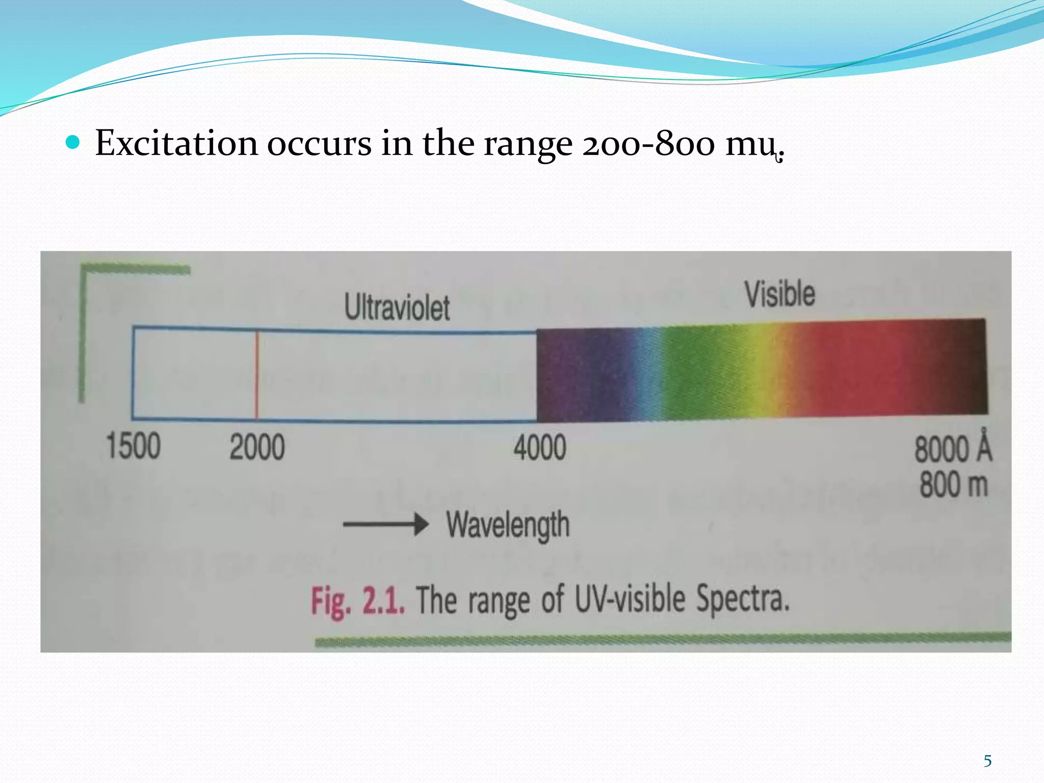
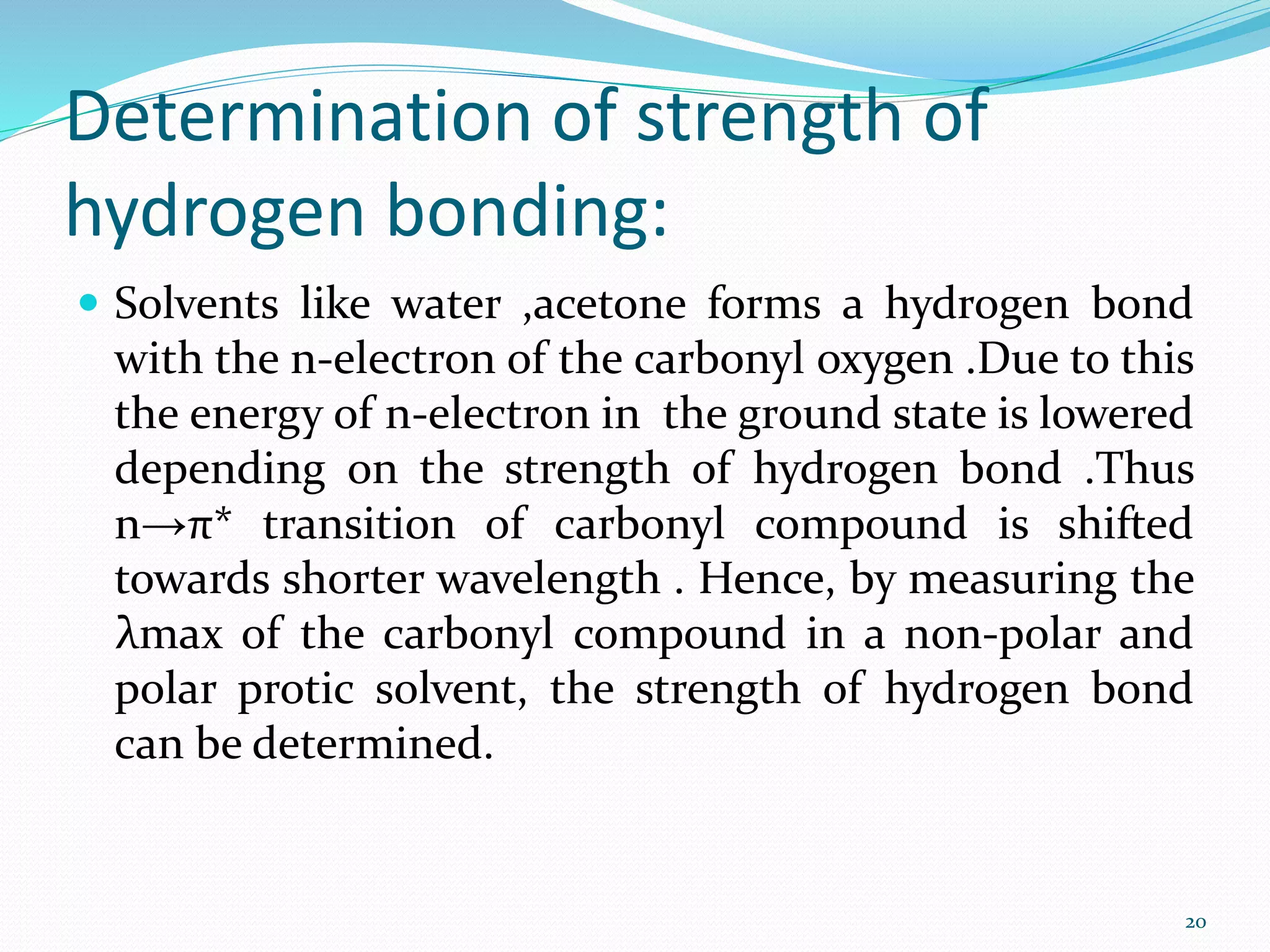
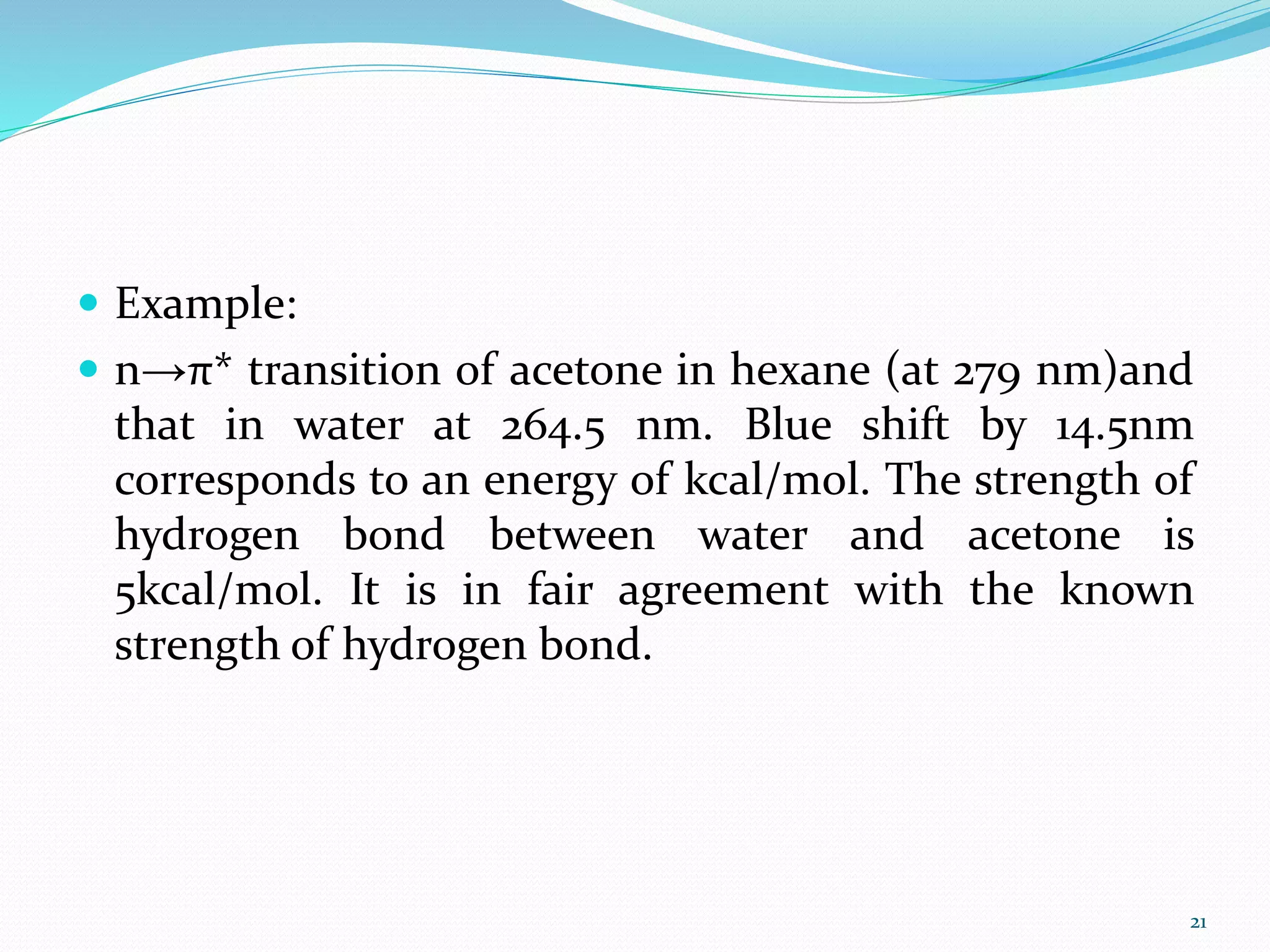
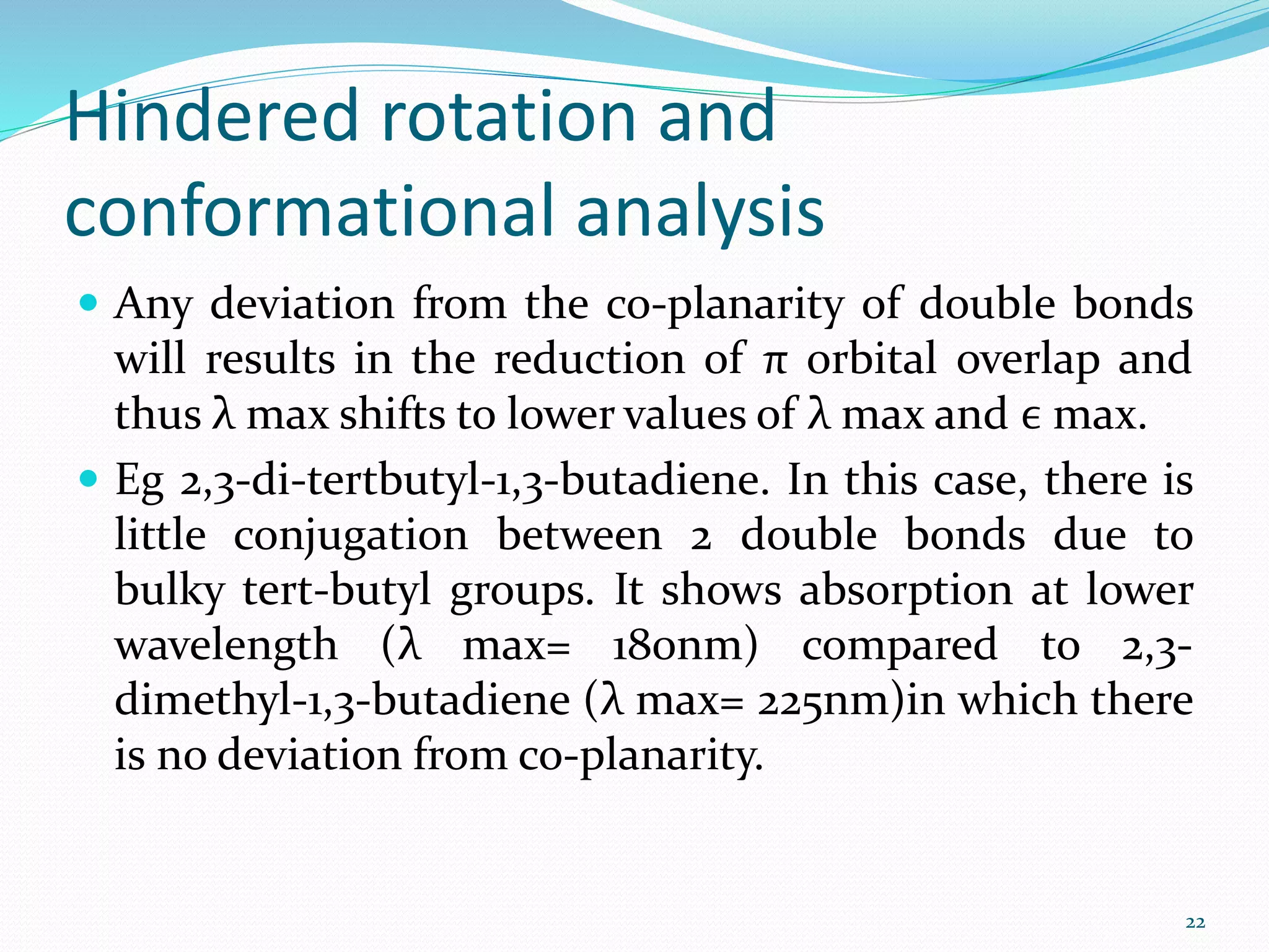
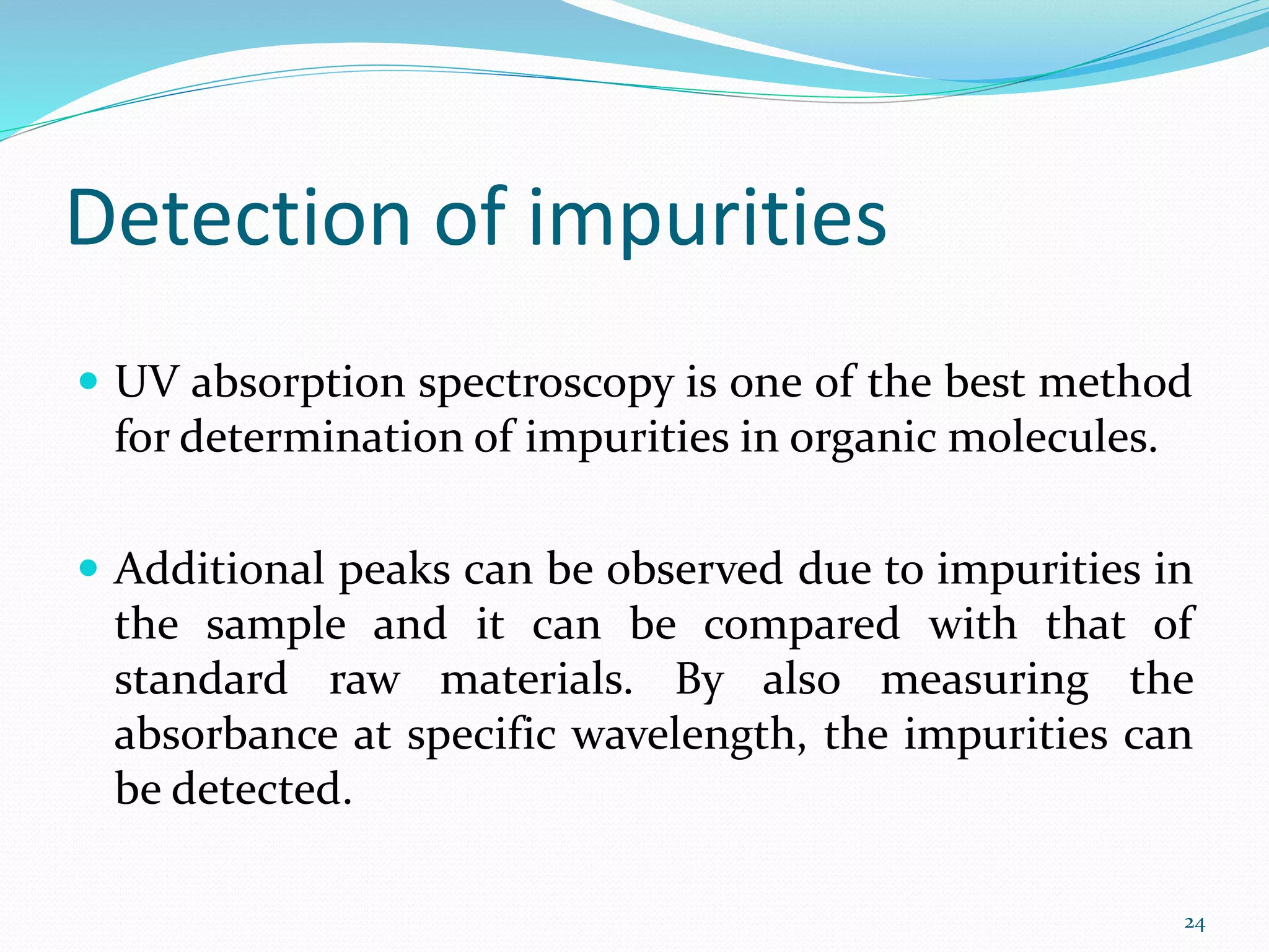
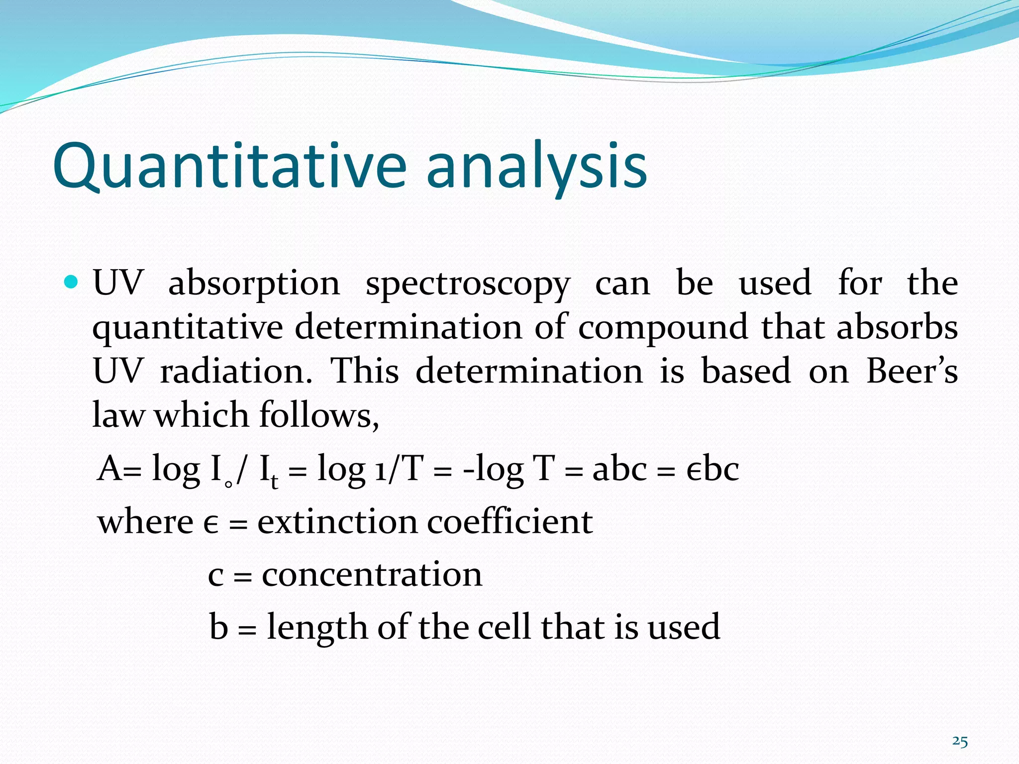
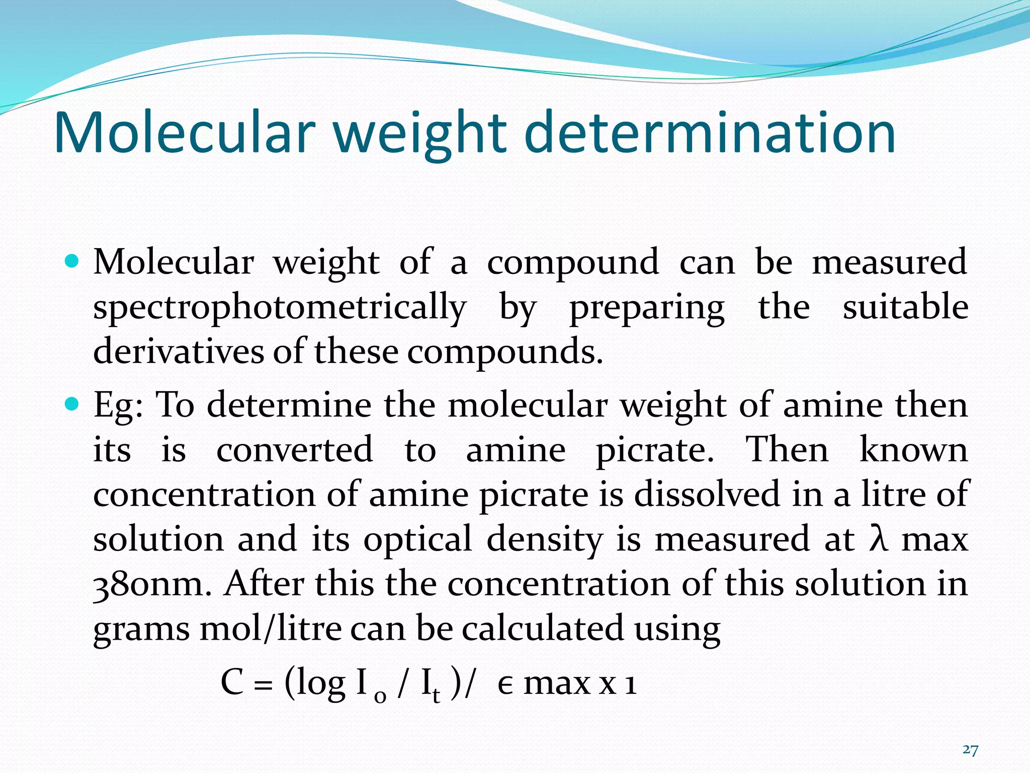
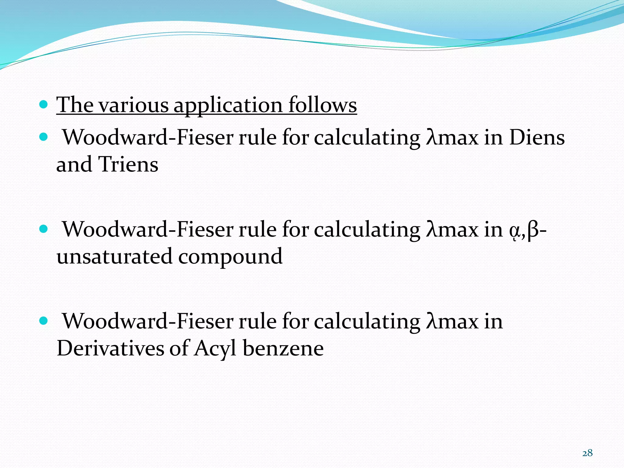
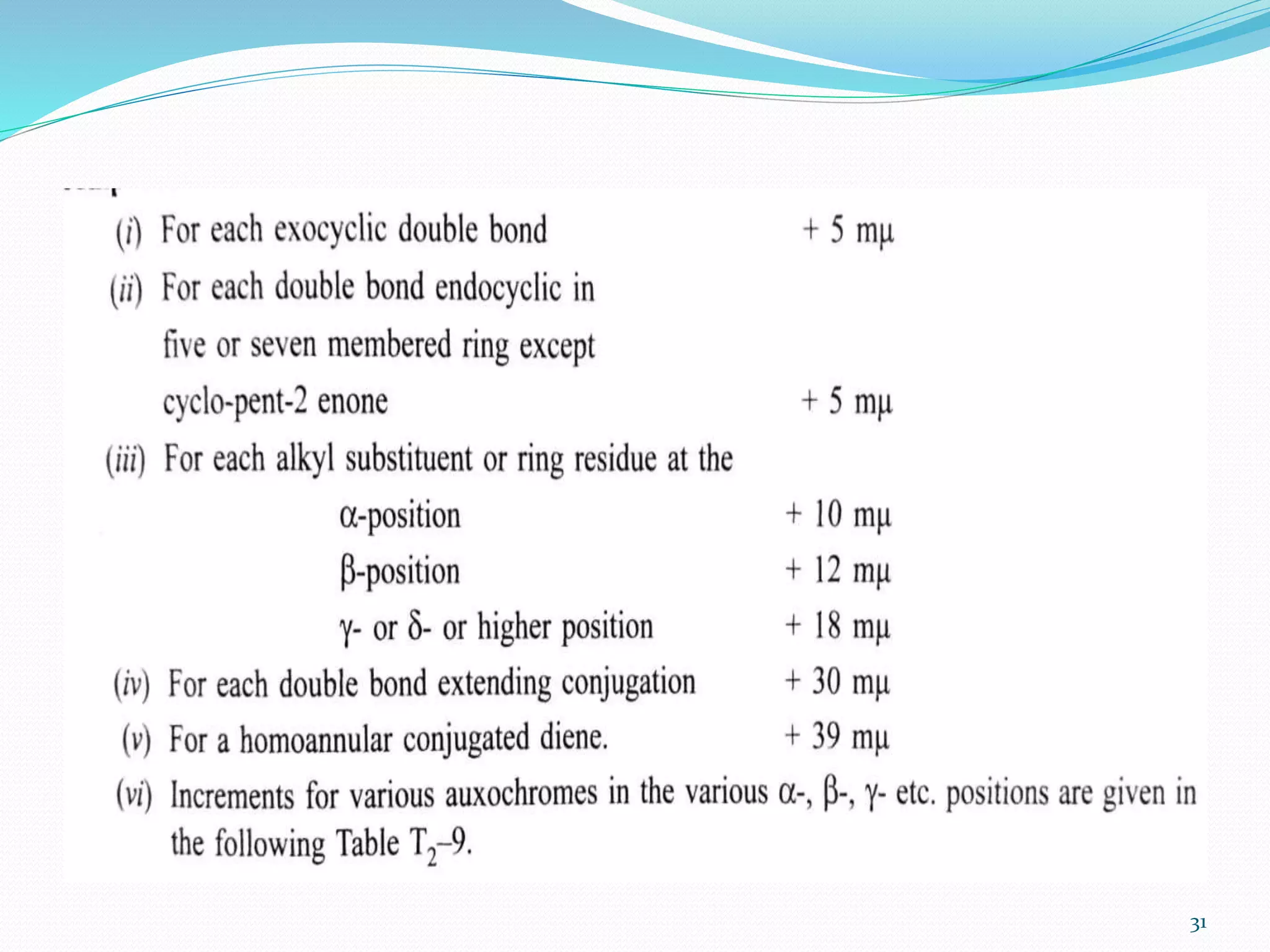
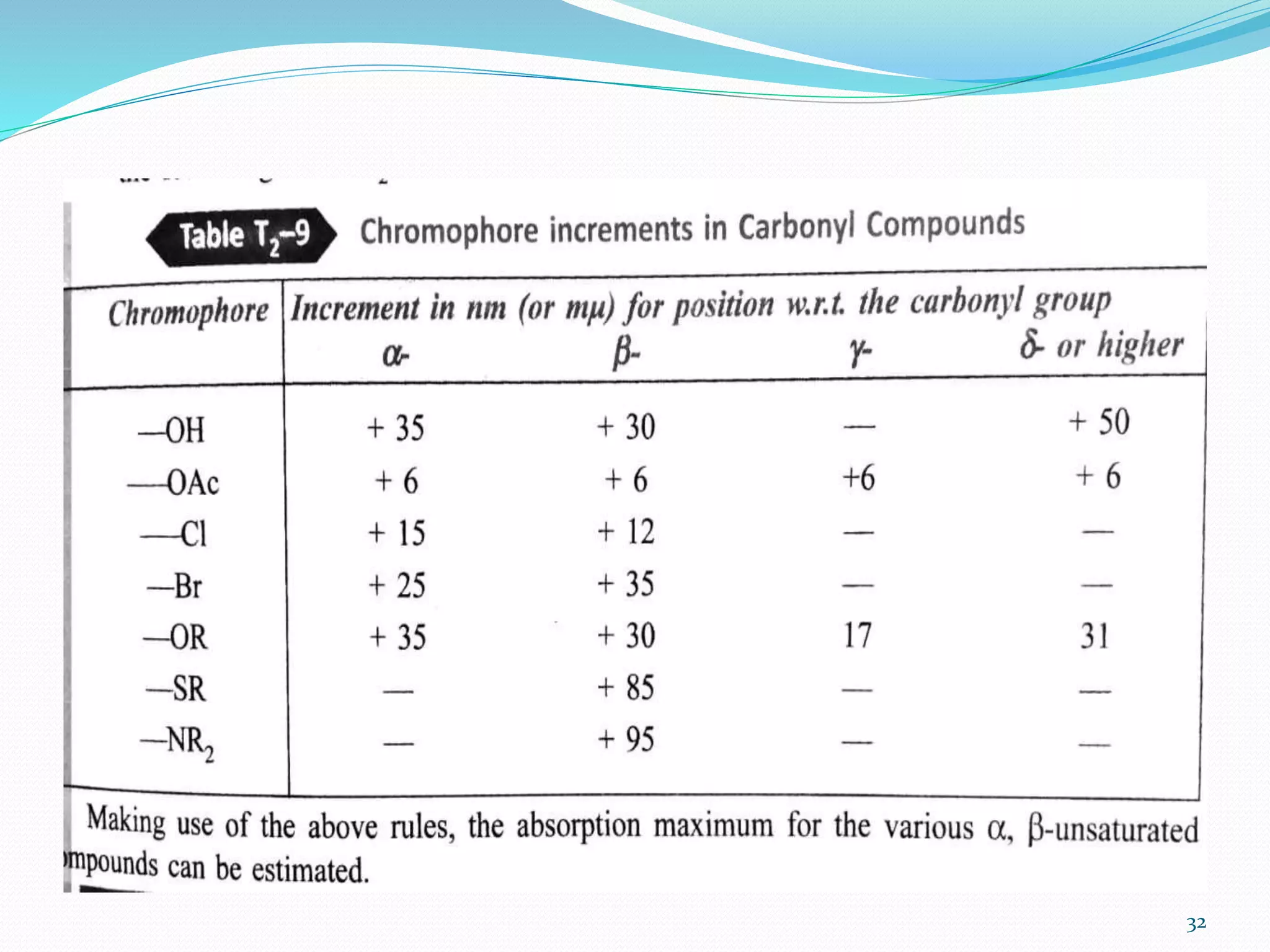
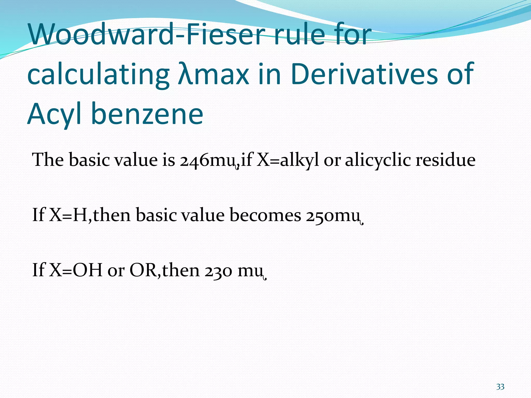
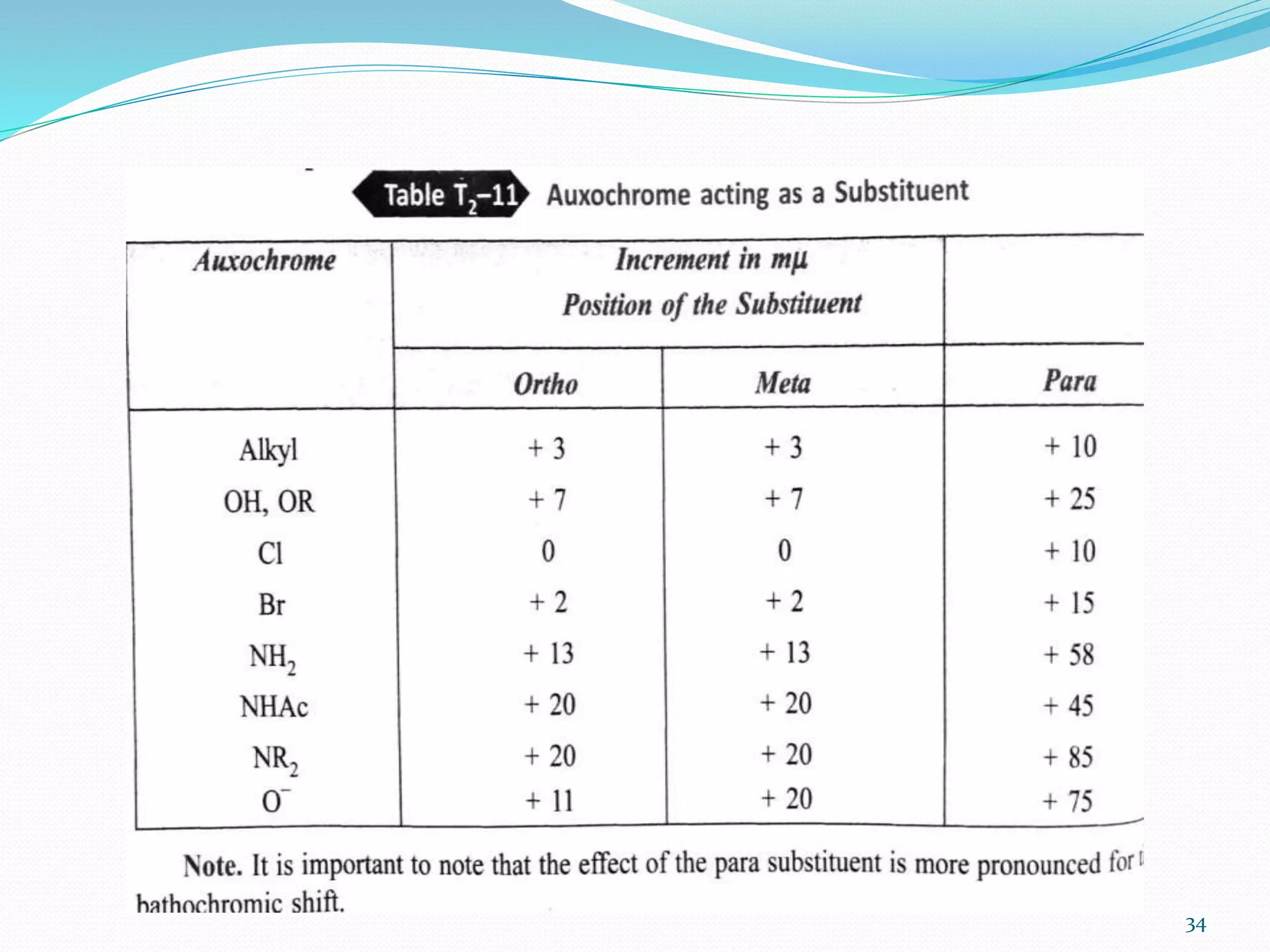
SINDHOORA D
First M.pharm
Department of
Pharmacology
1
CONTENT:
 Introduction to Uv-visible spectroscopy.
 Applications of Uv-visible spectroscopy.
 Brief introduction to woodward –fieser rules.
2
Introduction to UV-Visible
spectroscopy
 UV-Visible spectroscopy is also known as electronic
spectroscopy.In which the amount of light absorbed at
each wavelength of UV and Visible region of
electromagnetic spectrum is measured.
 This absorption of electromagnetic radiation by the
molecules leads to molecular excitation.
3
 This is called Electronic spectroscopy since it involves
the promotion of electron from the ground state to the
higher energy state.
 It is very useful to measure the number of conjugated
double bond and also aromatic conjugation within the
various molecule.
 It is also distinguishes between conjugated and non-
conjugated system;ᾳ,β-Unsaturated carbonyl
compound from β,ϒ-analogues; homoannular and
heteroannular conjugated diens etc.
4
 Excitation occurs in the range 200-800 mᶙ.
5
APPLICATION OF UV- VISIBLE
SPECTROCOPY
 UV –Visible spectroscopy has been mainly applied for the ,
Detection of unknown compound.
Detect the extent of conjugation
Detection of polynuclear compounds by comparison etc.
6
Extent of conjugation :
 The extent of conjugation in polyene can be estimated,
R-(CH=CH)n-R .
 Addition in unsaturation with the increase in the no.of
double bond ( increase in the value of n)shifts the
absorption to longer wavelength .
 It is found that the absorption occurs in the visible
region ,i.e at about 420mᶙ.If n=8 in the above polyene,
such an alkene appears colouerd to human eye.
7
Distinguish in conjugated and
non conjugated compounds
 It is also distinguish between a conjugated and an non
conjugated compounds.
8
 The forbidden n→π* band for the carbonyl group in
the compound (i)will appear at longer wavelength
compared to that for the compound (ii)
 The alkyl substitution in an alkene causes a
bathochromic shift.
9
Identification of an unknown
compound:
 A unknown compound can be identified by comparing
its spectrum with the known spectra
 If the two spectra coincide , the two compound must
be identical.
 If the two spectra do not coincide, then the expected
structure is different from the known compound.
10
Examination of polynuclear
hydrocarbon :
 Benzene and polynuclear hydrocarbon have
charactersitics spectra in the UV –Visible region .
 Thus the identification of the polynuclear
hydrocarbon can be made by comparision with the
spectra of known polynucler compound .
 The presence of substituent of on the ring , generally
,shifts the absorption maximum to longer wavelength.
11
Identification of a compound in
different solvent:
 Structure of the compound changes in the solvent
.Chloral hydrate shows an absorption maxima at
290mᶙ in hexane while the absorption decreases in the
aqueous solution.
 The compound in the hexane contains a carbonyl
group (a) where in aqueous solvent as (b)
 (a)CCl3.CHO.H2O, (b)CCl3.CH(OH)2.
12
Elucidation of the structure of
vitamin A and K:
 Elucidation of the structure of vitamin K1& K2 and
vitamin A1&A2.
 The UV spectra of vitamin K1&K2 are due to the
presence of the same chromophore ,i.e,2,3 dimethyl
naphtha –quinone. The absorption maxima of this
compound are 243,249,260,269 and 330mᶙ .
 The elucidation of the structure of vitamin A1&A2 are
possible by this method .
13
 Vitamin A1 absorbs at 325 mᶙ and for Vitamin A2 at
287 and 351 mᶙ.
 The absorption maxima occurs at longer wavelength
for Vitamin A2 due to presence of additional ethylenic
bond.
14
15
Preference over to tautomeric
form:
 Consider 2-hydroxy pyridine which exists in equilibrium
with its tautomeric form,pyridone -2.
 The spectra of these two compound were found to favour
pyridone -2 which is an ᾳ,β-unsaturated ketone and
clearly ,the equilibrium is shifted towards the right ,i.e.,
pyridone-2.
16
17
Determination of configuration of
geometrical isomers:
 The results of absorption shows that cis-alkene
absorbs at different wavelength as compared to their
corresponding trans-isomer
 The distinction is possible when one of the isomers is
forced to be non coplanar by steric hindrence.Thus ,cis
forms suffer distortion and absorption occurs at lower
wavelength.
18
19
Determination of strength of
hydrogen bonding:
 Solvents like water ,acetone forms a hydrogen bond
with the n-electron of the carbonyl oxygen .Due to this
the energy of n-electron in the ground state is lowered
depending on the strength of hydrogen bond .Thus
n→π* transition of carbonyl compound is shifted
towards shorter wavelength . Hence, by measuring the
λmax of the carbonyl compound in a non-polar and
polar protic solvent, the strength of hydrogen bond
can be determined.
20
 Example:
 n→π* transition of acetone in hexane (at 279 nm)and
that in water at 264.5 nm. Blue shift by 14.5nm
corresponds to an energy of kcal/mol. The strength of
hydrogen bond between water and acetone is
5kcal/mol. It is in fair agreement with the known
strength of hydrogen bond.
21
Hindered rotation and
conformational analysis
 Any deviation from the co-planarity of double bonds
will results in the reduction of π orbital overlap and
thus λ max shifts to lower values of λ max and ϵ max.
 Eg 2,3-di-tertbutyl-1,3-butadiene. In this case, there is
little conjugation between 2 double bonds due to
bulky tert-butyl groups. It shows absorption at lower
wavelength (λ max= 180nm) compared to 2,3-
dimethyl-1,3-butadiene (λ max= 225nm)in which there
is no deviation from co-planarity.
22
23
Detection of impurities
 UV absorption spectroscopy is one of the best method
for determination of impurities in organic molecules.
 Additional peaks can be observed due to impurities in
the sample and it can be compared with that of
standard raw materials. By also measuring the
absorbance at specific wavelength, the impurities can
be detected.
24
Quantitative analysis
 UV absorption spectroscopy can be used for the
quantitative determination of compound that absorbs
UV radiation. This determination is based on Beer’s
law which follows,
A= log I˳/ It = log 1/T = -log T = abc = ϵbc
where ϵ = extinction coefficient
c = concentration
b = length of the cell that is used
25
Other methods are:
 Calibration curve method
 Simultaneous multi component method
 Difference spectrophotometric method
 Derivative spectrophotometric method
26
Molecular weight determination
 Molecular weight of a compound can be measured
spectrophotometrically by preparing the suitable
derivatives of these compounds.
 Eg: To determine the molecular weight of amine then
its is converted to amine picrate. Then known
concentration of amine picrate is dissolved in a litre of
solution and its optical density is measured at λ max
380nm. After this the concentration of this solution in
grams mol/litre can be calculated using
C = (log I o / It )/ ϵ max x 1
27
 The various application follows
 Woodward-Fieser rule for calculating λmax in Diens
and Triens
 Woodward-Fieser rule for calculating λmax in ᾳ,β-
unsaturated compound
 Woodward-Fieser rule for calculating λmax in
Derivatives of Acyl benzene
28
 Woodward-Fieser rule for calculating λmax in Diens
and Triens
29
Woodward-Fieser rule for
calculating λmax in ᾳ,β-unsaturated
compound
a) The basic value of ᾳ,β-unsaturated ketone as 215 mᶙ
CH-COX,If X=alkyl group is 215mᶙ,if X=H then
207mᶙ,if X=OH or OR then 193mᶙ.
b)If the double bond and the carbonyl group are
contained in a five membered ring,then the basic
value is 202mᶙ
30
31
32
Woodward-Fieser rule for
calculating λmax in Derivatives of
Acyl benzene
The basic value is 246mᶙ,if X=alkyl or alicyclic residue
If X=H,then basic value becomes 250mᶙ
If X=OH or OR,then 230 mᶙ
33
34
References:
1) Elementary Organic spectroscopy –Y.R
Sharma, Revised edition , S.Chand
publisher, New Delhi
2) Instrumental and biomedical analysis, by
Ravishankar ,Fifth edition
3) Quantitative analysis of Drugs in
pharmaceutical formulations –P.D. Sethi,
Third edition,CBS publisher ,New delhi
35
36

Applications of uv visible spectroscopy

  • 1.
  • 2.
    CONTENT:  Introduction toUv-visible spectroscopy.  Applications of Uv-visible spectroscopy.  Brief introduction to woodward –fieser rules. 2
  • 3.
    Introduction to UV-Visible spectroscopy UV-Visible spectroscopy is also known as electronic spectroscopy.In which the amount of light absorbed at each wavelength of UV and Visible region of electromagnetic spectrum is measured.  This absorption of electromagnetic radiation by the molecules leads to molecular excitation. 3
  • 4.
     This iscalled Electronic spectroscopy since it involves the promotion of electron from the ground state to the higher energy state.  It is very useful to measure the number of conjugated double bond and also aromatic conjugation within the various molecule.  It is also distinguishes between conjugated and non- conjugated system;ᾳ,β-Unsaturated carbonyl compound from β,ϒ-analogues; homoannular and heteroannular conjugated diens etc. 4
  • 5.
     Excitation occursin the range 200-800 mᶙ. 5
  • 6.
    APPLICATION OF UV-VISIBLE SPECTROCOPY  UV –Visible spectroscopy has been mainly applied for the , Detection of unknown compound. Detect the extent of conjugation Detection of polynuclear compounds by comparison etc. 6
  • 7.
    Extent of conjugation:  The extent of conjugation in polyene can be estimated, R-(CH=CH)n-R .  Addition in unsaturation with the increase in the no.of double bond ( increase in the value of n)shifts the absorption to longer wavelength .  It is found that the absorption occurs in the visible region ,i.e at about 420mᶙ.If n=8 in the above polyene, such an alkene appears colouerd to human eye. 7
  • 8.
    Distinguish in conjugatedand non conjugated compounds  It is also distinguish between a conjugated and an non conjugated compounds. 8
  • 9.
     The forbiddenn→π* band for the carbonyl group in the compound (i)will appear at longer wavelength compared to that for the compound (ii)  The alkyl substitution in an alkene causes a bathochromic shift. 9
  • 10.
    Identification of anunknown compound:  A unknown compound can be identified by comparing its spectrum with the known spectra  If the two spectra coincide , the two compound must be identical.  If the two spectra do not coincide, then the expected structure is different from the known compound. 10
  • 11.
    Examination of polynuclear hydrocarbon:  Benzene and polynuclear hydrocarbon have charactersitics spectra in the UV –Visible region .  Thus the identification of the polynuclear hydrocarbon can be made by comparision with the spectra of known polynucler compound .  The presence of substituent of on the ring , generally ,shifts the absorption maximum to longer wavelength. 11
  • 12.
    Identification of acompound in different solvent:  Structure of the compound changes in the solvent .Chloral hydrate shows an absorption maxima at 290mᶙ in hexane while the absorption decreases in the aqueous solution.  The compound in the hexane contains a carbonyl group (a) where in aqueous solvent as (b)  (a)CCl3.CHO.H2O, (b)CCl3.CH(OH)2. 12
  • 13.
    Elucidation of thestructure of vitamin A and K:  Elucidation of the structure of vitamin K1& K2 and vitamin A1&A2.  The UV spectra of vitamin K1&K2 are due to the presence of the same chromophore ,i.e,2,3 dimethyl naphtha –quinone. The absorption maxima of this compound are 243,249,260,269 and 330mᶙ .  The elucidation of the structure of vitamin A1&A2 are possible by this method . 13
  • 14.
     Vitamin A1absorbs at 325 mᶙ and for Vitamin A2 at 287 and 351 mᶙ.  The absorption maxima occurs at longer wavelength for Vitamin A2 due to presence of additional ethylenic bond. 14
  • 15.
  • 16.
    Preference over totautomeric form:  Consider 2-hydroxy pyridine which exists in equilibrium with its tautomeric form,pyridone -2.  The spectra of these two compound were found to favour pyridone -2 which is an ᾳ,β-unsaturated ketone and clearly ,the equilibrium is shifted towards the right ,i.e., pyridone-2. 16
  • 17.
  • 18.
    Determination of configurationof geometrical isomers:  The results of absorption shows that cis-alkene absorbs at different wavelength as compared to their corresponding trans-isomer  The distinction is possible when one of the isomers is forced to be non coplanar by steric hindrence.Thus ,cis forms suffer distortion and absorption occurs at lower wavelength. 18
  • 19.
  • 20.
    Determination of strengthof hydrogen bonding:  Solvents like water ,acetone forms a hydrogen bond with the n-electron of the carbonyl oxygen .Due to this the energy of n-electron in the ground state is lowered depending on the strength of hydrogen bond .Thus n→π* transition of carbonyl compound is shifted towards shorter wavelength . Hence, by measuring the λmax of the carbonyl compound in a non-polar and polar protic solvent, the strength of hydrogen bond can be determined. 20
  • 21.
     Example:  n→π*transition of acetone in hexane (at 279 nm)and that in water at 264.5 nm. Blue shift by 14.5nm corresponds to an energy of kcal/mol. The strength of hydrogen bond between water and acetone is 5kcal/mol. It is in fair agreement with the known strength of hydrogen bond. 21
  • 22.
    Hindered rotation and conformationalanalysis  Any deviation from the co-planarity of double bonds will results in the reduction of π orbital overlap and thus λ max shifts to lower values of λ max and ϵ max.  Eg 2,3-di-tertbutyl-1,3-butadiene. In this case, there is little conjugation between 2 double bonds due to bulky tert-butyl groups. It shows absorption at lower wavelength (λ max= 180nm) compared to 2,3- dimethyl-1,3-butadiene (λ max= 225nm)in which there is no deviation from co-planarity. 22
  • 23.
  • 24.
    Detection of impurities UV absorption spectroscopy is one of the best method for determination of impurities in organic molecules.  Additional peaks can be observed due to impurities in the sample and it can be compared with that of standard raw materials. By also measuring the absorbance at specific wavelength, the impurities can be detected. 24
  • 25.
    Quantitative analysis  UVabsorption spectroscopy can be used for the quantitative determination of compound that absorbs UV radiation. This determination is based on Beer’s law which follows, A= log I˳/ It = log 1/T = -log T = abc = ϵbc where ϵ = extinction coefficient c = concentration b = length of the cell that is used 25
  • 26.
    Other methods are: Calibration curve method  Simultaneous multi component method  Difference spectrophotometric method  Derivative spectrophotometric method 26
  • 27.
    Molecular weight determination Molecular weight of a compound can be measured spectrophotometrically by preparing the suitable derivatives of these compounds.  Eg: To determine the molecular weight of amine then its is converted to amine picrate. Then known concentration of amine picrate is dissolved in a litre of solution and its optical density is measured at λ max 380nm. After this the concentration of this solution in grams mol/litre can be calculated using C = (log I o / It )/ ϵ max x 1 27
  • 28.
     The variousapplication follows  Woodward-Fieser rule for calculating λmax in Diens and Triens  Woodward-Fieser rule for calculating λmax in ᾳ,β- unsaturated compound  Woodward-Fieser rule for calculating λmax in Derivatives of Acyl benzene 28
  • 29.
     Woodward-Fieser rulefor calculating λmax in Diens and Triens 29
  • 30.
    Woodward-Fieser rule for calculatingλmax in ᾳ,β-unsaturated compound a) The basic value of ᾳ,β-unsaturated ketone as 215 mᶙ CH-COX,If X=alkyl group is 215mᶙ,if X=H then 207mᶙ,if X=OH or OR then 193mᶙ. b)If the double bond and the carbonyl group are contained in a five membered ring,then the basic value is 202mᶙ 30
  • 31.
  • 32.
  • 33.
    Woodward-Fieser rule for calculatingλmax in Derivatives of Acyl benzene The basic value is 246mᶙ,if X=alkyl or alicyclic residue If X=H,then basic value becomes 250mᶙ If X=OH or OR,then 230 mᶙ 33
  • 34.
  • 35.
    References: 1) Elementary Organicspectroscopy –Y.R Sharma, Revised edition , S.Chand publisher, New Delhi 2) Instrumental and biomedical analysis, by Ravishankar ,Fifth edition 3) Quantitative analysis of Drugs in pharmaceutical formulations –P.D. Sethi, Third edition,CBS publisher ,New delhi 35
  • 36.