Facial fractures the upper face
•
163 likes•18,205 views

This document discusses maxillary and periorbital fractures. It begins by describing the classic tripod, orbital floor, and LeFort fractures, noting that precise anatomic reduction is key. It then covers the epidemiology, mechanisms of injury, clinical assessment, radiographic assessment, management principles, and various types of upper face fractures including nasal fractures, naso-orbital-ethmoidal fractures, frontal sinus fractures, and orbital fractures.

More Related Content
What's hot
What's hot (20)
Viewers also liked
Viewers also liked (20)
Similar to Facial fractures the upper face
Similar to Facial fractures the upper face (20)
More from Notre Dame De Chartres Hospital
More from Notre Dame De Chartres Hospital (20)
Recently uploaded
Recently uploaded (20)
Facial fractures the upper face
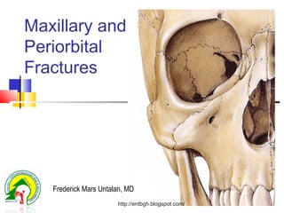
- 1. Maxillary and Periorbital Fractures Frederick Mars Untalan, MD http://entbgh.blogspot.com/
- 3. Facial Injuries Dental Terminology http://entbgh.blogspot.com/
- 4. Overview Classic tripod, orbital floor, LeFort fractures better thought of as orbitozygomaticomaxillary fractures Precise anatomic reduction is key Goal is functional and cosmetic rehabilitation http://entbgh.blogspot.com/
- 5. Epidemiology Males : Females -- 4:1 Predominantly in 20’s or 30’s Cause MVA > altercation > fall Site Nasal > Zygoma > other In altercations left zygoma fractured more often http://entbgh.blogspot.com/
- 6. History Mechanism of InjuryMechanism of Injury Previous facial injuries / Dental AbN Premorbid history Loss of consciousness Medications, Allergies, Tetanus Status Associated Injuries http://entbgh.blogspot.com/
- 7. Clinical Assessment of the Face SYMPTOMS Diplopia Abnormal Sensation Malocclusion Pain http://entbgh.blogspot.com/
- 8. Clinical Assessment of the Face GCS, Ocular exam, C-spine exam Cranial nerve exam, CSF rhinorrhea Inspect (including septum & TM) Palpate (including midface stability) Assess occlusion & intraoral exam http://entbgh.blogspot.com/
- 9. CSF rhinorrhea – Halo sign Septal hematoma Hemotympanum Malocclusion Ocular Findings Diplopia/restricted EOM, Subconj. Hem., hyphema, anisocoria, impaired VA Clinical Assessment of the Face http://entbgh.blogspot.com/
- 10. Clinical Assessment of the Face Reliable Physical signs Bony facial asymmetry True V2 numbness Malocclusion Clinical Assessment of the Face http://entbgh.blogspot.com/
- 11. Physical Exam Palpate for midface instability http://entbgh.blogspot.com/
- 12. Physical Exam Often edema, swelling, or patient’s mental status make physical exam difficult CT is modality of choice -- axial and coronal http://entbgh.blogspot.com/
- 13. Physical Exam Midface asymmetry may indicate zygoma fracture http://entbgh.blogspot.com/
- 15. Emergency Management C-Spine Injury 10% C-Spine Fracture Head Injury 50% - Loss of Consciousness 5% Significant Intracranial Injury Ocular Injury 25% - Some Degree of Injury http://entbgh.blogspot.com/
- 16. Emergency Management Hemorrhage Local Pressure Dressings / Packing Reduction Of Facial Fractures Endovascular Consultation Ligation Of Vessels IMAX http://entbgh.blogspot.com/
- 17. Radiographic Assessment of the Face Water’s view Panorex (Orthopantomogram) Mandible views CT scans Standard of care for major facial trauma http://entbgh.blogspot.com/
- 18. Water’s view The “W” system PA view for visualization of maxillary sinuses, maxilla, orbits, and zygomatic arches May also see nasal bone fractures Radiographic Assessment of the Face http://entbgh.blogspot.com/
- 19. CT areas to evaluate Vertical buttresses Zygomatic arch Orbital walls Bony palate Mandibular condyles http://entbgh.blogspot.com/
- 20. Signs And Symptoms Pain / Tenderness Crepitus From Bony Fractures Hypoesthesia Paralysis Malocclusion Visual Disturbances Deformity Obstructive Respiration Lacerations Bleeding Contusions Facial Asymmetry http://entbgh.blogspot.com/
- 21. Soft Tissue Injuries Tetanus Prophylaxis Structures Facial Nerve Trigeminal Nerve Parotid Duct Lacrimal System http://entbgh.blogspot.com/
- 22. Principles Of Craniomaxillofacial Fracture Management Precise anatomic diagnosis Direct fracture exposure Reduction / rigid internal fixation Mandible fracture stabilization Reconstruction of horizontal and vertical facial buttresses Primary bone grafting Periosteal and soft-tissue suspension and repair http://entbgh.blogspot.com/
- 23. Facial Fractures The Upper Face http://entbgh.blogspot.com/
- 24. Outline Facial Fracture Basics Nasal Fractures Naso-orbital-ethmoidal Fractures Frontal Sinus Fractures Zygomatic Fractures Maxillary Fractures Orbital Fractures http://entbgh.blogspot.com/
- 26. Physical Exam Edema Crepitus Periorbital Ecchymosis Epistaxis Internal And External Lacerations Widened Nasal Bridge Septal Hematoma http://entbgh.blogspot.com/
- 27. Classification Stranc and Robertson Lateral Impact Injuries Unilateral vs. Bilateral Frontal Impact Injuries Plane I Plane II Plane III http://entbgh.blogspot.com/
- 28. Treatment Lateral Impact Injuries Early Versus Delayed Treatment Closed Reduction (Local vs. General) Drainage of Septal Hematoma Simple Repositioning of Deviated Nasal Bones and Septum Completion Of The Fracture Internal Packing and External Splint http://entbgh.blogspot.com/
- 29. Treatment Frontal Impact Injuries Plane I edema / ecchymosis distal nasal bridge and tip possible septal distortion closed reduction with internal support possibly may require secondary septorhinoplasty http://entbgh.blogspot.com/
- 30. Treatment Frontal Impact Injuries Plane II Increased Comminution of the Nasal Pyramid Bilateral Possible “Saddling” Initial Closed reduction May Require Delayed Septal Reconstruction With Grafts http://entbgh.blogspot.com/
- 31. Treatment Frontal Impact Injuries Plane III Extend Into Pyriform Aperture And Medial Orbital Rim ie. Naso-Orbital-Ethmoidal Fractures Open Reduction an Internal Fixation of Frontal Process of Maxilla Transnasal Reduction of Medial Canthal Ligaments http://entbgh.blogspot.com/
- 32. Complications Occur In Up To 70% Deviated Nasal Pyramid Nasal “Hump” Septal Deformity With Respiratory Obstruction http://entbgh.blogspot.com/
- 35. Naso-Orbital-Ethmoidal Fractures Interorbital “Space” two ethmoidal labyrinths superior and middle turbinates perpendicular plate of ethmoid Medial Orbital Wall anteriorly - lacrimal bone and lamina papyracea posteriorly - body of sphenoid http://entbgh.blogspot.com/
- 36. Naso-Orbital-Ethmoidal Fractures Interorbital space displaced backwards Medial Canthal Tendon and Lacrimal Apparatus frequently injured May extend into: cribriform plate and anterior cranial fossa optic foramen Associated Orbit and Midface Fractures Common http://entbgh.blogspot.com/
- 37. Naso-Orbital-Ethmoidal Fractures Flat nose Swollen medial canthal area Telecanthus (12-20%) Lack of skeletal support on palpation of nose CSF leak Positive eyelid traction test http://entbgh.blogspot.com/
- 38. Naso-Orbital-Ethmoidal Fractures Telecanthus Normal intercanthal distance (Stranc) White males: 33-34mm Females: 32-33mm Consider >35mm abnormal (Manson) http://entbgh.blogspot.com/
- 39. Naso-Orbital-Ethmoidal Fractures Classification Markowitz Type I - Single central segment Type II - Comminuted central segment Type III - Avulsed medial canthal tendon http://entbgh.blogspot.com/
- 41. Naso-Orbital-Ethmoidal Fractures Classification - Gruss 1 Isolated injury to bony naso-orbital region 2 Associated fractures of the central maxilla 3 Associated LeFort II and III 4 Naso-orbital fractures with orbital dystopia 5 Naso-orbital fractures with bone loss http://entbgh.blogspot.com/
- 42. Naso-Orbital-Ethmoidal Fractures Management Early open reduction Four Objectives: correct epicanthal folds restore bony contour reestablish lacrimal system continuity medial canthoplasty / canthopexy http://entbgh.blogspot.com/
- 43. Naso-Orbital-Ethmoidal Fractures Management Wide Exposure coronal incision “open sky” - transverse across root of nose vertical midline nasal subciliary buccal sulcus extend existing lacerations http://entbgh.blogspot.com/
- 44. Naso-Orbital-Ethmoidal Fractures Management Correct nasofrontal separation Elevate nasal bones Reduce comminuted nasal bones Bone graft where needed Explore septum Stabilize nasomaxillary buttresses http://entbgh.blogspot.com/
- 46. Frontal Sinus Embryology Begin to Develop At 2 Years of Age Extension of the Ethmoid Air Cells Radiographically Evident At ~ 8 Years Do Not Reach Adult Size Until 12 or Older 10% - Unilateral Development 4% - Absent All Together Drain Into Middle Meatushttp://entbgh.blogspot.com/
- 47. Frontal Sinus Anatomy Supraorbital / Temporal vs Frontal Sinus Anterior Wall and/or Posterior Wall Nasofrontal Duct http://entbgh.blogspot.com/
- 48. Frontal Sinus Diagnosis Signs And Symptoms Forehead Laceration CSF Rhinorrhea Supraorbital Nerve Anesthesia Depressed Frontal Region Subconjunctival Ecchymosis http://entbgh.blogspot.com/
- 49. Frontal Sinus Diagnosis X-Ray Air Fluid Levels CT Scan Axial and Coronal Images http://entbgh.blogspot.com/
- 51. Frontal Sinus Treatment Operative Indications Anterior Table Displacement With Contour Change Nasofrontal Duct Involvement Displaced Posterior Table http://entbgh.blogspot.com/
- 52. Frontal Sinus Treatment Nasofrontal Duct Injury Remove Mucosa Burr Inner Cortex Occlusion Of Duct Sinus Obliteration http://entbgh.blogspot.com/
- 53. Frontal Sinus Treatment Posterior Table Cranialization 1 Bicoronal Approach 2 Preserve Pericranial Flap 3 Dural Repair 4 Remove Sinus Mucosa 5 Obliterate Nasofrontal Duct 6 Remove Intersinus Septum And Posterior Wall 7 Pericranial Flap To Floor Of Sinus http://entbgh.blogspot.com/
- 54. Frontal Sinus Complications Early (within 6 months) Frontal Sinusitis Meningitis Late Mucocele Mucopyocele Brain Abcess Osteomyelitis http://entbgh.blogspot.com/
- 55. Frontal Sinus Complications Incidence Of Late Complications Freihofer 71 Fractures 2 Patients - Meningitis 1 Patient - Mucopyocele With Osteomyelitis Of Frontal Bone http://entbgh.blogspot.com/
- 58. Orbital Fractures Blowout Fractures Pure Blowout - only orbital floor or medial wall injured Impure Blowout - associated orbital rim fractures http://entbgh.blogspot.com/
- 60. Orbital Blowout Injury Usually inferior and/or medial wall Cone will become more spherical Leads to enophthalmos, inferior displacement Muscle entrapment causes diplopia http://entbgh.blogspot.com/
- 61. Orbital Fractures Physical Exam Diplopia Enophthalmos Inferior Displacement Palpebral Fissure Anesthesia of Infraorbital Nerve Orbital Emphysema http://entbgh.blogspot.com/
- 62. Orbital Fractures Physical Exam Diplopia Commonly on Upward Gaze Primary (Central Gaze) or Secondary (Perpheral) Mechanical (incarceration of infraorbital tissue) or Nonmechanical (paresis) Forced Duction Test http://entbgh.blogspot.com/
- 63. Orbital Fractures Forced Duction Test http://entbgh.blogspot.com/
- 64. Orbital Fractures Physical Exam Enophthalmos Inferior and Posterior Displacement of Globe and Intraorbital Soft Tissue Etiology Enlargement of the Bony Orbital Cavity Escape of Orbital Fat or Fat Necrosis Muscle Entrapment in Fracture Line Soft Tissue Scarring and Contracture http://entbgh.blogspot.com/
- 66. Orbital Fractures Physical Exam No Diplopia + No Enophthalmos ?Significant Fracture Diplopia + No Enophthalmos Incarceration Only No Diplopia + Enophthalmos Volume Discrepancy Only Diplopia + Enophthalmos Incarceration + Volume Discrepancy http://entbgh.blogspot.com/
- 67. Orbital Fractures Indications For Exploration Symptomatic Diplopia With Positive Forced Duction Test Xray evidence of Extraocular Muscle Entrapment Early Enophthalmos (>3mm) Large Orbital Floor Defect Abnormally Low Vertical Globe Level Associated Orbital Rim or Other Craniofacial Fractures http://entbgh.blogspot.com/
- 68. Orbital Rim Access A -- subciliary B -- lower eyelid C -- infraorbital http://entbgh.blogspot.com/
- 70. Orbital Fractures Management Grafts Autologous Bone Cartilage Fascia lata Alloplastic Implants Teflon Silastic Titanium http://entbgh.blogspot.com/
- 71. Orbital Floor Dotted line shows anatomic goal of restoration http://entbgh.blogspot.com/
- 72. Orbital Fractures Complications Infection Implant problems Persistent Diplopia (2-50%) Persistent Enophthalmos (15-22%) Ectropion (1%) Blindness http://entbgh.blogspot.com/
- 73. Orbital Fractures Complications Superior Orbital Fissure Syndrome extension of fracture into SOF ophthalmoplegia with injury to III, IV or VI anesthesia in V1 plus loss of corneal reflex ptosis and proptosis parasympathetic block fixed, dilated pupil http://entbgh.blogspot.com/
- 74. Orbital Fractures Complications Orbital Apex Syndrome same as superior orbital fissure syndrome plus blindness http://entbgh.blogspot.com/
- 75. Transconjunctival Approach Conjunctiva is being used to protect globe http://entbgh.blogspot.com/
- 76. Materials for reconstruction Autogenous tissues Avoid risk of infected implant Additional operative time, donor site morbidity , graft absorption Calvarial bone, iliac crest, rib, septal or auricular cartilage http://entbgh.blogspot.com/
- 77. Orbital Floor Bone Grafting Need to support floor full 4 cm http://entbgh.blogspot.com/
- 78. Alloplastic implants Decreased operative time, easily available, no donor site morbidity, can provide stable support Risk of infection 0.4-7% Gelfilm, polygalactin film, silastic, marlex mesh, teflon, prolene, polyethylene, titanium http://entbgh.blogspot.com/
- 79. Orbital Floor Materials Marlex mesh needs 360 degree support better for concave anterior floor only Medpor needs medial/ lateral support can use for anterior/posterior defect Calvarial bone graft Titanium mesh http://entbgh.blogspot.com/
- 82. Orbital Roof Uncommon due to high levels of force needed to fracture orbital roof Commonly with intracranial problems http://entbgh.blogspot.com/
- 84. Orbital Roof Repair Repair roof higher on frontal bar http://entbgh.blogspot.com/
- 85. From: Strong EB, Sykes JM. Zygoma Complex Fractures. Facial Plastic Surgery 1990;14(1):109. http://entbgh.blogspot.com/
- 86. Periorbital & Zygomatic bone Fractures http://entbgh.blogspot.com/
- 87. Zygoma Fractures Results from lateral forceshttp://entbgh.blogspot.com/
- 88. Zygoma Anatomy Tetrapod Structure Frontal Bone Temporal Bone Maxilla Greater Wing Of Sphenoid http://entbgh.blogspot.com/
- 90. Zygoma Anatomy Muscular Attachments Masseter Temporalis Zygomaticus Zygomatic Head of Quadratus Labii Superioris http://entbgh.blogspot.com/
- 91. Zygoma Physical Exam Circumorbital Swelling / Ecchymosis Subconjunctival Hemorrage Abnormal Sensation V2 Distribution Diplopia or Globe Displacement Increased Facial Width Depressed Malar Prominence Palpable Step Deformities Unilateral Epistaxis Hematoma Upper Buccal Sulcus Trismus Due To Coronoid Process Impingement http://entbgh.blogspot.com/
- 94. Zygoma Fracture Classification Knight 1 Undisplaced 2 Arch Fractures 3 Unrotated Body Fractures 4 Medially Rotated Body Fractures 5 Laterally Rotated Body Fractures 6 Complex Fractures - Additional Fractures Across Zygoma http://entbgh.blogspot.com/
- 95. Zygoma Fracture Classification Manson Low Energy minimal displacement do not require operative reduction Middle Energy High Energy often part of panfacial fractures http://entbgh.blogspot.com/
- 96. Zygoma Radiology X-Ray Water’s View Most Useful CT Scan Coronal Cuts For Orbital Anatomy http://entbgh.blogspot.com/
- 97. Zygoma Fractures Impacted zygoma may mask orbital floor defect http://entbgh.blogspot.com/
- 98. Treatment of Zygomaticomaxillary Complex fractures Restore pre-injury facial configuration Prevent cosmetic deformity Prevent delayed visual disturbances Repair within 5-7 days allows edema to decrease and avoids shortening of masseter with lateral and inferior rotation http://entbgh.blogspot.com/
- 99. Zygoma Ideally done between 5-7 days for resolution of edema Pre- or intra- operative steroids can help with edema After 10 days masseter begins to shorten http://entbgh.blogspot.com/
- 100. Zygoma reduction only - Minimally displaced, non comminuted plating of lateral antrum, orbital rim, ZF suture, and even the zygomatic arch - for Increasing amounts of displacement & comminution One can wire the ZF suture first to assist with reduction, then plate it after other areas stabilized http://entbgh.blogspot.com/
- 102. Zygoma Management Undisplaced Nonoperative Displaced Isolated Zygomatic Arch - Gilles Elevation Orbitozygomatic Fractures - Open reduction and Stabilization http://entbgh.blogspot.com/
- 107. Surgical Approaches Coronal Sublabial Transconjunctival Lateral Brow http://entbgh.blogspot.com/
- 110. Coronal Approach Supraorbital nerve may be released for more exposure http://entbgh.blogspot.com/
- 112. Lateral Brow Incision Avoid shaving brow hairs Goal is the ZF suturehttp://entbgh.blogspot.com/
- 113. Sublabial Approach Leave mucosa to sew to later Identify and preserve V2 http://entbgh.blogspot.com/
- 118. Zygoma Complications - Early Bleeding Infection Exacerbation of Sinus Disease Malfunction of Extraocular Muscles Blindness http://entbgh.blogspot.com/
- 119. Zygoma Complications - Late Nonunion / Malunion Diplopia (10% initial, 5% permanent) Persistent V2 Anesthesia (24%) Orbital Dystopia Chronic Maxillary Sinusitis (4-7%) Scarring Ectropion Problems With Mandible Motion Enophthalmos (3%) Soft Tissue Descent With Loss of Malar Prominence http://entbgh.blogspot.com/
- 120. Treatment of Zygomaticomaxillary Complex fractures Restore pre-injury facial configuration Prevent cosmetic deformity Prevent delayed visual disturbances Repair within 5-7 days allows edema to decrease and avoids shortening of masseter with lateral and inferior rotation http://entbgh.blogspot.com/
- 121. Soft diet and malar protection Closed reduction ORIF with plating of one to four buttresses Provide fixation as necessary for stable reduction http://entbgh.blogspot.com/
- 123. Types LeFort or Maxillary fractures Zygomaticomaxillary complex fractures Orbitozygomaticomaxillary complex fractures http://entbgh.blogspot.com/
- 124. Anatomy of the Maxilla Paired embryologically Functionally acts with palatine bone http://entbgh.blogspot.com/
- 125. Anatomy of the Maxilla http://entbgh.blogspot.com/
- 126. LeFort fractures Rene LeFort 1901 in cadaver skulls Based on the most superior level Frequently different levels on either side LeFort I LeFort II LeFort III http://entbgh.blogspot.com/
- 127. Facial Buttress system From: Celin SE. Fractures of the Upper Facial and Midfacial Skeleton. In: Myers EN ed., Operative Otolaryngology Head and Neck Surgery, Philadelphia, WB Saunders Company 1997:1143-1192. http://entbgh.blogspot.com/
- 128. From: Dolan KD, Jacoby CG, Smoker WR. Radiology of Facial Injury. New York, MacMillian Publishing Company 1988, pg76. http://entbgh.blogspot.com/
- 132. LeFort fractures Rene LeFort 1901 in cadaver skulls Based on the most superior level Frequently different levels on either side LeFort I LeFort II LeFort III http://entbgh.blogspot.com/
- 133. LeFort Fractures Experimentally determined weak points Can be in combinations bilaterally Useful descriptor Results from anterior forces http://entbgh.blogspot.com/
- 134. Forces of mastication From: Banks P, Brown A. Fractures of the Facial Skeleton, Oxford, Wright 2001 pg.6 http://entbgh.blogspot.com/
- 135. Facial Buttress system From :Stanley RB. Maxillary and Periorbital Fractures. In :Bailey BJ ed., Head and Neck Surgery-Otolaryngology, third edition, Philadelphia, Lippincott Williams & Wilkins 2001, pg 777.http://entbgh.blogspot.com/
- 136. From: Marciani RD. Management of Midface Fractures: fifty years later. J Oral Maxillofac Surg 1993;51:962. http://entbgh.blogspot.com/
- 137. From: Dolan KD, Jacoby CG, Smoker WR. Radiology of Facial Injury. New York, MacMillian Publishing Company 1988, pg76. http://entbgh.blogspot.com/
- 138. Maxillary Fractures LeFort Fractures LeFort I Transverse Fracture That Separates Maxillary Alveolus From Midface Skeleton Runs Above Roots of Maxillary Teeth, Across Lower Pyriform Aperature, and Severs Pterygoid Process http://entbgh.blogspot.com/
- 140. Maxillary Fractures LeFort Fractures Lefort II “Pyramidal” Fracture of Maxilla Separates Nasomaxillary Segment from Zygomatic and Upper Lateral Midface Fracture Line May Go Above or Beneath Medial Canthal Ligament Insertion Lacrimal System May Be Involved http://entbgh.blogspot.com/
- 142. Maxillary Fractures LeFort Fractures LeFort III Craniofacial Dysjunction Zygomaticofrontal Junction, Traverses Lateral, Inferior, and Medial Orbit, Separates Frontal Process of Maxilla From Frontal Bones http://entbgh.blogspot.com/
- 145. Modified LeFort Classification From: Marciani RD. Management of Midface Fractures: fifty years later. J Oral Maxillofac Surg 1993;51:962. http://entbgh.blogspot.com/
- 146. Donat, Endress, Mathog classification From: Donat TL et al. Facial Fracture Classification According to Skeletal Support Mechanisms. Arch Otolaryngol Head Neck Surg 1998;124:1306-1314.http://entbgh.blogspot.com/
- 147. Maxillary Fractures Examination and Diagnosis Epistaxis Ecchymosis (periorbital, conjunctival, and scleral) Malocclusion With Anterior Open Bite Buccal Mucosa Hematoma Tear in Intraoral Soft Tissues Elongated, Retruded Appearance “Donkey-Like” Facies CSF Leak in 25-50% of LeFort II and III http://entbgh.blogspot.com/
- 148. Maxillary Fractures Radiology X-Rays Bilateral Maxillary Sinus Opacification Pterygoid Plate Fracture On Lateral Projection Fracture Through ZF and Nasofrontal Suture http://entbgh.blogspot.com/
- 149. From: Som PM, Curtin HD. Head and Neck Imaging;. Fourth Edition; St. Louis, Mosby 2003, pg 386. http://entbgh.blogspot.com/
- 150. From: Som PM, Curtin HD. Head and Neck Imaging;. Fourth Edition; St. Louis, Mosby 2003, pg 387. http://entbgh.blogspot.com/
- 151. From: Som PM, Curtin HD. Head and Neck Imaging;. Fourth Edition; St. Louis, Mosby 2003, pg 393.http://entbgh.blogspot.com/
- 152. Maxillary Fractures Complications Early Extensive Hemorrhage Airway Obstruction Infection CSF Leak Blindness http://entbgh.blogspot.com/
- 153. Maxillary Fractures Complications Late Palpable Hadware Non-Union / Malunion Plate Exposure Lacrimal System Obstruction V2 Anesthesia Devitalized Teeth Extra-Occular Muscle Imbalance http://entbgh.blogspot.com/
- 154. Maxillary Fractures Complications Late Diplopia Enophthalmos Orbital Dystopia Change In Facial Appearance Facial height and width Nasal Obstruction Malocclusion http://entbgh.blogspot.com/
- 155. Maxillary Fractures Management Goals re-establish midfacial height and projection establish occlusal relationship maintain integrity of nose and orbits http://entbgh.blogspot.com/
- 156. Maxillary Fractures Management Intermaxillary Fixation Open Reduction LeFort I Bilateral Buccal Sulcus Incisions LeFort II and III Coronal and Lower Eyelid Incisions http://entbgh.blogspot.com/
- 158. Treatment Goal is functional and cosmetic restoration Treatment must be individualized Various factors can affect management strategies Multi-trauma Concomitant mandible injury Only-seeing eye http://entbgh.blogspot.com/
- 159. Maxillary Fractures Management Rigid Internal Fixation Frontal Bone as a Guide Mandibuar Ramus Dictates Facial Height Stabilize Vertical Buttresses Bone Grafts If Necessary http://entbgh.blogspot.com/
- 160. Maxillary Fractures Palatal Fractures 8% of LeFort fractures Younger vs. Older <30 years midline fracture >30 years sagittal fractures adjacent to midline or alveolus http://entbgh.blogspot.com/
- 161. Maxillary Fractures Palatal Fractures Stabilize before IMF Open reduction of palatal roof Pyriform aperture plate to unite maxillary segements Dental splints to prevent occlusion http://entbgh.blogspot.com/
- 164. Associated Injuries Brandt et al 1991 59% caused by MVA had intracranial injury 10% caused by fall/beating had intracranial injury http://entbgh.blogspot.com/
- 165. Associated injuries Haug et al 1990 402 patients Zygoma fractures: Lacerations 43% Orthopedic injuries 32% Additional facial fractures 22% Neurologic injury 27% Pulmonary, abdominal, cardiac 7%, 4.1%, 1% http://entbgh.blogspot.com/
- 166. Maxillary fractures: Lacerations and abrasions 75% Orthopedic injury 51% Other facial fractures 42% Neurologic injury 51% Pulmonary 13%, abdominal 5.7%, cardiac 3.8% http://entbgh.blogspot.com/
- 167. Midface “Rigid” fixation misnomer with small plates and thin bones Semirigid fixation (wire) sometimes preferable Early function can be achieved with soft diet only http://entbgh.blogspot.com/
- 169. Midface Disimpaction May be necessary to restore facial dimensions before fixation http://entbgh.blogspot.com/
- 170. Palate Fracture Wire can be placed posteriorly for stabilization before triangular reduction http://entbgh.blogspot.com/
- 172. Order of Repairs Work from stable to unstable Use occlusion as guide Generally stabilize mandible, zygoma and palate before midface before orbit and NOE http://entbgh.blogspot.com/
- 174. Treatment of maxillary fractures Early repair Single-stage Extended access approaches Rigid fixation Immediate bone grafting Re-suspension of soft tissues http://entbgh.blogspot.com/
- 175. Maxillary fractures Steps of reconstruction-Rohrich and Shewmake Reestablish facial height and width IMF with ORIF of mandible Zygomatic arch reconstruction restores facial width and projection Reconstruction continues from stable bone to unstable and from lateral to medial http://entbgh.blogspot.com/
- 176. Internal fixation vs. traditional methods Klotch et al 1987 43 patients 22 treated with ORIF using AO miniplates 21 treated with combination of intermaxillary fixation, and/or interosseous wiring, and/or primary bone grafting http://entbgh.blogspot.com/
- 177. Most severe injuries in rigid internal fixation group Shorter IMF, early return to diet, lower percentage of tracheotomy No plate infections http://entbgh.blogspot.com/
- 178. Haug et al 1995 134 patients treated by maxillomandibular fixation or rigid internal fixation Postoperative problems in 60% vs 64% http://entbgh.blogspot.com/
- 179. Treatment of maxillary fractures Early repair Single-stage Extended access approaches Rigid fixation Immediate bone grafting Re-suspension of soft tissues http://entbgh.blogspot.com/
- 180. Approaches Circumvestibular Facial degloving Bicoronal Transconjuctival http://entbgh.blogspot.com/
- 181. From: Haug RH, Buchbinder D. Incisions For Access to Craniomaxillofacial Fractures. Atlas of the Oral and Maxillofacial Surgery Clinics of North America 1993;1(2):23. http://entbgh.blogspot.com/
- 182. From: Haug RH, Buchbinder D. Incisions For Access to Craniomaxillofacial Fractures. Atlas of the Oral and Maxillofacial Surgery Clinics of North America 1993;1(2):25. http://entbgh.blogspot.com/
- 183. Bicoronal approach From: Celin SE. Fractures of the Upper Facial and Midfacial Skeleton. In: Myers EN ed., Operative Otolaryngology Head and Neck Surgery, Philadelphia, WB Saunders Company 1997:1143-1192.http://entbgh.blogspot.com/
- 184. From: Celin SE. Fractures of the Upper Facial and Midfacial Skeleton. In: Myers EN ed., Operative Otolaryngology Head and Neck Surgery, Philadelphia, WB Saunders Company 1997:1143-1192. http://entbgh.blogspot.com/
- 185. Cutting Edge Topics Bioresorbable plates Intraoperative CT 3-D CT reconstruction Endoscopic assistance http://entbgh.blogspot.com/
- 186. Conclusion Goal is functional and cosmetic rehabilitation Precise anatomic restoration key Treatment tailored to each individual Knowledge of anatomy and techniques will lead to superior results http://entbgh.blogspot.com/
- 187. Conclusions High index of suspicion for associated injuries- especially ocular Assessment of buttress system Wide exposure via cosmetically acceptable incisions Rigid fixation Soft tissue resuspension http://entbgh.blogspot.com/
- 189. 30 yo WF MVA PMH unknown http://entbgh.blogspot.com/
- 192. Maxillary and Periorbital Fractures Frederick Mars Untalan, MD http://entbgh.blogspot.com/