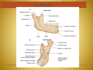
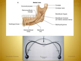
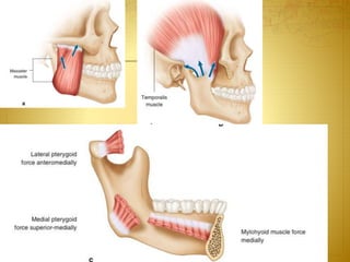
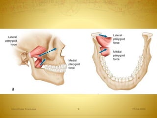
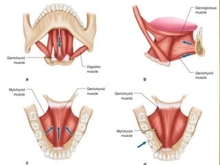
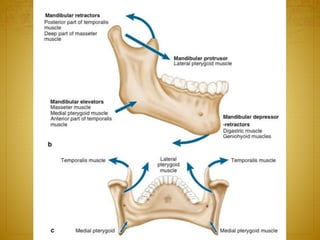
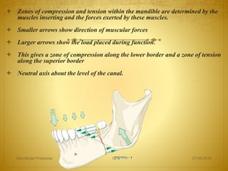
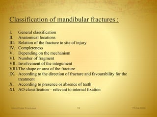
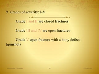
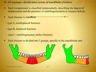
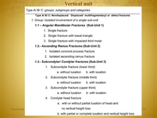
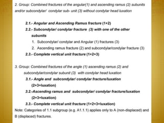
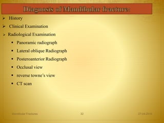
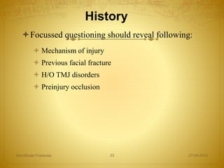
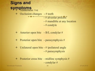
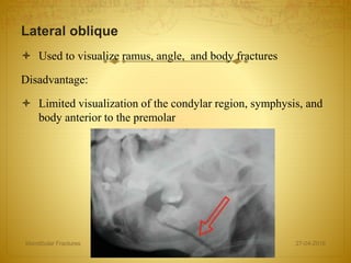
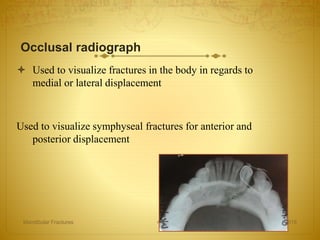
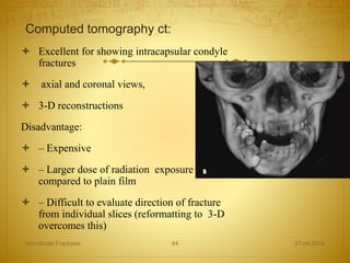
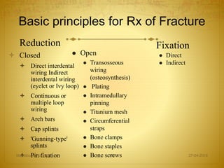
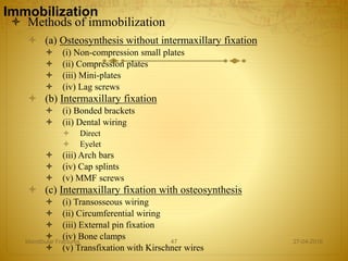
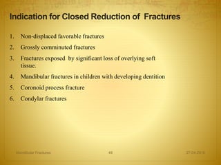
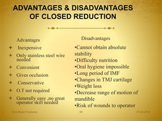
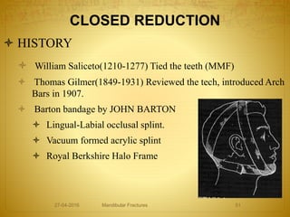
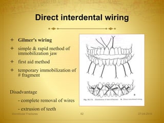
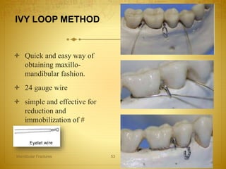
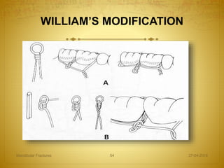
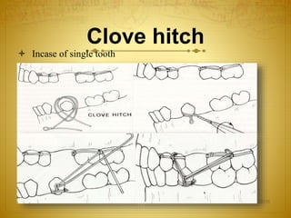
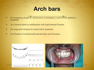
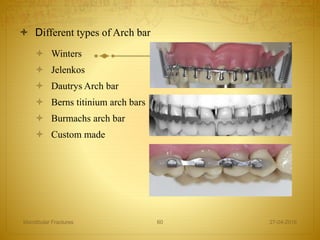
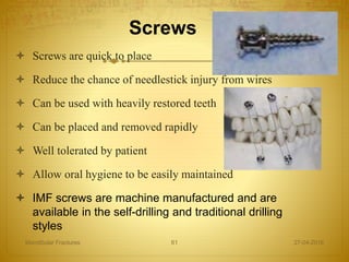
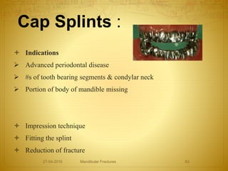
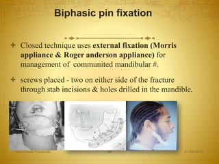
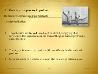
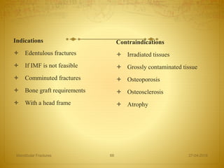
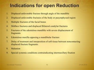
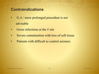
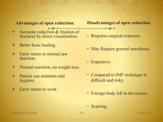
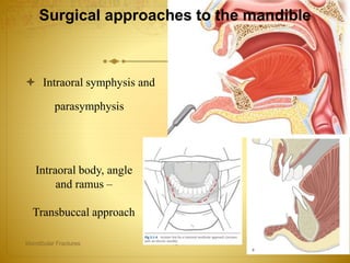
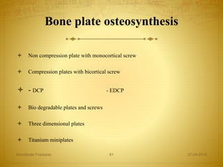
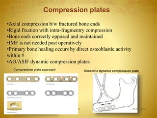
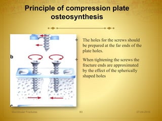
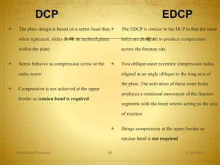
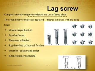
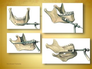
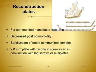
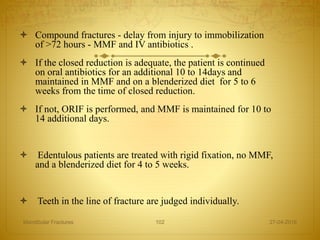
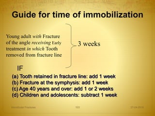
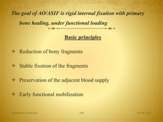
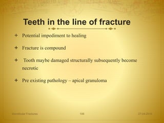
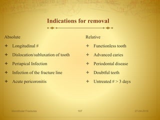
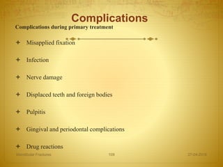
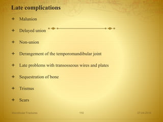
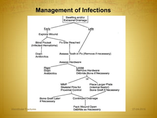
The document discusses the history, anatomy, classification, examination, diagnosis, and treatment of mandibular fractures. It traces the history from descriptions in ancient texts to modern techniques. The mandible has a unique anatomy as a bent bone that provides both compression and tension zones. Fractures are classified in various ways including by location, number of fragments, involvement of surrounding tissues, and relation to occlusion. Clinical examination involves inspection, palpation, and neurological and range of motion tests while radiographs help confirm and characterize fractures.
Dr. Ahmed M. Adawy, Professor Emeritus, Dep. Oral & Maxillofacial Surgery. Former Dean, Faculty of Dental Medicine, Al-Azhar University. Mandibular angle fractures account for 23% to 42% of all facial fractures. Fracture of mandibular angle can be classified as (A) Vertical favorable or unfavorable, (B) Horizontally favorable of unfavorable. Traditionally, mandibular angle fractures have been treated with either closed reduction and inter-maxillary fixation or open reduction and internal fixation with or without inter-maxillary fixation. Patients treated with inter-maxillary fixation have a restricted airway and loose excess weight. Rigid internal fixation and early return to function have eliminated the use of wire osteosenthysis and prolonged use of inter-maxillary fixation. The principal of rigid fixation, however, have inherent set of disadvantages including damage to the inferior alveolar nerve and the marginal mandibular branch of facial nerve. Postoperative malocclusion rates are also high. With the introduction of semi-rigid technique fracture of the mandibular angle could be treated according to Champy’s Ideal lines of osteosenthysis. The technique involves placement of a single monocortial miniplate on the superior border of the mandible. However, some studies suggested using a second miniplate along the inferior border. Wether one or two miniplates should be used is still debatable. The application of 3D plates may provide additional stability in 3 dimension and good resistance against torque forces.
Odontogenic keratocyst (OKC) is the cyst arising from the cell rests of dental lamina. It can occur anywhere in the jaw, but commonly seen in the posterior part of the mandible. Radiographically, most OKCs are unilocular when presented at the periapex and can be mistaken for radicular or lateral periodontal cyst.
The lower jaw frequently breaks due to accidents, assaults or sometimes due to underlying disease. Just as with other bones in the body, there are a various methods for repairing the mandible.
orthognathic surgery is very intresting and well knowing branch in oral surgery ....this presentation is dealing with jaw correction surgery in upper jaw.
The Indian Dental Academy is the Leader in continuing dental education , training dentists in all aspects of dentistry and offering a wide range of dental certified courses in different formats.
Indian dental academy provides dental crown & Bridge,rotary endodontics,fixed orthodontics,
Dental implants courses.for details pls visit www.indiandentalacademy.com ,or call
00919248678078
Le fort fracture by Dr. Amit Suryawanshi .Dentist in Kolhapur (MDS). Oral &...All Good Things
Description:
Hi. This is Dr. Amit T. Suryawanshi. Dentist in Kolhapur (MDS) Oral & Maxillofacial surgeon from Kolhapur, India. I am here on slideshare.com to share some of my own presentations presented at various levels in the field of OMFS. Hope this would somehow be helpful to you making your presentations. All the best & your replies are welcomed!
Suture Materials and Suturing Techniques - Presented by Dr. Prasanjit Das and group as a part of Dhaka Dental College, OMS Department weekly presentation program.
Mandibular fractures
Dr. Ahmed M. Adawy
Professor Emeritus, Dept. Oral & Maxillofacial Surg.
Former Dean, Faculty of Dental Medicine
Al-Azhar University
Fractures of the mandible are a common form of facial injury in adults and occur most frequently in males during the third decade of life. The main causes of mandibular fractures are road traffic accidents, interpersonal violence, falls and sport injuries. Mandibular fractures are classified according to various criteria. The three main factors to consider are the cause of the fracture, the type of fracture and the site of the fracture. Clinical diagnosis as well as radiographic examinations are presented. Treatment modalities are discussed. Moreover, treatment-related complications are given.
The Indian Dental Academy is the Leader in continuing dental education , training dentists in all aspects of dentistry and offering a wide range of dental certified courses in different formats.
Indian dental academy provides dental crown & Bridge,rotary endodontics,fixed orthodontics,
Dental implants courses.for details pls visit www.indiandentalacademy.com ,or call
00919248678078
Maxillofacial trauma mandible /certified fixed orthodontic courses by Indian ...Indian dental academy
The Indian Dental Academy is the Leader in continuing dental education , training dentists in all aspects of dentistry and offering a wide range of dental certified courses in different formats.
Indian dental academy provides dental crown & Bridge,rotary endodontics,fixed orthodontics,
Dental implants courses.for details pls visit www.indiandentalacademy.com ,or call
0091-9248678078
The Indian Dental Academy is the Leader in continuing dental education , training dentists in all aspects of dentistry and offering a wide range of dental certified courses in different formats.
Indian dental academy provides dental crown & Bridge,rotary endodontics,fixed orthodontics,
Dental implants courses.for details pls visit www.indiandentalacademy.com ,or call
00919248678078
Fractures of The Body of The Mandible In Maxillofacial SurgeryShahdHIbrahim
Fractures of the body of the mandible:
Introduction
Classification
History
Presentation
Examination
Radiography
Management
Complications
Post-operative Care
Dr. Ahmed M. Adawy, Professor Emeritus, Dep. Oral & Maxillofacial Surgery. Former Dean, Faculty of Dental Medicine, Al-Azhar University. Condylar fractures represent one of the most controversial issues in maxillofacial traumatology regarding classification, diagnoses and therapeutic management. Classification systems of condylar fracture is discussed. Diagnosis is usually based on history clinical examination and radiographic finding. Treatment ranges from observation, jaw exercises to closed or opened interventions. For years closed reduction was thought to be essentially complication-free. Several serious complications however have been reported including temporomandibular joint ankyloses, malocclusion, mandibular deviation and the generative joint pathology. The absolute and relative indications for open reduction is given. The debate between supporters of open or closed reduction is still continuing and the issue has not been resolved. However, the final choice treatment modality should takes into account the location of the fracture, age of the patient, presence or absence of other associated injuries, cosmetic impact of the surgery and presence of other systemic medical conditions.
Zygomatic Complex Fractures
Dr. Ahmed M. Adawy
Professor Emeritus, Dept. Oral & Maxillofacial Surg.
Former Dean, Faculty of Dental Medicine
Al-Azhar University
The term “zygomatic complex” refers to zygomatic bone and parts of maxilla, frontal, temporal and sphenoid bone. Fracture of the zygomatic complex, also known as a quadripod fracture, and formerly referred to as a tripod fracture, varies in severity from a simple crack to major disruption. The etiology, clinical presentations, and radiographic findings are presented. Classification systems are mentioned. The management of zygomatic complex fracture depends on the degree of displacement and the resultant esthetical and functional deficit. As a general rule, non- displaced or minimal displaced fracture can usually be treated conservatively. On the other hand, open reduction and internal fixation is applied in all dislocated, instable, and comminuted fractures of the zygomatic bone. Different surgical approaches and fixation methods are discussed.
The Indian Dental Academy is the Leader in continuing dental education , training dentists in all aspects of dentistry and
offering a wide range of dental certified courses in different formats.for more details please visit
www.indiandentalacademy.com
Dentist in pune.(BDS) MDS- OMFS - Dr. Amit T. Suryawanshi.. Mandibular fractu...All Good Things
Dentist in pune. (BDS. MDS) - Dr. Amit T. Suryawanshi. Seminar-Canine Impaction.
Email ID- amitsuryawanshi999@gmail.com
Contact -Ph no.-9405622455
Subscribe our channel on youtube - https://www.youtube.com/channel/UC_gylEXTrjmEbbOTSXjuZ4Q/videos?view_as=public
Follow us on slideshare
We all have good and bad thoughts from time to time and situation to situation. We are bombarded daily with spiraling thoughts(both negative and positive) creating all-consuming feel , making us difficult to manage with associated suffering. Good thoughts are like our Mob Signal (Positive thought) amidst noise(negative thought) in the atmosphere. Negative thoughts like noise outweigh positive thoughts. These thoughts often create unwanted confusion, trouble, stress and frustration in our mind as well as chaos in our physical world. Negative thoughts are also known as “distorted thinking”.
Synthetic Fiber Construction in lab .pptxPavel ( NSTU)
Synthetic fiber production is a fascinating and complex field that blends chemistry, engineering, and environmental science. By understanding these aspects, students can gain a comprehensive view of synthetic fiber production, its impact on society and the environment, and the potential for future innovations. Synthetic fibers play a crucial role in modern society, impacting various aspects of daily life, industry, and the environment. ynthetic fibers are integral to modern life, offering a range of benefits from cost-effectiveness and versatility to innovative applications and performance characteristics. While they pose environmental challenges, ongoing research and development aim to create more sustainable and eco-friendly alternatives. Understanding the importance of synthetic fibers helps in appreciating their role in the economy, industry, and daily life, while also emphasizing the need for sustainable practices and innovation.
How to Make a Field invisible in Odoo 17Celine George
It is possible to hide or invisible some fields in odoo. Commonly using “invisible” attribute in the field definition to invisible the fields. This slide will show how to make a field invisible in odoo 17.
The Roman Empire A Historical Colossus.pdfkaushalkr1407
The Roman Empire, a vast and enduring power, stands as one of history's most remarkable civilizations, leaving an indelible imprint on the world. It emerged from the Roman Republic, transitioning into an imperial powerhouse under the leadership of Augustus Caesar in 27 BCE. This transformation marked the beginning of an era defined by unprecedented territorial expansion, architectural marvels, and profound cultural influence.
The empire's roots lie in the city of Rome, founded, according to legend, by Romulus in 753 BCE. Over centuries, Rome evolved from a small settlement to a formidable republic, characterized by a complex political system with elected officials and checks on power. However, internal strife, class conflicts, and military ambitions paved the way for the end of the Republic. Julius Caesar’s dictatorship and subsequent assassination in 44 BCE created a power vacuum, leading to a civil war. Octavian, later Augustus, emerged victorious, heralding the Roman Empire’s birth.
Under Augustus, the empire experienced the Pax Romana, a 200-year period of relative peace and stability. Augustus reformed the military, established efficient administrative systems, and initiated grand construction projects. The empire's borders expanded, encompassing territories from Britain to Egypt and from Spain to the Euphrates. Roman legions, renowned for their discipline and engineering prowess, secured and maintained these vast territories, building roads, fortifications, and cities that facilitated control and integration.
The Roman Empire’s society was hierarchical, with a rigid class system. At the top were the patricians, wealthy elites who held significant political power. Below them were the plebeians, free citizens with limited political influence, and the vast numbers of slaves who formed the backbone of the economy. The family unit was central, governed by the paterfamilias, the male head who held absolute authority.
Culturally, the Romans were eclectic, absorbing and adapting elements from the civilizations they encountered, particularly the Greeks. Roman art, literature, and philosophy reflected this synthesis, creating a rich cultural tapestry. Latin, the Roman language, became the lingua franca of the Western world, influencing numerous modern languages.
Roman architecture and engineering achievements were monumental. They perfected the arch, vault, and dome, constructing enduring structures like the Colosseum, Pantheon, and aqueducts. These engineering marvels not only showcased Roman ingenuity but also served practical purposes, from public entertainment to water supply.
The French Revolution, which began in 1789, was a period of radical social and political upheaval in France. It marked the decline of absolute monarchies, the rise of secular and democratic republics, and the eventual rise of Napoleon Bonaparte. This revolutionary period is crucial in understanding the transition from feudalism to modernity in Europe.
For more information, visit-www.vavaclasses.com
How to Create Map Views in the Odoo 17 ERPCeline George
The map views are useful for providing a geographical representation of data. They allow users to visualize and analyze the data in a more intuitive manner.
Palestine last event orientationfvgnh .pptxRaedMohamed3
An EFL lesson about the current events in Palestine. It is intended to be for intermediate students who wish to increase their listening skills through a short lesson in power point.
Unit 8 - Information and Communication Technology (Paper I).pdfThiyagu K
This slides describes the basic concepts of ICT, basics of Email, Emerging Technology and Digital Initiatives in Education. This presentations aligns with the UGC Paper I syllabus.
This is a presentation by Dada Robert in a Your Skill Boost masterclass organised by the Excellence Foundation for South Sudan (EFSS) on Saturday, the 25th and Sunday, the 26th of May 2024.
He discussed the concept of quality improvement, emphasizing its applicability to various aspects of life, including personal, project, and program improvements. He defined quality as doing the right thing at the right time in the right way to achieve the best possible results and discussed the concept of the "gap" between what we know and what we do, and how this gap represents the areas we need to improve. He explained the scientific approach to quality improvement, which involves systematic performance analysis, testing and learning, and implementing change ideas. He also highlighted the importance of client focus and a team approach to quality improvement.
Read| The latest issue of The Challenger is here! We are thrilled to announce that our school paper has qualified for the NATIONAL SCHOOLS PRESS CONFERENCE (NSPC) 2024. Thank you for your unwavering support and trust. Dive into the stories that made us stand out!
2. HISTORY
INTRODUCTION
ANATOMY
CLASSIFICATION
EXAMINATION AND DIAGNOSIS
TREATMENT
27-04-2016Mandibular Fractures 2
3. The pre-Christian era
The first description of mandibular fractures dates to the 17th Century
BC in the ‘Edwin Smith papyrus’,
Hippocrates – direct reapproximation of # segments with the use of
circum dental wires
1180, Textbook written in Salerno, Italy – importance of establishing a
proper occlusion.
1492, the book Cyrurgia by Guglielmo Salicetti – first mention of the use
of maxillomandibular fixation in treatment of mandibular #.
27-04-2016Mandibular Fractures 3
4. History
1887, Gilmer reintroduced MMF in United States.
Buck & Kinlock- first to do ORIF using wires.
1888 Schede- First to use stainless steel plate & screws.
1960, Luhr- first to use Vitallium compression plate
1970, Spiessl through AO/ASIF introduced principles of rigid internal fixation.
1970, Michelet- introduced small bendable, non compression plates- these were
further modified by Champy.
1987 – M.S. Leonard first to report use of lag screws
Late 1990s – introduction of use of bioresorbable plates
27-04-2016Mandibular Fractures 4
5. • A tubular long bone, which is bent into a blunt V-shape.
• Mandible is strongest anteriorly in midline with
progressively less strength towards condyle .
• dentition
• Muscle attachments.
• Mandible is one of the strongest bones, the energy
required to # it being of the order of 44.6 –74.4 Kg /
M(425Lb), which is about same as zygoma and about ½
that of frontal bone
27-04-2016Mandibular Fractures 5
Mandible is embryologically a membrane bent bone although, resembles
physically long bone .
12. Zones of compression and tension within the mandible are determined by the
muscles inserting and the forces exerted by these muscles.
Smaller arrows show direction of muscular forces
Larger arrows show the load placed during function.
This gives a zone of compression along the lower border and a zone of tension
along the superior border
Neutral axis about the level of the canal.
27-04-2016Mandibular Fractures 12
13. FRACTURE :
Definition :
Fracture is defined as break in the continuity of the
bone.
Mandibular fractures :
Fractures of the mandible are common in patients, who
sustain facial trauma.
SEX :
Most mandibular fractures are seen to occur in male patients.
Ratio is approximately 4.5 : 1
AGE :
35 % of mandibular fractures occur between the ages of
20 to 30 years.
27-04-2016Mandibular Fractures 13
14. ETIOLOGY OF MANDIBULAR FRACTURES
Vehicular accidents
Altercation,assaults,
interpersonel violence
Fall
Sporting accidents
Industrial mishaps or work
accidents
Pathological fractures or
miscellaneous
27-04-2016Mandibular Fractures 14
15. Factors influencing displacement
of fracture
Degree of force
Resistance to the force offered by the facial bones
Direction of force
Point of application of force
Cross-sectional area of the agent or object struck
Attached muscles
27-04-2016Mandibular Fractures 15
16. # SYMPHYSIS AND PARASYMPHYSIS:-
Mylohyoid constitues a diaphragm b/w hyoid bone &
mylohyoid ridge on inner aspect of mandible
• Oblique # in this region tends to overlaps -- genio &
mylohyoid diaphragm
27-04-2016Mandibular Fractures 16
17. Bucket handle displacement
27-04-2016Mandibular Fractures 17
•B/L # of parasymphysis results from
force which disrupts the periosteum.
• displaced posteriorly under the
influence of genioglossus /
geniohyoid muscle
•Often removes attachment of tongue
& allows
TONGUE FALL BACK
18. Classification of mandibular fractures :
I. General classification
II. Anatomical locations
III. Relation of the fracture to site of injury
IV. Completeness
V. Depending on the mechanism
VI. Number of fragment
VII. Involvement of the integument
VIII.The shape or area of the fracture
IX. According to the direction of fracture and favourability for the
treatment
X. According to presence or absence of teeth
XI. AO classification – relevant to internal fixation
27-04-2016Mandibular Fractures 18
19. 1. Kruger's general classification
• Simple or Closed Fracture
• Compound or Open
• Comminuted
• Complicated or complex
• Impacted
• Greenstick fracture
• Pathological
27-04-2016Mandibular Fractures 19
20. 2. Rowe & Killey classification
• Fractures not involving basal bone
• Fractures involving basal bone of the mandible. Subdivided into following:
Single Unilateral
Double unilateral
Bilateral
Multiple
3. Dingman & Natvig classification
• Midline
• Parasymphyseal
• Symphysis
• Body
• Angle
• Ramus
• Condylar process
• Coronoid process
• Alveolar process 27-04-2016Mandibular Fractures 20
21. 4. Kruger & Schilli classification
I. Relation to the external environment
• Simple Or closed
• Compound or open
II. Types of fracture
• Incomplete
• Greenstick
• Complete
• Comminuted
III. Dentition of the jaw with reference to the use of splint
• Sufficiently dentulous patient
• Edentulous or insufficiently dentulous patient
• Primary and Mixed dentition
IV. Localization
• Fractures of the symphysis region between canines
• Fractures of the canine region
• Fractures of the body of the mandible
• Fractures of the angle
• Fractures of the mandibular ramus
• Fractures of the coronoid process
• Fractures of the condyle
27-04-2016Mandibular Fractures 21
22. 5. Kazanjian classification
Class – III : Patient is edentulolus
Class – I : teeth are present on both sides of
the fracture line
Class – II : Teeth are present on only one side of
fracture line
27-04-2016Mandibular Fractures 22
24. 7. Relation of the fracture to the site of injury
Direct fracture
Indirect fracture
27-04-2016Mandibular Fractures 24
25. 8. AO Classification(relevant to internal fixation):
1) F: Number of fracture or fragments
2) L: Location (site) of fracture
3) O: Status of occlusion
4) S: Soft tissue involvement
5) A: Associated fractures of facial skeleton
27-04-2016Mandibular Fractures 25
26. 9. Grades of severity: I-V
Grade I and II are closed fractures
Grade III and IV are open fractures
Grade V open fracture with a bony defect
(gunshot)
27-04-2016Mandibular Fractures 26
27. 10. AO-analogue classification system of mandibular fractures
Each compartment is classified independently, describing the degree of
displacement and the presence of multifragmentation or osseous defects.
Each fracture is classified:
- type A, nondisplaced fractures
- type B, displaced fractures
- type C, multifragmentary/defect fractures
Each fracture is divided into 3 groups, specific to the mandibular unit.
27-04-2016Mandibular Fractures 27
33. History
Focussed questioning should reveal following:
Mechanism of injury
Previous facial fracture
H/O TMJ disorders
Preinjury occlusion
27-04-2016Mandibular Fractures 33
34. Clinical examination
Examination of pt with # of mandible takes place in 3
stages:
A. Immediate assessment and treatment of any
condition constituting a threat to life.
B. General clinical examination of pt.
C. Local examination of mandibular #.
27-04-2016Mandibular Fractures 34
35. • Change in occlusion
• Anesthesia, Paresthesia or Dysesthesia of
lower lip
• Abnormal mandibular movements
• Change in facial contour and mandibular arch
form
• Laceration, Hematoma and Ecchymosis
• Loose teeth and crepitation on palpation
Clinical Examination
27-04-2016Mandibular Fractures 35
38. Signs and
symptoms Tenderness +ve
Occlusion changes - # teeth
- # alveolar process
- # mandible at any location
- # condyle
Anterior open bite - B/L condylar #
Posterior open bite - parasymphysis #
Unilateral open bite - # ipsilateral angle
- # parasymphysis
Posterior cross bite - midline symphysis #
- condylar #
27-04-2016Mandibular Fractures 38
39. Radiological examination
Ideally need 2 radiographic views of the fracture that are
oriented 90’ from one another to properly work up
fractures
Single view can lead to misdiagnosis and complications
with treatment
27-04-2016Mandibular Fractures 39
40. OPG
Most informative
Shows entire mandible and direction of fracture (horizontal favorable,
unfavorable)
Disadvantages:
– Patient must sit up up-right
– Difficult to determine buccal/lingual bone and medial condylar
displacement
– Some detail is lost/blurred in the symphysis, TMJ and dentoalveolar
regions
27-04-2016Mandibular Fractures 40
41. Posteroanterior (pa) radiograph:
Shows displacement of fractures in the ramus, angle, body,
and symphysis region
Disadvantage:
Cannot visualize the condylar
region
27-04-2016Mandibular Fractures 41
42. Lateral oblique
Used to visualize ramus, angle, and body fractures
Disadvantage:
Limited visualization of the condylar region, symphysis, and
body anterior to the premolar
27-04-2016Mandibular Fractures 42
43. Occlusal radiograph
Used to visualize fractures in the body in regards to
medial or lateral displacement
Used to visualize symphyseal fractures for anterior and
posterior displacement
27-04-2016Mandibular Fractures 43
44. Computed tomography ct:
Excellent for showing intracapsular condyle
fractures
axial and coronal views,
3-D reconstructions
Disadvantage:
– Expensive
– Larger dose of radiation exposure
compared to plain film
– Difficult to evaluate direction of fracture
from individual slices (reformatting to 3-D
overcomes this)
27-04-2016Mandibular Fractures 44
45. 1. The patient’s general physical status
2. Diagnosis and treatment of mandibular fractures should be
approached methodically not with an “emergency-type” mentality
3. Dental injuries should be evaluated and treated concurrently with
treatment of mandibular fractures
4. Re-establishment of occlusion is the primary goal in the treatment of
mandibular fracture.
5. With multiple facial fracture mandibular fracture should be treated
first.
6. Intermaxillary fixation time should vary according to the type,
location, number severity of the mandibular fracture as well as the
patient’s age and health.
7. Prophylactic antibiotics should be used for compound fractures.
General principles in the treatment of
mandibular fracture
27-04-2016Mandibular Fractures 45
46. Basic principles for Rx of Fracture
Reduction
Closed
Direct interdental
wiring Indirect
interdental wiring
(eyelet or Ivy loop)
Continuous or
multiple loop
wiring
Arch bars
Cap splints
'Gunning-type'
splints
Pin fixation 27-04-2016Mandibular Fractures 46
Open
Transosseous
wiring
(osteosynthesis)
Plating
Intramedullary
pinning
Titanium mesh
Circumferential
straps
Bone clamps
Bone staples
Bone screws
Fixation
Direct
Indirect
47. Immobilization
Methods of immobilization
(a) Osteosynthesis without intermaxillary fixation
(i) Non-compression small plates
(ii) Compression plates
(iii) Mini-plates
(iv) Lag screws
(b) Intermaxillary fixation
(i) Bonded brackets
(ii) Dental wiring
Direct
Eyelet
(iii) Arch bars
(iv) Cap splints
(v) MMF screws
(c) Intermaxillary fixation with osteosynthesis
(i) Transosseous wiring
(ii) Circumferential wiring
(iii) External pin fixation
(iv) Bone clamps
(v) Transfixation with Kirschner wires
27-04-2016Mandibular Fractures 47
49. 1. Non-displaced favorable fractures
2. Grossly comminuted fractures
3. Fractures exposed by significant loss of overlying soft
tissue.
4. Mandibular fractures in children with developing dentition
5. Coronoid process fracture
6. Condylar fractures
Indication for Closed Reduction of Fractures
27-04-2016Mandibular Fractures 49
50. ADVANTAGES & DISADVANTAGES
OF CLOSED REDUCTION
Advantages
Inexpensive
Only stainless steel wire
needed
Convenient
Gives occlusion
Conservative
O.T not required
Generally easy ,no great
operator skill needed
Disadvantages
27-04-2016Mandibular Fractures 50
•Cannot obtain absolute
stability
•Difficulty nutrition
•Oral hygiene impossible
•Long period of IMF
•Changes in TMJ cartilage
•Weight loss
•Decrease range of motion of
mandible
•Risk of wounds to operator
51. CLOSED REDUCTION
HISTORY
William Saliceto(1210-1277) Tied the teeth (MMF)
Thomas Gilmer(1849-1931) Reviewed the tech, introduced Arch
Bars in 1907.
Barton bandage by JOHN BARTON
Lingual-Labial occlusal splint.
Vacuum formed acrylic splint
Royal Berkshire Halo Frame
27-04-2016 Mandibular Fractures 51
52. Direct interdental wiring
Gilmer's wiring
simple & rapid method of
immobilization jaw
first aid method
temporary immobilization of
# fragment
Disadvantage
- complete removal of wires
- extrusion of teeth
27-04-2016Mandibular Fractures 52
53. IVY LOOP METHOD
Quick and easy way of
obtaining maxillo-
mandibular fashion.
24 gauge wire
simple and effective for
reduction and
immobilization of #
27-04-2016Mandibular Fractures 53
56. Button Wiring
Leonard (1977) considers that eyelet
wires have several drawbacks.
He described the use of titanium buttons
of 8mm diameter, inclusive of a 1mm
rim, and 2mm deep.
27-04-2016Mandibular Fractures 56
59. Arch bars
For temporary fragment stabilization in emergency cases before definitive
treatment
As a tension band in combination with rigid internal fixation
For long-term fixation in conservative treatment
For fixation of avulsed teeth and alveolar crest fractures
27-04-2016Mandibular Fractures 59
60. Different types of Arch bar
Winters
Jelenkos
Dautrys Arch bar
Berns titinium arch bars
Burmachs arch bar
Custom made
27-04-2016Mandibular Fractures 60
61. Screws
Screws are quick to place
Reduce the chance of needlestick injury from wires
Can be used with heavily restored teeth
Can be placed and removed rapidly
Well tolerated by patient
Allow oral hygiene to be easily maintained
IMF screws are machine manufactured and are
available in the self-drilling and traditional drilling
styles
27-04-2016Mandibular Fractures 61
62. Monocortical in nature
Once a screw loosens, it must be removed and replaced, or an
alternative method of reduction of the fracture should be
considered
Do not allow for any dynamic movement, and occlusal
discrepancies may not be adjusted as with arch bars and
elastics.
27-04-2016Mandibular Fractures 62
Disadvantage
63. Cap Splints :
Indications
Advanced periodontal disease
#s of tooth bearing segments & condylar neck
Portion of body of mandible missing
Impression technique
Fitting the splint
Reduction of fracture
27-04-2016 Mandibular Fractures 63
64. Biphasic pin fixation
Closed technique uses external fixation (Morris
appliance & Roger anderson appliance) for
management of communited mandibular #.
screws placed - two on either side of the fracture
through stab incisions & holes drilled in the mandible.
27-04-2016Mandibular Fractures 64
65. Once external pins are in position,
the fracture segments are manipulated to
achieve reduction.
Then the pins are locked in reduced position by applying of an
acrylic mix that is placed over the ends of the pins that are protruding
out of the skin.
The acrylic is allowed to harden while mandible is held in reduced
position.
Steinmann pins or Kirshner wires can also be used as external pins
27-04-2016Mandibular Fractures 65
66. Indications
Edentulous fractures
If IMF is not feasible
Comminuted fractures
Bone graft requirements
With a head frame
Contraindications
Irradiated tissues
Grossly contaminated tissue
Osteoporosis
Osteosclerosis
Atrophy
27-04-2016Mandibular Fractures 66
67. Advantages
Control of the edentulous
fragments without involving
the fracture lines.
under LA.
avoidance of the need for
surgery at the fracture site,
minimum operative time
Simple surgical technique.
Disadvantages
Conspicuous uncomfortable
uncooperative or cerebrally
irritated patient.
Difficulty with washing and
shaving
scars caused- pinholes
risk of infection.
27-04-2016Mandibular Fractures 67
68. • Used in edentulous jaw fractures
• Acrylic splints take the form of modified dentures with bite
block in place of molar teeth & space in the incisor area to
facilitate feeding
Gunning splints
27-04-2016Mandibular Fractures 68
69. INDICATION
unilateral / bilateral # edentulous mandible
CONTRAINDICATIONS
unfavorable displaced #s lying out side denture
bearing areas
severe posterior displacement of #s of the anterior
part of mandible
27-04-2016Mandibular Fractures 69
72. 1. Displaced unfavorable fracture through angle of the mandible
2. Displaced unfavorable fractures of the body or pasymphyseal region
3. Multiple fractures of the facial bones
4. Midface fractures and displaced Bilateral condylar fractures
5. Fractures of the edentulous mandible with severe displacement of
fragments
6. Edentulous maxilla opposing a mandibular fracture
7. Delay of treatment and interposition of soft tissue between noncontacting
displaced fracture fragments.
8. Malunion
9. Special systemic conditions contraindicating intermaxillary fixation
Indications for open Reduction
27-04-2016Mandibular Fractures 72
73. Contraindications
• G.A / more prolonged procedure is not
advisable
• Gross infections at the # site
• Severe comminution with loss of soft tissue
• Patients with difficult to control seizures
27-04-2016Mandibular Fractures 73
74. Advantages of open reduction.
Accurate reduction & fixation of
fractures by direct visualization.
Better bone healing.
Early return to normal jaw
function.
Normal nutrition, no weight loss.
Patient can maintain oral
hygiene.
Early return to work.
27-04-2016Mandibular Fractures 74
Disadvantages of open reduction.
• Requires surgical exposure.
• May Require general anesthesia.
• Expensive.
• Compared to IMF technique is
difficult and risky
• Foreign body left in the tissues.
• Scarring.
75. Surgical approaches to the mandible
Intraoral symphysis and
parasymphysis
27-04-2016Mandibular Fractures 75
Intraoral body, angle
and ramus –
Transbuccal approach
78. Transalveolar / upper border wiring
Sir Williams Kelsey Fry
To control the posterior fragment
Use – vertically and horizontally unfavorable #
Horizontal mattress wiring
27-04-2016Mandibular Fractures 78
79. Transosseous / lower border wiring
Hayton Williams 1958
# fragments expose extraorally
posterior fragment hole higher level then anterior
fragment
both wires passes simultaneously through same hole
1973 Obwegeser :-
Combined direct and figure of ‘8’ wiring with single
stand of wire
27-04-2016Mandibular Fractures 79
81. Bone plate osteosynthesis
Non compression plate with monocortical screw
Compression plates with bicortical screw
- DCP - EDCP
Bio degradable plates and screws
Three dimensional plates
Titanium miniplates
27-04-2016Mandibular Fractures 81
82. Compression plates
•Axial compression b/w fractured bone ends
•Rigid fixation with intra-fragmentry compression
•Bone ends correctly opposed and maintained
•IMF is not needed post operatively
•Primary bone healing occurs by direct osteoblastic activity
within #
•AO/ASIF dynamic compression plates
Compression plate approach Eccentric dynamic compression plate
27-04-2016Mandibular Fractures 82
83. Principle of compression plate
osteosynthesis
The holes for the screws should
be prepared at the far ends of the
plate holes.
When tightening the screws the
fracture ends are approximated
by the effect of the spherically
shaped holes
27-04-2016Mandibular Fractures 83
84. DCP EDCP
The plate design is based on a screw head that,
when tightened, slides down an inclined plane
within the plate.
Screw behaves as compression screw or the
static screw
Compression is not achieved at the upper
border so tension band is required
The EDCP is similar to the DCP in that the inner
holes are designed to produce compression
across the fracture site
Two oblique outer eccentric compression holes
aligned at an angle oblique to the long axis of
the plate. The activation of these outer holes
produces a rotational movement of the fracture
segments with the inner screws acting as the axis
of rotation
Brings compression at the upper border so
tension band is not required
27-04-2016Mandibular Fractures 84
85. Mini plate Osteosynthesis :-
1973 MICHELET
1975 CHAMPY MODIFIED
- Under physiological strain, forces of tension along the alveolar border &
forces of compression along the lower border of the mandible.
- With in the body of the mandible these forces produce, predominantly,
moments of flexion – angle strong & weak in PM region.
- with in the symphysis – torsional moments
- Champy et al analysed these moments using a mathematical model of the
mandible – ideal line of osteosynthesis.
# symphysis 2 plates
# angle 1 plate
Monocortical screws 2 mm diameter and 5 to 10 mm length
Plate 2cm long, 0.9mm thick and 6mm wide
27-04-2016Mandibular Fractures 85
87. Advantages of monocortical
miniplate osteosynthesis over
bicortical compression plates.
Monocortical
Requires minimal dissection.
Less technique sensitive
Less chances of complications
Bicortical
Extra oral approach
Nerve injury
Difficult to adapt
27-04-2016Mandibular Fractures 87
88. Compression plate Miniplates
Bicortical plates
Bulky and difficult to use
Applied extraorally
Cannot be used at the upper border of
the mandible
Provides rigid fixation
No interfragmentary movement
allowed
Monocortical plates
Easy to use
Applied intraorally, small incision , less
soft tissue dissection , less likely to be
palpable
Can be used without any associated
complication
Provides functionally stable fixation
Little interfragmentary movement
present, torsional movement seen under
functional loading
27-04-2016Mandibular Fractures 88
91. 3-D plate ostesynthesis
Titanium 3-D plating system was developed by Farmand to meet
the requirements of semi-rigid fixation with lesser complications.
The 3-D miniplate is a misnomer as the plates are not three
dimensional, but hold the fracture fragments rigidly by resisting
the forces in three dimensions, namely, shearing, bending, and
torsional forces.
The basic concept of 3-D fixation as explained by Farmand
is that a geometrically closed quadrangular plate secured with bone
screws creates stability in three dimensions. The stability is gained
over a defined surface area and is achieved by its configuration
and not by its thickness or length.
27-04-2016Mandibular Fractures 91
92. ADVANTAGES
The large free areas between the plate arms and minimal
dissection permit good blood supply to the bone.
The 3-D plating system uses fewer plates and screws as
compared to the conventional miniplates, to stabilize the bone
fragments. Thus, it uses lesser foreign material, and reduces
the operation time and overall cost of the treatment
The 3-D plating system has a compact design and is easy to
use. The 1.0-mm-thick 3-D plate is as stable as the much
thicker 2.0 mm miniplate.
This offers better bending stability and more resistance to out-
of plane movement or torque.
27-04-2016Mandibular Fractures 92
95. Bioabsorbable Plates
Bioresorbable materials used for rigid fixation
Polydioxanone
Polyglycolic acid
Polylactic acid
Strength inadequate to provide clinically acceptable rigid fixation.
Use of poly-L-lactide (PLLA) in 69 fractures by Kim et al
12% complication
8% infection
No malunion
27-04-2016Mandibular Fractures 95
96. Bioresorbable plates & screws
Advantages:
Provides the proper strength
when necessary and then
harmlessly degrades over
time.
No need for an additional
removal operation.
Reduce the total treatment &
rehabilitation time of the
patient.
No bending pliers are
necessary. 27-04-2016Mandibular Fractures 96
97. Lag screw
Compress fracture fragments without the use of bone plate
Two sound bony cortices are required -- Shares the loads with the bone
Uses:
absolute rigid fixation
Less hardware
More cost effective
Rigid method of internal fixation
Insertion -quicker and easier
Reduction more accurate
27-04-2016Mandibular Fractures 97
98. Lag screws
Placed in direction that is perpendicular to the line of
fracture to prevent overriding & displacement during
tightening of the screws.
INDICATIONS
• #s in edentulous parts
• Concomittant #s of body & condyle
• IMF contraindicated
• Saggital/oblique fractures
• Non/malunion
27-04-2016Mandibular Fractures 98
100. Reconstruction
plates
27-04-2016Mandibular Fractures 100
For communited mandibular fractures
Decreased post op morbidity
Stabilization of entire communited complex
2.0 mm plate with bicortical screw used in
conjunction with lag screws or miniplates
101. Protocol for treatment of mandibular fractures
Simple fractures of the condylar process and ramus -
closed reduction. MMF for 48 to72 hours - training
elastics and close observation
No MMF is required for coronoid fractures; archbars
and training elastics are used only if a malocclusion is
present.
Simple or compound fractures with a time delay from
injury to immobilization of < 72 hours are treated by a
closed reduction (CR) or, if indicated, open reduction
with rigid fixation (ORIF).
27-04-2016Mandibular Fractures 101
102. Compound fractures - delay from injury to immobilization
of >72 hours - MMF and IV antibiotics .
If the closed reduction is adequate, the patient is continued
on oral antibiotics for an additional 10 to 14days and
maintained in MMF and on a blenderized diet for 5 to 6
weeks from the time of closed reduction.
If not, ORIF is performed, and MMF is maintained for 10 to
14 additional days.
Edentulous patients are treated with rigid fixation, no MMF,
and a blenderized diet for 4 to 5 weeks.
Teeth in the line of fracture are judged individually.
27-04-2016Mandibular Fractures 102
103. Young adult with Fracture
of the angle receiving Early
treatment in which Tooth
removed from fracture line
3 weeks
Guide for time of immobilization
27-04-2016Mandibular Fractures 103
(a) Tooth retained in fracture line: add 1 week
(b) Fracture at the symphysis: add 1 week
(c) Age 40 years and over: add 1 or 2 weeks
(d) Children and adolescents: subtract 1 week
IF
105. The goal of AO/ASIF is rigid internal fixation with primary
bone healing, under functional loading
Basic principles
Reduction of bony fragments
Stable fixation of the fragments
Preservation of the adjacent blood supply
Early functional mobilization
27-04-2016Mandibular Fractures 105
106. Teeth in the line of fracture
Potential impediment to healing
Fracture is compound
Tooth maybe damaged structurally subsequently become
necrotic
Pre existing pathology – apical granuloma
27-04-2016Mandibular Fractures 106
107. Absolute
Longitudinal #
Dislocation/subluxation of tooth
Periapical Infection
Infection of the fracture line
Acute pericoronitis
Relative
Functionless tooth
Advanced caries
Periodontal disease
Doubtful teeth
Untreated # > 3 days
27-04-2016Mandibular Fractures 107
Indications for removal
108. Management of teeth retained in fracture line
Intra-oral periapical radiograph
Systemic antibiotic therapy
Splinting of tooth if mobile
Endodontic therapy if pulp exposed
Immediate extraction if fracture becomes infected
Follow-up for 1 yr with endodontic therapy if there is demonstrable loss of
vitality.
27-04-2016Mandibular Fractures 108
109. Complications
Complications during primary treatment
Misapplied fixation
Infection
Nerve damage
Displaced teeth and foreign bodies
Pulpitis
Gingival and periodontal complications
Drug reactions
27-04-2016Mandibular Fractures 109
110. Late complications
Malunion
Delayed union
Non-union
Derangement of the temporomandibular joint
Late problems with transosseous wires and plates
Sequestration of bone
Trismus
Scars
27-04-2016Mandibular Fractures 110
112. Reference
Maxillofacial injuries – N.L. Rowe, J Williams, Vol 1
Ied.
Oral & maxillofacial trauma – Raymond J Fonseca 4th
ed
Journal of Cranio-Maxillofacial Surgery 2008; 36: e251 -
e259
Subodh et al, Clinical Study An Epidemiological Study on
Pattern and Incidence of Mandibular Fractures, Hindawi
Publishing Corporation Plastic Surgery International,
Volume 2012, Article ID 834364,7pages
27-04-2016Mandibular Fractures 112
113. A comprehensive classification of mandibular fractures: a
preliminary agreement validation studyC. H. Buitrago-Tellez, L.
Audige, B. Strong, P. Gawelin, J. Hirsch Int. J. Oral Maxillofac.
Surg. 2008; 37: 1080–1088.
A. H. Kamboozia, A. Punnia-Moorthy: The fate of teeth in
mandibular fracture lines. A clinical and radiographic follow-up
study. Int. J. Oral Maxillofac. Surg 1993; 22. 9~101.
Atlas Oral Maxillofacial Surg Clin N Am 17 (2009) 81–
91,Fractures of the Growing Mandible;George M. Kushner, Paul
S. Tiwana.
Protocol for treatment of mandibular fractures
Philip L. Maloney,J Oral Maxillofac Surg,59:879-884, 2001.
27-04-2016Mandibular Fractures 113
114. 3-D plate osteosynthesis; Dental Research Journal /Mar 2012 /Vol 9 /
Issue 2
R. Mukerji , G. Mukerji , M. McGurk Mandibular fractures: Historical
perspective British Journal of Oral and Maxillofacial Surgery 44 (2006)
222–228
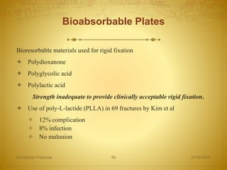
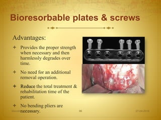
Bioresorbable plates & screws[Robert M. Laughlin JOMS
2007;65:89-96]
Killey & kay textbook of mandibular fractures.
27-04-2016Mandibular Fractures 114
[Swearingen 1965, Hodgson 1967, Nahum 1975a, Luce et al 1979]
Mylohyoid, Geniohyoid, Genioglossus & Anterior belly of omohyoid – postero-medial & inferior displacement of # fracture fragment.
Pterygomassetric sling – Supero-medial & anterior displacement of fractured lesser fragment.
Lateral Pterygoid muscle- Antero-medial displacement of fractured condyle.
Temporalis – postero-superior displacement of fractured coronoid process.
Panoramic view showing the vertical mandibular units (green), the lateral horizontal units (orange) and the central mandibular unit (red)
should be carefully evaluated and monitored prior to any consideration of treating mandibular fracture.
A 24-gauge wire, approximately 4 to 6 in long, is twisted into a 2- to 3-mm loop directly in the middle of the wire.
3mm diameter 5 cm length
A small 2-mm incision can be made in the mucosa and down through periosteum
poly(L-lactide) (PLLA)
Biodegradable materials usually degrade in vivo through a two-phase process.
During phase 1, water molecules hydrolytically attack the chemical bonds, cutting long polymer chains to many short chains
Phase 2 involves the cellular response whereby macrophages and giant cells metabolize the products of phase 1 degrada- tion into substances, such as water and carbon dioxide
In contrast, a true lag screw has threads only at its termi- nal end. When used, the threads engage the distant cortex and the head sits against the proximal cortex, resulting in compression and mechanical resthe Eckelt technique for treatment of condylar neck
The Krenkel technique for treatment of condylar neck fractures.