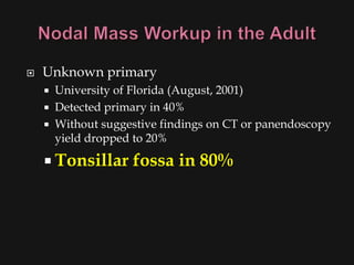
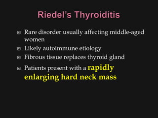
Evaluation of the Neck:
  The Thyroid Gland


      Frederick Untalan MD
   Common clinical finding – NECK   MASS
   All age groups
   Very complex differential
    diagnosis!
   Systematic approach essential
   Prominent
    landmarks
   Triangles of
    the neck
   Carotid bulb
   Lymphatic
    levels
   Prominent
    landmarks
   Triangles of the
    neck
   Carotid bulb
   Lymphatic levels
   Prominent
    landmarks
   Triangles of the
    neck
   Carotid bulb
   Lymphatic levels
   Patient age
       Pediatric (0 – 15 years): 90% benign
       Young adult (16 – 40 years): similar to pediatric
       Late adult (>40 years): “rule of 80s”
   Location
       Congenital masses: consistent in location
       Metastatic masses: key to primary lesion
Metastasis Location according
to Various Primary Lesions
   History
       Developmental time course
       Associated symptoms (dysphagia, otalgia, voice)
       Personal habits (tobacco, alcohol)
       Previous irradiation or surgery
   Physical Examination
       Complete head and neck exam (visualize & palpate)
       Emphasis on location, mobility and consistency
 Inflammatory mass suspected
 Two week trial of antibiotics
   Follow-up for further investigation
   Fine needle aspiration biopsy (FNAB)
   Computed tomography (CT)
   Magnetic resonance imaging (MRI)
   Ultrasonography
   Radionucleotide scanning
   Standard of diagnosis
   Indications
     Any neck mass that is not an obvious
      abscess
     Persistence after a 2 week course of
      antibiotics
   Small gauge needle
     Reduces bleeding
     Seeding of tumor – not a concern
   No contraindications (vascular ?)
   Distinguish cystic from
    solid
   Extent of lesion
   Vascularity (with contrast)
   Detection of unknown
    primary (metastatic)
   Pathologic node (lucent,
    >1.5cm, loss of shape)
   Avoid contrast in thyroid
    lesions
   Similar
    informati
    on as CT
   Better for
    upper
    neck and
    skull
    base
   Vascular
    delineati
    on with
    infusion
   Less important
    now with FNAB
   Solid versus cystic
    masses
   Congenital cysts
    from solid
    nodes/tumors
   Noninvasive
    (pediatric)
   Salivary and thyroid
    masses
   Location – glandular
    versus extra-glandular
   Functional information
   FNAB now preferred for
    for thyroid nodules
     Solitary nodules
     Multinodular goiter
       with new increasing
       nodule
     Hashimoto’s with new
       nodule
   Any solid asymmetric mass MUST
                                be
    considered a metastatic neoplastic
    lesion until proven otherwise
   Asymptomatic cervical mass
       12% of cancer
   ~ 80% of these are SCCa
   Ipsilateral otalgia with normal otoscopy –
    direct attention to tonsil, tongue base,
    supraglottis and hypopharynx
   Unilateral serous otitis – direct examination
    of nasopharynx
   Panendoscopy
     FNAB positive with no primary on repeat exam
     FNAB equivocal/negative in high risk patient
   Directed Biopsy
     All suspicious mucosal lesions
     Areas of concern on CT/MRI
     None observed – nasopharynx, tonsil
      (ipsilateral tonsillectomy for jugulodigastric
      nodes), base of tongue and piriforms
   Synchronous primaries (10 to 20%)
   Unknown primary
       University of Florida (August, 2001)
       Detected primary in 40%
       Without suggestive findings on CT or panendoscopy
        yield dropped to 20%

     Tonsillar      fossa in 80%
   Open excisional biopsy
     Only if complete workup negative
     Occurs in ~5% of patients
     Be prepared for a complete neck dissection
     Frozen section results (complete node
      excision)
       Inflammatory or granulomatous – culture
       Lymphoma or adenocarcinoma – close
        wound
   Congenital masses
   Benign lesions
   Vascular and lymphatic malformations
   Infectious and inflammatory conditions
   Malignant lesions
   Thyroid mass         Carotid body
   Lymphoma              and glomus
   Salivary tumors       tumors
   Lipoma               Neurogenic
                          tumors
   Leading cause of anterior
    neck masses
   Children
     Most common neoplastic
      condition
     Male predominance
     Higher incidence of
      malignancy
   Adults
       Female predominance
     Mostly benign
   Lymph node metastasis
     Initial symptom in 15% of papillary
      carcinomas
     40% with malignant nodules
     Histologically (microscopic) in >90%
   FNAB has replaced USG and radionucleotide
    scanning
     Decreases # of patients with surgery
     Increased # of malignant tumors found at
      surgery
     Doubled the # of cases followed up
     Unsatisfactory aspirate – repeat in 1 month
Nodular goiter


                 Goiter displacing the trachea
   Benign Nontoxic Conditions
     Diffuse and Nodular Goiter
   Benign Toxic Conditions
     Toxic Multinodular Goiter
     Graves’ Disease
     Toxic Adenoma
   Inflammatory Conditions
     Chronic (Hashimoto’s) Thyroiditis
     Subacute (De Quervain’s) Thyroiditis
     Riedel’s Thyroiditis
   Usually picked up on routine physical exam or
    as incidental finding
   Patients may have clinical or subclinical
    thyrotoxicosis
   Patients may have compressive symptoms:
    tracheal, vascular, esophageal, recurrent
    laryngeal nerve
   Suppressive Therapy
   Antithyroid Medications: Propylthiouracil and
    Methimazole
   I-131
   Surgical Therapy
   Antithyroid Drugs
     May require prolonged therapy
   Radioactive iodine
     May worsen ophthalmopathy unless
      followed by steroids
   Surgery
     Make patient euthyroid prior to surgery
     Potassium iodide two weeks prior to surgery
      can decrease the vascularity of the gland
   Autonomously functioning thyroid nodule
    hypersecreting T3 and T4 resulting in
    thyrotoxicosis (Plummer’s disease)
   Almost never malignant
   Manage with antithyroid drugs followed by
    either I-131 or surgery
   Also known as Hashimoto’s disease
   Probably the most common cause of
    hypothyroidism
   Autoantibodies include: thyroglobulin
    antibody, thyroid peroxidase antibody, TSH
    receptor blocking antibody
   Painless goiter in a patient who is either
    euthyroid or mildly hypothyroid
   Low incidence of permanent hypothyroidism
   May have periods of thyrotoxicosis
   Treat with levothyroxine
   Also known as De   Quervain's
    thyroiditis
   Most common cause of thyroid pain and
    tenderness
   Acute inflammatory disease most likely due to
    viral infection
   Transient hyperthyroidism followed by
    transient hypothyroidism; permanent
    hypothyroidism or relapses are uncommon
   Symptomatic: NSAIDS or a
    glucocorticoid
   Beta-blockers indicated if there are
    signs of thyrotoxicosis
   Levothyroxine may be given during
    hypothyroid phase
   Rare disorder usually affecting middle-aged
    women
   Likely autoimmune etiology
   Fibrous tissue replaces thyroid gland
   Patients present with a rapidly
    enlarging hard neck mass
   1.0%-1.5% of all new cancer cases
     tenfold less than that of lung,
      breast, or colorectal cancer
   8,000-14,000 new cases diagnosed each year
    (Sessions, 1993, Geopfert, 1998)
   3% of patients who die of other causes have
    occult thyroid cancer and 10% have microscopic
    cancers (Robbins, 1991)
   35% of thyroid gland at autopsy in some studies
    have papillary carcinomas (<1.0cm) (Mazzaferri,
    1993)
   1,000-1,200 patients in the U.S. die each year of
    thyroid cancer (Goldman, 1996)
   4%-7% of adults in North America have
    palpable thyroid nodules (Mazzaferri, 1993, Vander,
    1968)

   4:1 women to men (Mazzaferri, 1993)
   Overall, fewer than 5% of nodules are malignant
    (Mazzaferri, 1988)
   Symptoms
   The most common presentation of a thyroid
    nodule, benign or malignant, is a painless mass
    in the region of the thyroid gland (Goldman, 1996).
   Symptoms consistent with malignancy
         Pain
         dysphagia
         Stridor
         hemoptysis
         rapid enlargement
         hoarseness
   Risk factors
   Thyroid exposure to irradiation
       low or high dose external irradiation (40-50 Gy
        [4000-5000 rad])
       especially in childhood for:
         large thymus, acne, enlarged tonsils, cervical
          adenitis, sinusitis, and malignancies
       30%-50% chance of a thyroid nodule to be
        malignant (Goldman, 1996)
       Schneider and co-workers (1986) studied, with
        long term F/U, 3000 patients who underwent
        childhood irradiation.
         1145 had thyroid nodules
         318/1145 had thyroid cancer (mostly papillary)
   Family History
       History of family member with medullary
        thyroid carcinoma
       History of family member with other endocrine
        abnormalities (parathyroid, adrenals)
       History of familial polyposis (Gardner’s
        syndrome)
   Examination of the thyroid nodule:
       consistency - hard vs. soft
       size - < 4.0 cm
       Multinodular vs. solitary nodule
         multi nodular - 3% chance of malignancy
          (Goldman, 1996)
         solitary nodule - 5%-12% chance of
          malignancy (Goldman, 1996)
       Mobility with swallowing
       Mobility with respect to surrounding tissues
       Well circumscribed vs. ill defined borders
   Examine for ectopic thyroid tissue
   Indirect or fiberoptic laryngoscopy
       vocal cord mobility
       evaluate airway
       preoperative documentation of any
        unrelated abnormalities
   Systematic palpation of the neck
       Metastatic adenopathy commonly found:
         in the central compartment (level VI)
         along middle and lower portion of the
          jugular vein (regions III and IV) and
   Attempt to elicit Chvostek’s sign
   Thyroid function tests
       thyroxine (T4)
       triiodothyronin (T3)
       thyroid stimulating hormone (TSH)
   Serum Calcium
   Thyroglobulin (TG)
   Calcitonin
   Advantages
   Most sensitive procedure or identifying lesions
    in the thyroid (2-3mm)
   90% accuracy in categorizing nodules as solid,
    cystic, or mixed (Rojeski, 1985)
   Best method of determining the volume of a
    nodule (Rojeski, 1985)
   Can detect the presence of lymph node
    enlargement and calcifications
   Noninvasive and inexpensive
   Currently considered to be the best first-line
    diagnostic procedure in the evaluation of the thyroid
    nodule:
   Advantages:
        Safe
        Cost-effective
        Minimally invasive
        Leads to better selection of patients for
         surgery than any other test (Rojeski, 1985)
   Mechanism/Rationale
      Exogenous thyroid hormone feeds back to
       the pituitary to decrease the production of
       TSH
      Cancer is autonomous and does not require
       TSH for growth whereas benign processes do
      Thyroid masses that shrink with suppression
       therapy are more likely to be benign
      Thyroid masses that continue to enlarge are
       likely to be malignant
Limitations:
     16% of malignant nodules are suppressible
     Only 21% of benign nodules are suppressible
     Provides little use in distinguishing benign from
      malignant nodules


                                            (Geopfert, 1998)
   Uses:
     Preoperative
       patients with nonoxyphilic follicular neoplasms
       patients with solitary benign nodules that are
        nontoxic (particularly men and premenopausal
        women)
       women with repeated nondiagnostic tests
     Postoperative
       Use in follicular, papillary and Hurthle cell
        carcinomas
                      (Geopfert, 1998, Mazzaferri, 1993)
   How to use thyroid suppression
      administer levothyroxine
      maintain TSH levels at <0.1 mIU/L
      use ultrasound to monitor size of nodule
        if the nodule shrinks, continue L-thyroxine
         maintaining TSH at low-normal levels
        if the nodule has remained the same size after 3
         months, reaspirate
        if the nodule has increased in size, excise it

                                     (Geopfert, 1998,)
   Papillary carcinoma         Medullary Carcinoma
       Follicular variant      Miscellaneous
                                    Sarcoma
       Tall cell
                                    Lymphoma
       Diffuse sclerosing          Squamous cell carcinoma
       Encapsulated                Mucoepidermoid
   Follicular carcinoma             carcinoma
       Overtly invasive            Clear cell tumors
                                    Pasma cell tumors
       Minimally invasive
                                    Metastatic
   Hurthle cell carcinoma
                                      Direct extention
   Anaplastic carcinoma
                                      Kidney
       Giant cell                    Colon
       Small cell                    Melanoma
   Pathogenesis - unknown
   Papillary has been associated with the RET proto-
    oncogene but no definitive link has been proven
    (Geopfert, 1998)
   Certain clinical factors increase the likelihood of
    developing thyroid cancer
       Irradiation - papillary carcinoma
       Prolonged elevation of TSH (iodine deficiency) -
        follicular carcinoma (Goldman, 1996)
          relationship not seen with papillary carcinoma
          mechanism is not known
   60%-80% of all thyroid cancers (Geopfert, 1998, Merino, 1991)
   Histologic subtypes
       Follicular variant
       Tall cell
       Columnar cell
       Diffuse sclerosing
       Encapsulated
   Prognosis is 80% survival at 10 years (Goldman, 1996)
   Females > Males
   Mean age of 35 years (Mazzaferri, 1994)
   Lymph node involvement is common
      Major route of metastasis is   lymphatic
      46%-90% of patients have lymph node involvement
       (Goepfert, 1998, Scheumann, 1984, De Jong, 1993)
      Clinically undetectable lymph node involvement does not
       worsen prognosis (Harwood, 1978)
   20% of all thyroid malignancies
   Women > Men (2:1 - 4:1) (Davis, 1992, De Souza, 1993)
   Mean age of 39 years (Mazzaferri, 1994)
   Prognosis - 60% survive to 10 years (Geopfert, 1994)
   Metastasis - angioinvasion and hematogenous
               spread
       15% present with distant metastases to bone and lung
   Lymphatic involvement is seen in 13% (Goldman,
    1996)
   Variant of follicular carcinoma
   First described by Askanazy
       “Large, polygonal, eosinophilic thyroid follicular cells
       with abundant granular cytoplasm and numerous
       mitochondria” (Goldman, 1996)
   Definition (Hurthle cell neoplasm) - an
    encapsulated group of follicular cells with at least
    a 75% Hurthle cell component
   Carcinoma requires evidence of vascular and
    capsular invasion
   4%-10% of all thyroid malignancies (Sessions, 1993)
   Depending on variables, patients are categorized
    in to one of the following three groups:

      1) Low risk group - men younger than 40
       years and women younger than 50 years
                         regardless of histologic
       type
                        - recurrence rate -11%
                     - death rate - 4%

                                         (Cady and Rossi, 1988)
 1) Intermediate risk group - Men older than 40
  years and women older than 50 years who have
 papillary carcinoma
  - recurrence rate - 29%
   - death rate - 21%
 2) High risk group - Men older than 40 years and
  women older than 50 years who have follicular
        carcinoma
   - recurrence rate - 40%
   - death rate - 36%
   10% of all thyroid malignancies
   1000 new cases in the U.S. each year
   Arises from the parafollicular cell or C-cells of the
    thyroid gland
       derivatives of neural crest cells of the branchial arches
       secrete calcitonin which plays a role in calcium
       metabolism
   Familial MTC:
         Autosomal dominant transmission
         Not associated with any other endocrinopathies
         Mean age of 43
         Multifocal and bilateral
         Has the best prognosis of all types of MTC
         100% 15 year survival
                                    (Farndon, 1986)
   Multiple endocrine neoplasia IIa (Sipple’s
    Syndrome):
       MTC, Pheochromocytoma, parathyroid
        hyperplasia
       Autosomal dominant transmission
       Mean age of 27
       100% develop MTC (Cance, 1985)
       85%-90% survival at 15 years (Alexander, 1991,
        Brunt, 1987)
   Multiple endocrine neoplasia IIb (Wermer’s
    Syndrome, MEN III, mucosal syndrome):

       Pheochromocytoma, multiple mucosal
        neuromas, marfanoid body habitus
       90% develop MTC by the age of 20
       Most aggressive type of MTC
       15 year survival is <40%-50%

                                     (Carney, 1979)
   Highly lethal form of thyroid cancer
   Median survival <8 months (Jereb, 1975, Junor, 1992)
   1%-10% of all thyroid cancers (Leeper, 1985, LiVolsi, 1987)
   Affects the elderly (30% of thyroid cancers in
    patients >70 years) (Sou, 1996)
   Mean age of 60 years (Junor, 1992)
   53% have previous benign thyroid disease
    (Demeter, 1991)

   47% have previous history of WDTC (Demeter, 1991)
   Surgery is the definitive management of thyroid
    cancer, excluding most cases of ATC and lymphoma
   Types of operations:
     lobectomy with isthmusectomy - minimal operation
      required for a potentially malignant thyroid nodule
     total thyroidectomy - removal of all thyroid tissue
                  - preservation of the contralateral
            parathyroid glands
     subtotal thyroidectomy - anything less than a total
                                   thyroidectomy
   Rationale for total thyroidectomy
       1) 30%-87.5% of papillary carcinomas involve
        opposite lobe (Hirabayashi, 1961, Russell, 1983)
       2) 7%-10% develop recurrence in the contralateral
             lobe (Soh, 1996)
       3) Lower recurrence rates, some studies show
        increased survival (Mazzaferri, 1991)
       4) Facilitates earlier detection and tx for recurrent
        or metastatic carcinoma with iodine (Soh, 1996)
       5) Residual WDTC has the potential to
        dedifferentiate to ATC
   Rationale for subtotal thyroidectomy
       1) Lower incidence of complications
          Hypoparathyroidism (1%-29%) (Schroder, 1993)
          Recurrent laryngeal nerve injury (1%-2%)
          (Schroder, 1993)
           Superior laryngeal nerve injury
       2) Long term prognosis is not improved by
        total                  thyroidectomy (Grant, 1988)
   Indications for total thyroidectomy
       1) Patients older than 40 years with papillary or follicular
        carcinoma
       2) Anyone with a thyroid nodule with a history of
        irradiation
       3) Patients with bilateral disease
   Postoperative therapy/follow-up
       Radioactive iodine (administration)
         Scan at 4-6 weeks postop
         repeat scan at 6-12 months after ablation
         repeat scan at 1 year then...
         every 2 years thereafter
   Postoperative therapy/follow-up
       Thyroglobulin (TG) (Gluckman)
       measure serum levels every 6 months
       Level >30 ng/ml are abnormal
     Thyroid hormone suppression (control TSH
      dependent cancer) (Goldman, 1996)
         should be done in - 1) all total thyroidectomy
         patients
                        2) all patients who have had
                        radioactive ablation of any
                        remaining thyroid tissue
   Most have extensive extrathyroidal
    involvement at the time of diagnosis
       surgery is limited to biopsy and tracheostomy
   Current standard of care is:
       maximum surgical debulking, possible
       adjuvant radiotherapy and chemotherapy
        (Jereb and Sweeney, 1996)

Evaluation of the neck

  • 1.
    Evaluation of theNeck: The Thyroid Gland Frederick Untalan MD
  • 2.
    Common clinical finding – NECK MASS  All age groups  Very complex differential diagnosis!  Systematic approach essential
  • 4.
    Prominent landmarks  Triangles of the neck  Carotid bulb  Lymphatic levels
  • 5.
    Prominent landmarks  Triangles of the neck  Carotid bulb  Lymphatic levels
  • 6.
    Prominent landmarks  Triangles of the neck  Carotid bulb  Lymphatic levels
  • 9.
    Patient age  Pediatric (0 – 15 years): 90% benign  Young adult (16 – 40 years): similar to pediatric  Late adult (>40 years): “rule of 80s”  Location  Congenital masses: consistent in location  Metastatic masses: key to primary lesion
  • 10.
    Metastasis Location according toVarious Primary Lesions
  • 11.
    History  Developmental time course  Associated symptoms (dysphagia, otalgia, voice)  Personal habits (tobacco, alcohol)  Previous irradiation or surgery  Physical Examination  Complete head and neck exam (visualize & palpate)  Emphasis on location, mobility and consistency
  • 12.
     Inflammatory masssuspected  Two week trial of antibiotics  Follow-up for further investigation
  • 13.
    Fine needle aspiration biopsy (FNAB)  Computed tomography (CT)  Magnetic resonance imaging (MRI)  Ultrasonography  Radionucleotide scanning
  • 14.
    Standard of diagnosis  Indications  Any neck mass that is not an obvious abscess  Persistence after a 2 week course of antibiotics  Small gauge needle  Reduces bleeding  Seeding of tumor – not a concern  No contraindications (vascular ?)
  • 16.
    Distinguish cystic from solid  Extent of lesion  Vascularity (with contrast)  Detection of unknown primary (metastatic)  Pathologic node (lucent, >1.5cm, loss of shape)  Avoid contrast in thyroid lesions
  • 17.
    Similar informati on as CT  Better for upper neck and skull base  Vascular delineati on with infusion
  • 18.
    Less important now with FNAB  Solid versus cystic masses  Congenital cysts from solid nodes/tumors  Noninvasive (pediatric)
  • 19.
    Salivary and thyroid masses  Location – glandular versus extra-glandular  Functional information  FNAB now preferred for for thyroid nodules  Solitary nodules  Multinodular goiter with new increasing nodule  Hashimoto’s with new nodule
  • 20.
    Any solid asymmetric mass MUST be considered a metastatic neoplastic lesion until proven otherwise  Asymptomatic cervical mass  12% of cancer  ~ 80% of these are SCCa
  • 21.
    Ipsilateral otalgia with normal otoscopy – direct attention to tonsil, tongue base, supraglottis and hypopharynx  Unilateral serous otitis – direct examination of nasopharynx
  • 22.
    Panendoscopy  FNAB positive with no primary on repeat exam  FNAB equivocal/negative in high risk patient  Directed Biopsy  All suspicious mucosal lesions  Areas of concern on CT/MRI  None observed – nasopharynx, tonsil (ipsilateral tonsillectomy for jugulodigastric nodes), base of tongue and piriforms  Synchronous primaries (10 to 20%)
  • 23.
    Unknown primary  University of Florida (August, 2001)  Detected primary in 40%  Without suggestive findings on CT or panendoscopy yield dropped to 20%  Tonsillar fossa in 80%
  • 24.
    Open excisional biopsy  Only if complete workup negative  Occurs in ~5% of patients  Be prepared for a complete neck dissection  Frozen section results (complete node excision)  Inflammatory or granulomatous – culture  Lymphoma or adenocarcinoma – close wound
  • 25.
    Congenital masses  Benign lesions  Vascular and lymphatic malformations  Infectious and inflammatory conditions  Malignant lesions
  • 26.
    Thyroid mass  Carotid body  Lymphoma and glomus  Salivary tumors tumors  Lipoma  Neurogenic tumors
  • 27.
    Leading cause of anterior neck masses  Children  Most common neoplastic condition  Male predominance  Higher incidence of malignancy  Adults  Female predominance  Mostly benign
  • 28.
    Lymph node metastasis  Initial symptom in 15% of papillary carcinomas  40% with malignant nodules  Histologically (microscopic) in >90%  FNAB has replaced USG and radionucleotide scanning  Decreases # of patients with surgery  Increased # of malignant tumors found at surgery  Doubled the # of cases followed up  Unsatisfactory aspirate – repeat in 1 month
  • 30.
    Nodular goiter Goiter displacing the trachea
  • 31.
    Benign Nontoxic Conditions  Diffuse and Nodular Goiter  Benign Toxic Conditions  Toxic Multinodular Goiter  Graves’ Disease  Toxic Adenoma  Inflammatory Conditions  Chronic (Hashimoto’s) Thyroiditis  Subacute (De Quervain’s) Thyroiditis  Riedel’s Thyroiditis
  • 32.
    Usually picked up on routine physical exam or as incidental finding  Patients may have clinical or subclinical thyrotoxicosis  Patients may have compressive symptoms: tracheal, vascular, esophageal, recurrent laryngeal nerve
  • 33.
    Suppressive Therapy  Antithyroid Medications: Propylthiouracil and Methimazole  I-131  Surgical Therapy
  • 34.
    Antithyroid Drugs  May require prolonged therapy  Radioactive iodine  May worsen ophthalmopathy unless followed by steroids  Surgery  Make patient euthyroid prior to surgery  Potassium iodide two weeks prior to surgery can decrease the vascularity of the gland
  • 35.
    Autonomously functioning thyroid nodule hypersecreting T3 and T4 resulting in thyrotoxicosis (Plummer’s disease)  Almost never malignant  Manage with antithyroid drugs followed by either I-131 or surgery
  • 36.
    Also known as Hashimoto’s disease  Probably the most common cause of hypothyroidism  Autoantibodies include: thyroglobulin antibody, thyroid peroxidase antibody, TSH receptor blocking antibody
  • 37.
    Painless goiter in a patient who is either euthyroid or mildly hypothyroid  Low incidence of permanent hypothyroidism  May have periods of thyrotoxicosis  Treat with levothyroxine
  • 38.
    Also known as De Quervain's thyroiditis  Most common cause of thyroid pain and tenderness  Acute inflammatory disease most likely due to viral infection  Transient hyperthyroidism followed by transient hypothyroidism; permanent hypothyroidism or relapses are uncommon
  • 39.
    Symptomatic: NSAIDS or a glucocorticoid  Beta-blockers indicated if there are signs of thyrotoxicosis  Levothyroxine may be given during hypothyroid phase
  • 40.
    Rare disorder usually affecting middle-aged women  Likely autoimmune etiology  Fibrous tissue replaces thyroid gland  Patients present with a rapidly enlarging hard neck mass
  • 41.
    1.0%-1.5% of all new cancer cases  tenfold less than that of lung, breast, or colorectal cancer  8,000-14,000 new cases diagnosed each year (Sessions, 1993, Geopfert, 1998)  3% of patients who die of other causes have occult thyroid cancer and 10% have microscopic cancers (Robbins, 1991)  35% of thyroid gland at autopsy in some studies have papillary carcinomas (<1.0cm) (Mazzaferri, 1993)
  • 42.
    1,000-1,200 patients in the U.S. die each year of thyroid cancer (Goldman, 1996)  4%-7% of adults in North America have palpable thyroid nodules (Mazzaferri, 1993, Vander, 1968)  4:1 women to men (Mazzaferri, 1993)  Overall, fewer than 5% of nodules are malignant (Mazzaferri, 1988)
  • 43.
    Symptoms  The most common presentation of a thyroid nodule, benign or malignant, is a painless mass in the region of the thyroid gland (Goldman, 1996).  Symptoms consistent with malignancy  Pain  dysphagia  Stridor  hemoptysis  rapid enlargement  hoarseness
  • 44.
    Risk factors  Thyroid exposure to irradiation  low or high dose external irradiation (40-50 Gy [4000-5000 rad])  especially in childhood for:  large thymus, acne, enlarged tonsils, cervical adenitis, sinusitis, and malignancies  30%-50% chance of a thyroid nodule to be malignant (Goldman, 1996)  Schneider and co-workers (1986) studied, with long term F/U, 3000 patients who underwent childhood irradiation.  1145 had thyroid nodules  318/1145 had thyroid cancer (mostly papillary)
  • 45.
    Family History  History of family member with medullary thyroid carcinoma  History of family member with other endocrine abnormalities (parathyroid, adrenals)  History of familial polyposis (Gardner’s syndrome)
  • 46.
    Examination of the thyroid nodule:  consistency - hard vs. soft  size - < 4.0 cm  Multinodular vs. solitary nodule  multi nodular - 3% chance of malignancy (Goldman, 1996)  solitary nodule - 5%-12% chance of malignancy (Goldman, 1996)  Mobility with swallowing  Mobility with respect to surrounding tissues  Well circumscribed vs. ill defined borders
  • 47.
    Examine for ectopic thyroid tissue  Indirect or fiberoptic laryngoscopy  vocal cord mobility  evaluate airway  preoperative documentation of any unrelated abnormalities  Systematic palpation of the neck  Metastatic adenopathy commonly found:  in the central compartment (level VI)  along middle and lower portion of the jugular vein (regions III and IV) and  Attempt to elicit Chvostek’s sign
  • 48.
    Thyroid function tests  thyroxine (T4)  triiodothyronin (T3)  thyroid stimulating hormone (TSH)  Serum Calcium  Thyroglobulin (TG)  Calcitonin
  • 49.
    Advantages  Most sensitive procedure or identifying lesions in the thyroid (2-3mm)  90% accuracy in categorizing nodules as solid, cystic, or mixed (Rojeski, 1985)  Best method of determining the volume of a nodule (Rojeski, 1985)  Can detect the presence of lymph node enlargement and calcifications  Noninvasive and inexpensive
  • 50.
    Currently considered to be the best first-line diagnostic procedure in the evaluation of the thyroid nodule:  Advantages:  Safe  Cost-effective  Minimally invasive  Leads to better selection of patients for surgery than any other test (Rojeski, 1985)
  • 51.
    Mechanism/Rationale  Exogenous thyroid hormone feeds back to the pituitary to decrease the production of TSH  Cancer is autonomous and does not require TSH for growth whereas benign processes do  Thyroid masses that shrink with suppression therapy are more likely to be benign  Thyroid masses that continue to enlarge are likely to be malignant
  • 52.
    Limitations:  16% of malignant nodules are suppressible  Only 21% of benign nodules are suppressible  Provides little use in distinguishing benign from malignant nodules (Geopfert, 1998)
  • 53.
    Uses:  Preoperative  patients with nonoxyphilic follicular neoplasms  patients with solitary benign nodules that are nontoxic (particularly men and premenopausal women)  women with repeated nondiagnostic tests  Postoperative  Use in follicular, papillary and Hurthle cell carcinomas (Geopfert, 1998, Mazzaferri, 1993)
  • 54.
    How to use thyroid suppression  administer levothyroxine  maintain TSH levels at <0.1 mIU/L  use ultrasound to monitor size of nodule  if the nodule shrinks, continue L-thyroxine maintaining TSH at low-normal levels  if the nodule has remained the same size after 3 months, reaspirate  if the nodule has increased in size, excise it (Geopfert, 1998,)
  • 55.
    Papillary carcinoma  Medullary Carcinoma  Follicular variant  Miscellaneous  Sarcoma  Tall cell  Lymphoma  Diffuse sclerosing  Squamous cell carcinoma  Encapsulated  Mucoepidermoid  Follicular carcinoma carcinoma  Overtly invasive  Clear cell tumors  Pasma cell tumors  Minimally invasive  Metastatic  Hurthle cell carcinoma  Direct extention  Anaplastic carcinoma  Kidney  Giant cell  Colon  Small cell  Melanoma
  • 56.
    Pathogenesis - unknown  Papillary has been associated with the RET proto- oncogene but no definitive link has been proven (Geopfert, 1998)  Certain clinical factors increase the likelihood of developing thyroid cancer  Irradiation - papillary carcinoma  Prolonged elevation of TSH (iodine deficiency) - follicular carcinoma (Goldman, 1996)  relationship not seen with papillary carcinoma  mechanism is not known
  • 57.
    60%-80% of all thyroid cancers (Geopfert, 1998, Merino, 1991)  Histologic subtypes  Follicular variant  Tall cell  Columnar cell  Diffuse sclerosing  Encapsulated  Prognosis is 80% survival at 10 years (Goldman, 1996)  Females > Males  Mean age of 35 years (Mazzaferri, 1994)
  • 58.
    Lymph node involvement is common  Major route of metastasis is lymphatic  46%-90% of patients have lymph node involvement (Goepfert, 1998, Scheumann, 1984, De Jong, 1993)  Clinically undetectable lymph node involvement does not worsen prognosis (Harwood, 1978)
  • 59.
    20% of all thyroid malignancies  Women > Men (2:1 - 4:1) (Davis, 1992, De Souza, 1993)  Mean age of 39 years (Mazzaferri, 1994)  Prognosis - 60% survive to 10 years (Geopfert, 1994)  Metastasis - angioinvasion and hematogenous spread  15% present with distant metastases to bone and lung  Lymphatic involvement is seen in 13% (Goldman, 1996)
  • 60.
    Variant of follicular carcinoma  First described by Askanazy  “Large, polygonal, eosinophilic thyroid follicular cells with abundant granular cytoplasm and numerous mitochondria” (Goldman, 1996)  Definition (Hurthle cell neoplasm) - an encapsulated group of follicular cells with at least a 75% Hurthle cell component  Carcinoma requires evidence of vascular and capsular invasion  4%-10% of all thyroid malignancies (Sessions, 1993)
  • 61.
    Depending on variables, patients are categorized in to one of the following three groups: 1) Low risk group - men younger than 40 years and women younger than 50 years regardless of histologic type - recurrence rate -11% - death rate - 4% (Cady and Rossi, 1988)
  • 62.
     1) Intermediaterisk group - Men older than 40 years and women older than 50 years who have papillary carcinoma  - recurrence rate - 29%  - death rate - 21%  2) High risk group - Men older than 40 years and women older than 50 years who have follicular carcinoma  - recurrence rate - 40%  - death rate - 36%
  • 63.
    10% of all thyroid malignancies  1000 new cases in the U.S. each year  Arises from the parafollicular cell or C-cells of the thyroid gland  derivatives of neural crest cells of the branchial arches  secrete calcitonin which plays a role in calcium metabolism
  • 64.
    Familial MTC:  Autosomal dominant transmission  Not associated with any other endocrinopathies  Mean age of 43  Multifocal and bilateral  Has the best prognosis of all types of MTC  100% 15 year survival (Farndon, 1986)
  • 65.
    Multiple endocrine neoplasia IIa (Sipple’s Syndrome):  MTC, Pheochromocytoma, parathyroid hyperplasia  Autosomal dominant transmission  Mean age of 27  100% develop MTC (Cance, 1985)  85%-90% survival at 15 years (Alexander, 1991, Brunt, 1987)
  • 66.
    Multiple endocrine neoplasia IIb (Wermer’s Syndrome, MEN III, mucosal syndrome):  Pheochromocytoma, multiple mucosal neuromas, marfanoid body habitus  90% develop MTC by the age of 20  Most aggressive type of MTC  15 year survival is <40%-50% (Carney, 1979)
  • 67.
    Highly lethal form of thyroid cancer  Median survival <8 months (Jereb, 1975, Junor, 1992)  1%-10% of all thyroid cancers (Leeper, 1985, LiVolsi, 1987)  Affects the elderly (30% of thyroid cancers in patients >70 years) (Sou, 1996)  Mean age of 60 years (Junor, 1992)  53% have previous benign thyroid disease (Demeter, 1991)  47% have previous history of WDTC (Demeter, 1991)
  • 68.
    Surgery is the definitive management of thyroid cancer, excluding most cases of ATC and lymphoma  Types of operations:  lobectomy with isthmusectomy - minimal operation required for a potentially malignant thyroid nodule  total thyroidectomy - removal of all thyroid tissue - preservation of the contralateral parathyroid glands  subtotal thyroidectomy - anything less than a total thyroidectomy
  • 69.
    Rationale for total thyroidectomy  1) 30%-87.5% of papillary carcinomas involve opposite lobe (Hirabayashi, 1961, Russell, 1983)  2) 7%-10% develop recurrence in the contralateral lobe (Soh, 1996)  3) Lower recurrence rates, some studies show increased survival (Mazzaferri, 1991)  4) Facilitates earlier detection and tx for recurrent or metastatic carcinoma with iodine (Soh, 1996)  5) Residual WDTC has the potential to dedifferentiate to ATC
  • 70.
    Rationale for subtotal thyroidectomy  1) Lower incidence of complications  Hypoparathyroidism (1%-29%) (Schroder, 1993)  Recurrent laryngeal nerve injury (1%-2%) (Schroder, 1993)  Superior laryngeal nerve injury  2) Long term prognosis is not improved by total thyroidectomy (Grant, 1988)
  • 71.
    Indications for total thyroidectomy  1) Patients older than 40 years with papillary or follicular carcinoma  2) Anyone with a thyroid nodule with a history of irradiation  3) Patients with bilateral disease
  • 72.
    Postoperative therapy/follow-up  Radioactive iodine (administration)  Scan at 4-6 weeks postop  repeat scan at 6-12 months after ablation  repeat scan at 1 year then...  every 2 years thereafter
  • 73.
    Postoperative therapy/follow-up  Thyroglobulin (TG) (Gluckman)  measure serum levels every 6 months  Level >30 ng/ml are abnormal  Thyroid hormone suppression (control TSH dependent cancer) (Goldman, 1996)  should be done in - 1) all total thyroidectomy patients 2) all patients who have had radioactive ablation of any remaining thyroid tissue
  • 74.
    Most have extensive extrathyroidal involvement at the time of diagnosis  surgery is limited to biopsy and tracheostomy  Current standard of care is:  maximum surgical debulking, possible  adjuvant radiotherapy and chemotherapy (Jereb and Sweeney, 1996)