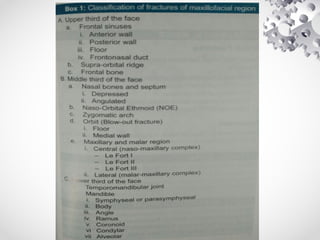
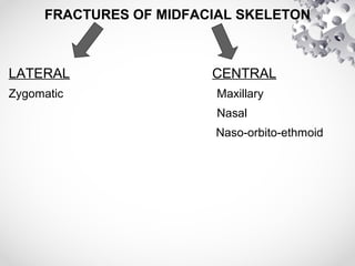
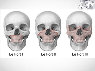
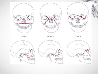
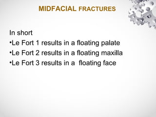
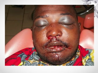
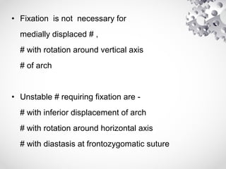
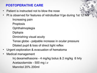
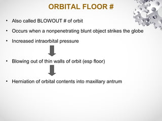
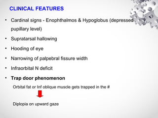
MAXILLO FACIAL
TRAUMA -
An Overview
Dr Dheeraj S
JR, ENT
INTRODUCTION
• Injuries of face are the reason for 10% of A&E attendance
• In the past, outcome of facial fracture management was
judged entirely on the basis final dental occlusion
• Restoration of vertical and transverse facial proportions is
equally important
• CSF Rhinorrhea & Retrobulbar hematoma must be
excluded in all middle third fractures
AETIOLOGY
• Low energy injuries - heal well
(1) Physical assault
(2) Sports injuries
(3) Animal bites
• High energy injuries - don't heal well
(1) Road traffic accidents
(2) Industrial injuries
(3) Attempted suicides - eg: jumping from height
(4) Military injuries - GSW
AETIOLOGY (...contd)
• Exact mechanism of injury must be ascertained
• Importance : Medicolegal reasons
Adequate screening to detect other injuries
Prognosis for stability of repair
PRIMARY SURVEY
ATLS protocols must be followed
• A - Airway - Secure the airway
• B - Breathing - spontaneous / assisted ventillation
• C - Circulation - control blood loss & maintain blood volume
• D - Disability - assessment of level of consciousness &
neurological dysfunction ; GCS
• E - Exposure - adequate exposure of all injuries
PRIMARY SURVEY (...contd)
Bleeding control
• Haemorrhage from facial fractures can be torrential & needs
immediate attention
• Ant & post nasal packing has been replaced by the use of
EPISTAT
• Inadvertent intubation of ant cranial fossa via cribriform plate
or medial wall of orbit must be avoided
• In case of intense shock rule out abdominal / thoracic / pelvic /
orthopaedic injuries
SECONDARY SURVEY
• Secondary survey is carried out to exclude other injuries & to
categorize the extent of facial injuries
• Soft tissues of face are carefully scrutinized
• Facial N function must be noted
• Parotid duct integrity must be assessed
• Ocular movements & visual acuity
• Palpate orbital margins, zygomatic projection, nasal skeleton,
mandibular outline
• Dental examination for any # or missing teeth, dental
occlusion and maxillary or mandibular instability
CLASSIFICATION
• For the sake of description and management, the injuries
of maxillofacial region can be divided into :
1. Bony injuries
2. Soft tissue injuries
FRACTURES OF MIDFACIAL SKELETON
LATERAL CENTRAL
Zygomatic Maxillary
Nasal
Naso-orbito-ethmoid
MIDFACIAL FRACTURES (...contd)
• Le Fort described 3 levels of midfacial fractures
• Pure Lefort # are accurate only for low energy injuries
• LeFort I (Transverse)
Runs just above the floor of nasal cavity through the nasal septum,
maxillary sinuses & inf parts of medial and lateral pterygoid plates
• Le Fort II (Pyramidal)
Runs from the floor of the maxillary sinuses superiorly to the
infraorbital margin & through the zygomaticomaxillary suture.Within
the orbit it passes across the lacrimal bone to the nasion
MIDFACIAL FRACTURES (...contd)
• Le Fort III (Craniofacial disjunction)
# line traverses the medial wall of orbit to the sup orbital
fissure and exits across the Gr wing of sphenoid &
zygomatic bone to the zygomaticofrontal suture.
Posteriorly # line runs inf to the optic foramen, across
the lesser wing of sphenoid to the pterygomaxillary
fissure & sphenopalatine foramen.
MIDFACIAL FRACTURES
In short
•Le Fort 1 results in a floating palate
•Le Fort 2 results in a floating maxilla
•Le Fort 3 results in a floating face
MAXILLARY FRACTURES
• Clinical features :
Epistaxis
Circumorbital ecchymosis (panda facies)
Facial edema
Emphysema
Lengthening of face
Anterior open bite in Le fort 2 & 3
Infra-orbital N sensory deficit
Bruising at the junction of soft and hard palate
MAXILLARY FRACTURES - Management
EMERGENCY MANAGEMENT
• Hemostasis using epistat or nasal packing
• Secure the airway
REDUCTION
• If there is retroposition of maxilla it can be pulled forward
using index and middle finger placed behind patient's
palate
• Maxilla can be mobilised by a combination of digital
pressure & traction on arch bars or interdental wires.
• Rowe maxillary disimpaction foreps can also be used
ROWE MAXILLARY DISIMPACTION FORCEPS
MAXILLARY FRACTURES -
Management
FIXATION
Maxilla can be stabilised by a number of methods
(1) Intermaxillary fixation
(2) External fixation
(3) Internal suspension
(4) Internal fixation
MIDFACIAL FRACTURES -
Management
INTERMAXILLARY FIXATION (IMF)
• Can be used when the # is at a lower level (Le fort 1) and not grossly
displaced
• Done by wiring the teeth together (using teeth as biological bone pins) -
dental wires, arch bars, leonard button
• Advantage - Easy technique
• Disadvantage - Period of immobilization was inconveniet &
uncomfortable
- Dietary restrictions
- Fixation is often inadequate esp if dentition is incomplete
- Needle stick injuries to doctor and nursing staff
EXTERNAL FIXATION
• Has been largely superseded by internal fixation
• Maxilla is fixed by attaching a fork to the teeth with a silver cap
splint and this is in turn anchored to stable bone above #
• Halo frame, Levant frame
• Is extremely rapid and allows surgeon to make fine adjustments
during the initial phase of bony healing (1st
2 weeks)
• Disadv - Nonaccurate reduction
- Poor facial appearance
INTERNAL SUSPENSION
• Provide rapid stabilization of maxilla without need for ext fixation
• Fractured maxillary elements are suspended from various points
on the craniofacial skeleton above # line - zygomatic arch, orbital
rim
• Simple & rapid
• Positioning of maxilla is difficult to achieve
• Relatively poor fixation
INTERNAL FIXATION
• 1.5 mm low-profile miniplates placed along buttresses provide
satisfactory stabilization
• # of ant & lat maxilla can be accessed through a gingivo-buccal
incision
• Posterior limit of incision should not beyond 1st permenant molar
• Infraorbital rim needs to be reduced & fixed in Le Fort 2 #,
zygomatic injuries, nasomaxillary #
• Diff types of skin incision can be used - Transconjuctival,
Infraorbital, Transconjuctival with transcaruncular extension etc
ZYGOMATIC COMPLEX FRACTURES
• Is the 2nd most common # in the maxillofacial region
• Blow to zygomatic bone causes either a depressed # of entire
zygomatic bone or a # of zygomatic arch
• Originally termed as TRIPOD #, because of disruption of 3
articulations - Fronto-zygomatic; Infraorbital rim; Zygomatico
maxillary buttress
• Classified according to their rotation about vertical & horizontal axes
Vertical axis - runs b/w frontozygomatic suture & 1st molar
Horizontal axis - plane of zygomatic arch
• Zygomatic arch tends to break at its weakest point - ie just posterior
to zygomatico temporal suture
CLINICAL FEATURES
• Subconjuctival haemorrhage - invariably present in zygomatic
body #
• Restricted eye movements particularly in upward gaze (if there
is orbital floor dehiscence)
• Step deformity of infraorbital margin
• Tenderness of frontozygomatic suture
• Palpable depression & limited mouth opening - in arch #
• Limited sensation on cheek - if zygomaticotemporal or
zygomaticobuccal N are damaged
MANAGEMENT
• Minimally displaced #
Conservative management
Strict instruction to avoid nose blowing for 2 - 3 weeks
Review after 10 days when the edema has subsided
• Displaced #
Reduction with / without fixation
• Methods for reduction -
(a) Gilles temporal approach (b) Dingman supraorbital approach
(c) Poswilo hook approach (d) Intraoral / Keen approach
(e) Coronal approach
• Fixation is not necessary for
medially displaced # ,
# with rotation around vertical axis
# of arch
• Unstable # requiring fixation are -
# with inferior displacement of arch
# with rotation around horizontal axis
# with diastasis at frontozygomatic suture
POSTOPERATIVE CARE
• Patient is instructed not to blow the nose
• Pt is observed for features of retrobulbar h'ge during 1st 12 hrs
Increasing pain
Proptosis
Ophthalmoplegia
Diplopia
Diminishing visual acuity
Tense globe - palpable increase in ocular pressure
Dilated pupil & loss of direct light reflex
• Urgent exploration & evacuation of hematoma
• Medical management
Inj dexamethasone - 4 mg/kg bolus & 2 mg/kg 6 hrly
Acetazolamide - 500 mg i.v
Mannitol 20% 200ml
ORBITAL FLOOR #
• Also called BLOWOUT # of orbit
• Occurs when a nonpenetrating blunt object strikes the globe
• Increased intraorbital pressure
• Blowing out of thin walls of orbit (esp floor)
• Herniation of orbital contents into maxillary antrum
CLINICAL FEATURES
• Cardinal signs - Enophthalmos & Hypoglobus (depressed
pupillary level)
• Supratarsal hallowing
• Hooding of eye
• Narrowing of palpebral fissure width
• Infraorbital N deficit
• Trap door phenomenon
Orbital fat or Inf oblique muscle gets trapped in the #
Diplopia on upward gaze
MANAGEMENT
• Significant orbital floor injury requires exploration & repair
• Approach is determined by size & position of blowout #
• All soft tissue components should be mobilized and supported
by a graft
• Smaller defects - Silastic or PDS grafts
Silastic has tendancy to become infected & extrude
• Larger blowout # - Bone from iliac crest / rib / calvarium
• TRANSANTRAL APPROACH
• INFRAORBITAL APPROACH
• ENDOSCOPIC TRANSANTRAL REPAIR
NASO-ORBITO-ETHMOID COMPLEX #
• Occurs when the force directly hits over the nasion
• Involves nasal bones, perpendicular plate of ethmoid, ethmoid air
cells, medial wall of orbit
• Medial canthal ligament can be avulsed
• Markowitz et al classified it in terms of its attachment to medial
canthal ligaments
TYPE I Single large central fragment bearing the canthal ligaments
TYPE II
Fragmentation of central fragment, medial canthal ligament
attached to bone
TYPE III
Comminution of the central frgment with no bone attached to
canthal ligaments
CLINICAL FEATURES
• Pug nose - Depression of nasal bridge & elevation of nasal tip
• Signs of medial canthal ligament disruption
TELECANTHUS - Widening of intercanthal distance > 35 mm
Narrowing of palpebral fissure
Epiphora
• Periorbital ecchymosis
• Orbital hematoma - d/t bleeding from Ant & Post ethmoidal arteries
• CSF rhinorrhoea - if there is # of cribriform plate
MANAGEMENT
• Closed reduction
Uncomplicated NOE # may be reduced with Ashe's forceps &
stabilized with a wire passed through # segments and septum
Intransal packing ; Splinting for 10 days
• Open reduction
All 3 types of # are stabilized using miniplates
In type 1 # - Coronal flap intraorally + subciliary incision
In type 2 & 3 - Transcanthal canthopexy to reduce telecanthus &
to hold medial canthal lig in place (using plate / wire)
Lacrimal duct injuries are best managed conservatively if there is
no open laceration to it
FRACTURES OF NASAL BONES & SEPTUM
• Nasal bone # is the most common # of maxillofacial region
• Isolated # of nasal pyramid - 40% of all facial #
• Relatively little force required to fracture nasal bone : 25-75 lb / in2
Grading on the basis of extent of deformity
Grade 0 - bones perfectly straight
Grade 1 - bones deviated < half the width of nasal bridge
Grade 2 - bones deviated half to one full width of nasal bridge
Grade 3 - bones deviated >one full width of nasal bridge
Grade 4 - bones almost touching the cheek
Classification on the basis of nature of injury
(a) # as a result of lateral force - nose is displaced away
from the midline on the side of the injury --> Angulated Nose
(b) # as a result of frontal blow - nasal bones are pushed up
and splayed so that the upper nose (bridge) appears broad,
but the height of the nose is collapsed --> Depressed Nose
Pattern of nasal #
• Nasal # can be subdivided into 3 broad categories that
characterise the patern of damage sustained with increasing force
• Class I Class II Class III
Class I
• Are the result of small degree of force & hence the extent of deformity is
not marked
• Are mostly depressed # of Nasal bone
• # line runs parallel to the dorsum of the nose and naso maxillary suture
and joins at a point where the nasal bone becomes thicker (ie about 2/3
of the way along its length)
• Class I fracture of nasal bone is purely a clinical diagnosis
Class II
• Are the result of moderate degree of force & are often associated
with significant cosmetic deformity
• In addition to nasal bones, frontal process of maxila and septal
structures are also involved
• For a successful result both the nasal bones as well as the septum
will have to be reduced
• Precise nature of the deformity depends on the direction of the
blow sustained
• The perpendicular plate of ethmoid is invariably involved in these
fractures, and is characteristically C shaped (Jarjaway fracture of
nasal septum).
Class III
• Are the most severe nasal injuries encountered & are caused by
high velocity trauma
• Also termed as Naso-orbito-ethmoid # & often have associated #
of maxilla
• Two types of naso ethmoidal fractures have been recognised:
Type I: In this group the anterior skull base, posterior wall of the
frontal sinus and optic canal remain intact
Type II: Here the posterior wall of the frontal sinus is disrupted
with multiple fractures involving the roof of ethmoid and orbit
CLINICAL FEATURES
• Pain
• Swelling over nose
• Epistaxis
• Nasal deformity
• Nasal tenderness and crepitus
• Periorbital ecchymosis
Management
• # of nasal bones, if left uncorrected, could lead to loss of structural
integrity and the soft tissue changes that follow may lead to both
unfavorable appearance and function
• Management is based solely on the clinical assessment of function
and appearance
• Bleeding must first be controlled by nasal packing.
• Patients may also have considerable amount of swelling .
Conservative management for 1-2 weeks for the oedema to
subside enable precise assessment of bony injury
• Fracture reduction should be accomplished when accurate
evaluation and manipulation of the mobile nasal bones can be
performed; this is usually within 5-10 days in adults and 3-7 days in
children.
Radiological investigations:
1. Plain xray nasal bones
2. Xray paranasal sinuses water's view
3. CT scan paranasal sinuses - This is a must in all cases of class
II and class III fractures of nasal bones for precise estimation of
damage.
Class I #
• Most class I # can be managed by closed reduction and
imobilisation by application of POP. Digital pressure alone
commonly does the job.
• If the fractured fragments are impacted then a Welsham's forceps
will have to be used to disimpact and reduce the fractured nasal
bone.
Class II
• Closed reduction do not give optimal results because the septal
fracture is not corrected.
• Since the fractured fragments of the perpendicular plate of
ethmoid and the septal cartilage fragments are not repositioned
the results of closed reduction are not satisfactory.
• In these patients closed manipulation of nasal bones should
always be accompanied by open correction of septal deformity
Class III
• Must be treated with open reduction and internal fixation
• The problem here is eventhough the nasal bones can be
reduced,the adjacent supporting bones (components of the
ethmoidal labyrinth) donot support the nasal bones due to their
brittleness
• It is always better to reconstruct and stabilise the anterior table of
the frontal bone so that other parts of nasal skeleton can derive
support from it
• Formerly transnasal wires were used to fix the nasal bones, but
now a days plates and screws are preferred
MANDIBULAR FRACTURES
• Mandiblular # can be due to direct or indirect blow
• # occur at points of potential weakness where the bone is
relatively thin
• It is common for the mandible to fracture in > 1 places
• Displacement depends on many factors, the important ones are
pull of attached muscles - Masseter, Pterygoids, Temporalis and
direction of # line
• Vertically favourable - runs forward from lingual to buccal
• Horizontally fav - runs forward superiorly to inferiorly
DINGMAN CLASSIFICATION OF MANDIBULAR
#
CLINICAL FEATURES
• # of Angle, Body & Symphysis
(1) Pain & paradoxical movement; crepitus on distraction of #
segments
(2) Blood stained saliva
(3) Anaesthesia of lower lip
(4) Step deformity palpable externally or intraorally
(5) Asymmetry of lower dental arch & malocclusion
(6) Hematoma in floor of mouth / buccal sulcus
CLINICAL FEATURES
• # of Condyle
(1) Tenderness over TM joint
(2) Trismus
(3) Jaw deviation to injured side, on mouth opening
(4) Dental malocclusion
CONDYLAR FRACTURE
FRACTURE OF ANGLE
FRACTURE OF BODY
MANAGEMENT
• Closed reduction
• External fixation
• Internal fixation
Closed Reduction
• Done by intermaxillary fixation (IMF)
• Limited role in modern maxillofacial surgery
• Has still a part to play in
(1) pts with undisplaced fracture & no neural deficits who don't want
surgery
(2) pts with unicondylar fracture
• Intact dental arch - Dental tie wire
• Incomplete arch - Arch bars, Intermaxillary bone pins, Cast silver splints
• Edentulous pts - Gunning splints
External Fixation
• Mostly replaced by rigid & semirigid internal fixators
• Only indicated for pts with gross tissue loss & pathological #
• Simplest technique - Placement of cortical screws and
connecting them with an external bar made of acrylic
• mini - Pennig orthopaedic fixator
Internal Fixation
• Bone plates should be placed through intra-oral route wherever
possible
• External incisions are used for lower border of mandible & condylar
neck; also preferred in grossly resorbed edentulous mandible
• In simple mandibular #, mono-cortical 2mm plates provide
adequate fixation
• In ant mandible 2 plates are used while posteriorly one plate is
sufficient
• In complicated # with gross comminution, tissue loss, infection a
load bearing osteosynthesis with rigid internal fixation & bicortical
screws are required
• Management of condylar # is controversial
• Bilat displaced condylar neck # & high intracapsular
# have poor outcome and are better managed by
Open reduction
FRACTURES OF FRONTAL SINUS
• # of frontal sinus may lead to deformity & be complicated by
infections
• Classified according to involvement of ant / post tables & whether
Frontonasal duct is involved or not
• Ant table # - no deformity --> conservative management
- depressed # --> reduction & fixation
• Post table #
commonly accompanied with dural tears, brain injury, CSF leak
demands neurosurgical assessment
dural tear repaired with temporalis fascia
small frontal sinus is obliterated with fat
SUPRAORBITAL RIDGE FRACTURE
• Clinical features :
(1) Periorbital ecchymosis
(2) Flattening of eye brow
(3) Proptosis
(4) Impacted bone fragment in orbit
• Managed by open reduction via an incision either in the brow
or in transverse skin line of forehead followed by internal
fixation
• Facial wounds should be closed as soon as possible provided they
are clean.
• Meticulous debridement with antiseptic solution is essential
• It is crucial to ensure that any underlying bony injury is repaired to
enable correct support & soft tissue draping.
• Identification & repair of key landmarks like vermillion border of lip,
eyebrows, alar margin of nose & eyelids is essential for a good
cosmetic result.
• Facial N & Parotid duct are frequently damaged in significant soft
tissue injury.
• Facial N is easiest to find & repair in the 1st
48 hrs while the
peripheral branches can still be stimulated.
• Use of deep sutures (vicryl) to take out tension out of the wound &
to avoid deadspace.
PAEDIATRIC FACIAL INJURIES
• Fortunately serious maxillofacial injuries are uncommon in
children.
• Minor injuries particularly soft tissue & dentoalveolar are frequent.
• Treatment concepts & protocols are similar to adult patients, but
most will require Gen anaesthesia or sedation.
• Consideration should be given to excision of wound margins and
the use of resorbable sutures to avoid unnecessary gen
anaesthesia for suture removal.
• Dental injuries to upper ant teeth are very common and hence
opinion of paedodontist or maxillofacial surgeon must be taken.
• Reimplant avulsed teeth as soon as possible
• # of condyle are usually treated conservatively as there is immense
remodelling capacity.
THANK
YOU

Maxillo facial trauma

  • 1.
    MAXILLO FACIAL TRAUMA - AnOverview Dr Dheeraj S JR, ENT
  • 2.
    INTRODUCTION • Injuries offace are the reason for 10% of A&E attendance • In the past, outcome of facial fracture management was judged entirely on the basis final dental occlusion • Restoration of vertical and transverse facial proportions is equally important • CSF Rhinorrhea & Retrobulbar hematoma must be excluded in all middle third fractures
  • 3.
    AETIOLOGY • Low energyinjuries - heal well (1) Physical assault (2) Sports injuries (3) Animal bites • High energy injuries - don't heal well (1) Road traffic accidents (2) Industrial injuries (3) Attempted suicides - eg: jumping from height (4) Military injuries - GSW
  • 4.
    AETIOLOGY (...contd) • Exactmechanism of injury must be ascertained • Importance : Medicolegal reasons Adequate screening to detect other injuries Prognosis for stability of repair
  • 5.
    PRIMARY SURVEY ATLS protocolsmust be followed • A - Airway - Secure the airway • B - Breathing - spontaneous / assisted ventillation • C - Circulation - control blood loss & maintain blood volume • D - Disability - assessment of level of consciousness & neurological dysfunction ; GCS • E - Exposure - adequate exposure of all injuries
  • 6.
    PRIMARY SURVEY (...contd) Bleedingcontrol • Haemorrhage from facial fractures can be torrential & needs immediate attention • Ant & post nasal packing has been replaced by the use of EPISTAT • Inadvertent intubation of ant cranial fossa via cribriform plate or medial wall of orbit must be avoided • In case of intense shock rule out abdominal / thoracic / pelvic / orthopaedic injuries
  • 9.
    SECONDARY SURVEY • Secondarysurvey is carried out to exclude other injuries & to categorize the extent of facial injuries • Soft tissues of face are carefully scrutinized • Facial N function must be noted • Parotid duct integrity must be assessed • Ocular movements & visual acuity • Palpate orbital margins, zygomatic projection, nasal skeleton, mandibular outline • Dental examination for any # or missing teeth, dental occlusion and maxillary or mandibular instability
  • 10.
    CLASSIFICATION • For thesake of description and management, the injuries of maxillofacial region can be divided into : 1. Bony injuries 2. Soft tissue injuries
  • 14.
    FRACTURES OF MIDFACIALSKELETON LATERAL CENTRAL Zygomatic Maxillary Nasal Naso-orbito-ethmoid
  • 15.
    MIDFACIAL FRACTURES (...contd) •Le Fort described 3 levels of midfacial fractures • Pure Lefort # are accurate only for low energy injuries • LeFort I (Transverse) Runs just above the floor of nasal cavity through the nasal septum, maxillary sinuses & inf parts of medial and lateral pterygoid plates • Le Fort II (Pyramidal) Runs from the floor of the maxillary sinuses superiorly to the infraorbital margin & through the zygomaticomaxillary suture.Within the orbit it passes across the lacrimal bone to the nasion
  • 16.
    MIDFACIAL FRACTURES (...contd) •Le Fort III (Craniofacial disjunction) # line traverses the medial wall of orbit to the sup orbital fissure and exits across the Gr wing of sphenoid & zygomatic bone to the zygomaticofrontal suture. Posteriorly # line runs inf to the optic foramen, across the lesser wing of sphenoid to the pterygomaxillary fissure & sphenopalatine foramen.
  • 19.
    MIDFACIAL FRACTURES In short •LeFort 1 results in a floating palate •Le Fort 2 results in a floating maxilla •Le Fort 3 results in a floating face
  • 20.
    MAXILLARY FRACTURES • Clinicalfeatures : Epistaxis Circumorbital ecchymosis (panda facies) Facial edema Emphysema Lengthening of face Anterior open bite in Le fort 2 & 3 Infra-orbital N sensory deficit Bruising at the junction of soft and hard palate
  • 22.
    MAXILLARY FRACTURES -Management EMERGENCY MANAGEMENT • Hemostasis using epistat or nasal packing • Secure the airway REDUCTION • If there is retroposition of maxilla it can be pulled forward using index and middle finger placed behind patient's palate • Maxilla can be mobilised by a combination of digital pressure & traction on arch bars or interdental wires. • Rowe maxillary disimpaction foreps can also be used
  • 23.
  • 26.
    MAXILLARY FRACTURES - Management FIXATION Maxillacan be stabilised by a number of methods (1) Intermaxillary fixation (2) External fixation (3) Internal suspension (4) Internal fixation
  • 27.
    MIDFACIAL FRACTURES - Management INTERMAXILLARYFIXATION (IMF) • Can be used when the # is at a lower level (Le fort 1) and not grossly displaced • Done by wiring the teeth together (using teeth as biological bone pins) - dental wires, arch bars, leonard button • Advantage - Easy technique • Disadvantage - Period of immobilization was inconveniet & uncomfortable - Dietary restrictions - Fixation is often inadequate esp if dentition is incomplete - Needle stick injuries to doctor and nursing staff
  • 30.
    EXTERNAL FIXATION • Hasbeen largely superseded by internal fixation • Maxilla is fixed by attaching a fork to the teeth with a silver cap splint and this is in turn anchored to stable bone above # • Halo frame, Levant frame • Is extremely rapid and allows surgeon to make fine adjustments during the initial phase of bony healing (1st 2 weeks) • Disadv - Nonaccurate reduction - Poor facial appearance
  • 33.
    INTERNAL SUSPENSION • Providerapid stabilization of maxilla without need for ext fixation • Fractured maxillary elements are suspended from various points on the craniofacial skeleton above # line - zygomatic arch, orbital rim • Simple & rapid • Positioning of maxilla is difficult to achieve • Relatively poor fixation
  • 35.
    INTERNAL FIXATION • 1.5mm low-profile miniplates placed along buttresses provide satisfactory stabilization • # of ant & lat maxilla can be accessed through a gingivo-buccal incision • Posterior limit of incision should not beyond 1st permenant molar • Infraorbital rim needs to be reduced & fixed in Le Fort 2 #, zygomatic injuries, nasomaxillary # • Diff types of skin incision can be used - Transconjuctival, Infraorbital, Transconjuctival with transcaruncular extension etc
  • 37.
    ZYGOMATIC COMPLEX FRACTURES •Is the 2nd most common # in the maxillofacial region • Blow to zygomatic bone causes either a depressed # of entire zygomatic bone or a # of zygomatic arch • Originally termed as TRIPOD #, because of disruption of 3 articulations - Fronto-zygomatic; Infraorbital rim; Zygomatico maxillary buttress • Classified according to their rotation about vertical & horizontal axes Vertical axis - runs b/w frontozygomatic suture & 1st molar Horizontal axis - plane of zygomatic arch • Zygomatic arch tends to break at its weakest point - ie just posterior to zygomatico temporal suture
  • 40.
    CLINICAL FEATURES • Subconjuctivalhaemorrhage - invariably present in zygomatic body # • Restricted eye movements particularly in upward gaze (if there is orbital floor dehiscence) • Step deformity of infraorbital margin • Tenderness of frontozygomatic suture • Palpable depression & limited mouth opening - in arch # • Limited sensation on cheek - if zygomaticotemporal or zygomaticobuccal N are damaged
  • 42.
    MANAGEMENT • Minimally displaced# Conservative management Strict instruction to avoid nose blowing for 2 - 3 weeks Review after 10 days when the edema has subsided • Displaced # Reduction with / without fixation • Methods for reduction - (a) Gilles temporal approach (b) Dingman supraorbital approach (c) Poswilo hook approach (d) Intraoral / Keen approach (e) Coronal approach
  • 44.
    • Fixation isnot necessary for medially displaced # , # with rotation around vertical axis # of arch • Unstable # requiring fixation are - # with inferior displacement of arch # with rotation around horizontal axis # with diastasis at frontozygomatic suture
  • 45.
    POSTOPERATIVE CARE • Patientis instructed not to blow the nose • Pt is observed for features of retrobulbar h'ge during 1st 12 hrs Increasing pain Proptosis Ophthalmoplegia Diplopia Diminishing visual acuity Tense globe - palpable increase in ocular pressure Dilated pupil & loss of direct light reflex • Urgent exploration & evacuation of hematoma • Medical management Inj dexamethasone - 4 mg/kg bolus & 2 mg/kg 6 hrly Acetazolamide - 500 mg i.v Mannitol 20% 200ml
  • 46.
    ORBITAL FLOOR # •Also called BLOWOUT # of orbit • Occurs when a nonpenetrating blunt object strikes the globe • Increased intraorbital pressure • Blowing out of thin walls of orbit (esp floor) • Herniation of orbital contents into maxillary antrum
  • 47.
    CLINICAL FEATURES • Cardinalsigns - Enophthalmos & Hypoglobus (depressed pupillary level) • Supratarsal hallowing • Hooding of eye • Narrowing of palpebral fissure width • Infraorbital N deficit • Trap door phenomenon Orbital fat or Inf oblique muscle gets trapped in the # Diplopia on upward gaze
  • 51.
    MANAGEMENT • Significant orbitalfloor injury requires exploration & repair • Approach is determined by size & position of blowout # • All soft tissue components should be mobilized and supported by a graft • Smaller defects - Silastic or PDS grafts Silastic has tendancy to become infected & extrude • Larger blowout # - Bone from iliac crest / rib / calvarium • TRANSANTRAL APPROACH • INFRAORBITAL APPROACH • ENDOSCOPIC TRANSANTRAL REPAIR
  • 52.
    NASO-ORBITO-ETHMOID COMPLEX # •Occurs when the force directly hits over the nasion • Involves nasal bones, perpendicular plate of ethmoid, ethmoid air cells, medial wall of orbit • Medial canthal ligament can be avulsed • Markowitz et al classified it in terms of its attachment to medial canthal ligaments TYPE I Single large central fragment bearing the canthal ligaments TYPE II Fragmentation of central fragment, medial canthal ligament attached to bone TYPE III Comminution of the central frgment with no bone attached to canthal ligaments
  • 53.
    CLINICAL FEATURES • Pugnose - Depression of nasal bridge & elevation of nasal tip • Signs of medial canthal ligament disruption TELECANTHUS - Widening of intercanthal distance > 35 mm Narrowing of palpebral fissure Epiphora • Periorbital ecchymosis • Orbital hematoma - d/t bleeding from Ant & Post ethmoidal arteries • CSF rhinorrhoea - if there is # of cribriform plate
  • 54.
    MANAGEMENT • Closed reduction UncomplicatedNOE # may be reduced with Ashe's forceps & stabilized with a wire passed through # segments and septum Intransal packing ; Splinting for 10 days • Open reduction All 3 types of # are stabilized using miniplates In type 1 # - Coronal flap intraorally + subciliary incision In type 2 & 3 - Transcanthal canthopexy to reduce telecanthus & to hold medial canthal lig in place (using plate / wire) Lacrimal duct injuries are best managed conservatively if there is no open laceration to it
  • 55.
    FRACTURES OF NASALBONES & SEPTUM • Nasal bone # is the most common # of maxillofacial region • Isolated # of nasal pyramid - 40% of all facial # • Relatively little force required to fracture nasal bone : 25-75 lb / in2 Grading on the basis of extent of deformity Grade 0 - bones perfectly straight Grade 1 - bones deviated < half the width of nasal bridge Grade 2 - bones deviated half to one full width of nasal bridge Grade 3 - bones deviated >one full width of nasal bridge Grade 4 - bones almost touching the cheek
  • 56.
    Classification on thebasis of nature of injury (a) # as a result of lateral force - nose is displaced away from the midline on the side of the injury --> Angulated Nose (b) # as a result of frontal blow - nasal bones are pushed up and splayed so that the upper nose (bridge) appears broad, but the height of the nose is collapsed --> Depressed Nose
  • 58.
    Pattern of nasal# • Nasal # can be subdivided into 3 broad categories that characterise the patern of damage sustained with increasing force • Class I Class II Class III Class I • Are the result of small degree of force & hence the extent of deformity is not marked • Are mostly depressed # of Nasal bone • # line runs parallel to the dorsum of the nose and naso maxillary suture and joins at a point where the nasal bone becomes thicker (ie about 2/3 of the way along its length) • Class I fracture of nasal bone is purely a clinical diagnosis
  • 60.
    Class II • Arethe result of moderate degree of force & are often associated with significant cosmetic deformity • In addition to nasal bones, frontal process of maxila and septal structures are also involved • For a successful result both the nasal bones as well as the septum will have to be reduced • Precise nature of the deformity depends on the direction of the blow sustained • The perpendicular plate of ethmoid is invariably involved in these fractures, and is characteristically C shaped (Jarjaway fracture of nasal septum).
  • 62.
    Class III • Arethe most severe nasal injuries encountered & are caused by high velocity trauma • Also termed as Naso-orbito-ethmoid # & often have associated # of maxilla • Two types of naso ethmoidal fractures have been recognised: Type I: In this group the anterior skull base, posterior wall of the frontal sinus and optic canal remain intact Type II: Here the posterior wall of the frontal sinus is disrupted with multiple fractures involving the roof of ethmoid and orbit
  • 63.
    CLINICAL FEATURES • Pain •Swelling over nose • Epistaxis • Nasal deformity • Nasal tenderness and crepitus • Periorbital ecchymosis
  • 64.
    Management • # ofnasal bones, if left uncorrected, could lead to loss of structural integrity and the soft tissue changes that follow may lead to both unfavorable appearance and function • Management is based solely on the clinical assessment of function and appearance • Bleeding must first be controlled by nasal packing. • Patients may also have considerable amount of swelling . Conservative management for 1-2 weeks for the oedema to subside enable precise assessment of bony injury • Fracture reduction should be accomplished when accurate evaluation and manipulation of the mobile nasal bones can be performed; this is usually within 5-10 days in adults and 3-7 days in children.
  • 65.
    Radiological investigations: 1. Plainxray nasal bones 2. Xray paranasal sinuses water's view 3. CT scan paranasal sinuses - This is a must in all cases of class II and class III fractures of nasal bones for precise estimation of damage. Class I # • Most class I # can be managed by closed reduction and imobilisation by application of POP. Digital pressure alone commonly does the job. • If the fractured fragments are impacted then a Welsham's forceps will have to be used to disimpact and reduce the fractured nasal bone.
  • 67.
    Class II • Closedreduction do not give optimal results because the septal fracture is not corrected. • Since the fractured fragments of the perpendicular plate of ethmoid and the septal cartilage fragments are not repositioned the results of closed reduction are not satisfactory. • In these patients closed manipulation of nasal bones should always be accompanied by open correction of septal deformity
  • 68.
    Class III • Mustbe treated with open reduction and internal fixation • The problem here is eventhough the nasal bones can be reduced,the adjacent supporting bones (components of the ethmoidal labyrinth) donot support the nasal bones due to their brittleness • It is always better to reconstruct and stabilise the anterior table of the frontal bone so that other parts of nasal skeleton can derive support from it • Formerly transnasal wires were used to fix the nasal bones, but now a days plates and screws are preferred
  • 70.
    MANDIBULAR FRACTURES • Mandiblular# can be due to direct or indirect blow • # occur at points of potential weakness where the bone is relatively thin • It is common for the mandible to fracture in > 1 places • Displacement depends on many factors, the important ones are pull of attached muscles - Masseter, Pterygoids, Temporalis and direction of # line • Vertically favourable - runs forward from lingual to buccal • Horizontally fav - runs forward superiorly to inferiorly
  • 72.
  • 73.
    CLINICAL FEATURES • #of Angle, Body & Symphysis (1) Pain & paradoxical movement; crepitus on distraction of # segments (2) Blood stained saliva (3) Anaesthesia of lower lip (4) Step deformity palpable externally or intraorally (5) Asymmetry of lower dental arch & malocclusion (6) Hematoma in floor of mouth / buccal sulcus
  • 74.
    CLINICAL FEATURES • #of Condyle (1) Tenderness over TM joint (2) Trismus (3) Jaw deviation to injured side, on mouth opening (4) Dental malocclusion
  • 75.
  • 76.
  • 77.
  • 78.
    MANAGEMENT • Closed reduction •External fixation • Internal fixation
  • 79.
    Closed Reduction • Doneby intermaxillary fixation (IMF) • Limited role in modern maxillofacial surgery • Has still a part to play in (1) pts with undisplaced fracture & no neural deficits who don't want surgery (2) pts with unicondylar fracture • Intact dental arch - Dental tie wire • Incomplete arch - Arch bars, Intermaxillary bone pins, Cast silver splints • Edentulous pts - Gunning splints
  • 81.
    External Fixation • Mostlyreplaced by rigid & semirigid internal fixators • Only indicated for pts with gross tissue loss & pathological # • Simplest technique - Placement of cortical screws and connecting them with an external bar made of acrylic • mini - Pennig orthopaedic fixator
  • 82.
    Internal Fixation • Boneplates should be placed through intra-oral route wherever possible • External incisions are used for lower border of mandible & condylar neck; also preferred in grossly resorbed edentulous mandible • In simple mandibular #, mono-cortical 2mm plates provide adequate fixation • In ant mandible 2 plates are used while posteriorly one plate is sufficient • In complicated # with gross comminution, tissue loss, infection a load bearing osteosynthesis with rigid internal fixation & bicortical screws are required
  • 84.
    • Management ofcondylar # is controversial • Bilat displaced condylar neck # & high intracapsular # have poor outcome and are better managed by Open reduction
  • 86.
    FRACTURES OF FRONTALSINUS • # of frontal sinus may lead to deformity & be complicated by infections • Classified according to involvement of ant / post tables & whether Frontonasal duct is involved or not • Ant table # - no deformity --> conservative management - depressed # --> reduction & fixation • Post table # commonly accompanied with dural tears, brain injury, CSF leak demands neurosurgical assessment dural tear repaired with temporalis fascia small frontal sinus is obliterated with fat
  • 87.
    SUPRAORBITAL RIDGE FRACTURE •Clinical features : (1) Periorbital ecchymosis (2) Flattening of eye brow (3) Proptosis (4) Impacted bone fragment in orbit • Managed by open reduction via an incision either in the brow or in transverse skin line of forehead followed by internal fixation
  • 89.
    • Facial woundsshould be closed as soon as possible provided they are clean. • Meticulous debridement with antiseptic solution is essential • It is crucial to ensure that any underlying bony injury is repaired to enable correct support & soft tissue draping. • Identification & repair of key landmarks like vermillion border of lip, eyebrows, alar margin of nose & eyelids is essential for a good cosmetic result. • Facial N & Parotid duct are frequently damaged in significant soft tissue injury. • Facial N is easiest to find & repair in the 1st 48 hrs while the peripheral branches can still be stimulated. • Use of deep sutures (vicryl) to take out tension out of the wound & to avoid deadspace.
  • 90.
    PAEDIATRIC FACIAL INJURIES •Fortunately serious maxillofacial injuries are uncommon in children. • Minor injuries particularly soft tissue & dentoalveolar are frequent. • Treatment concepts & protocols are similar to adult patients, but most will require Gen anaesthesia or sedation. • Consideration should be given to excision of wound margins and the use of resorbable sutures to avoid unnecessary gen anaesthesia for suture removal. • Dental injuries to upper ant teeth are very common and hence opinion of paedodontist or maxillofacial surgeon must be taken. • Reimplant avulsed teeth as soon as possible • # of condyle are usually treated conservatively as there is immense remodelling capacity.
  • 91.