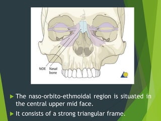
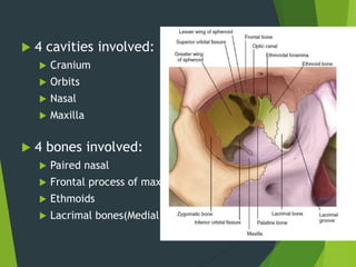
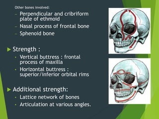
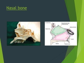
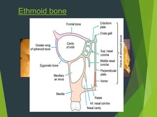
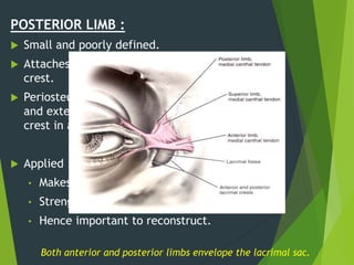
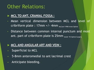
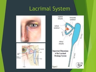
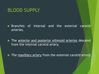
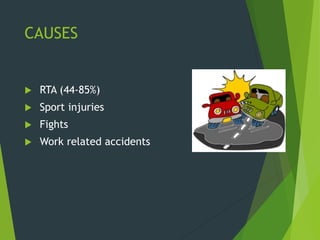
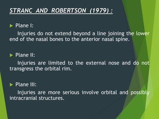
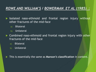
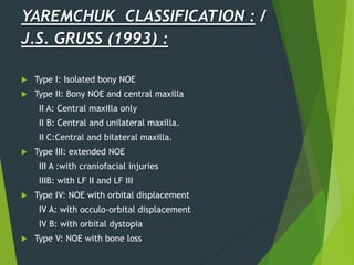
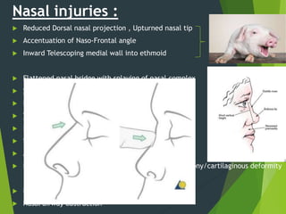
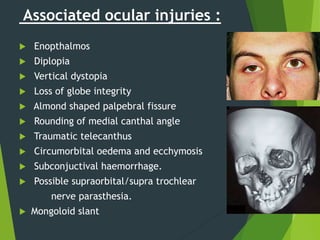
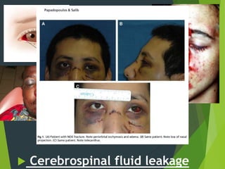
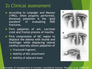
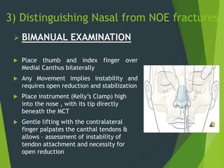
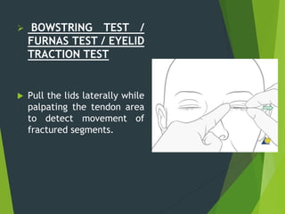
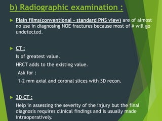
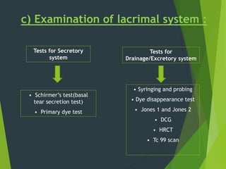
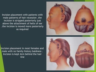
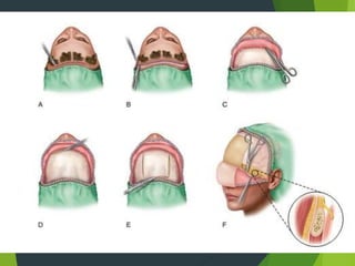
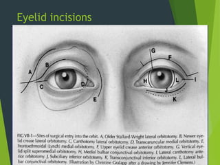
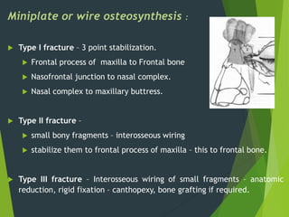
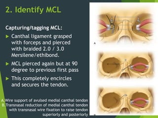
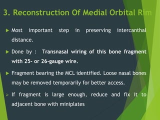
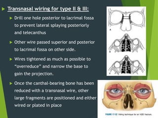
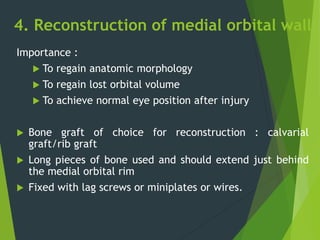
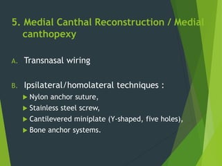
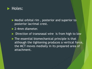
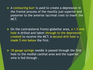
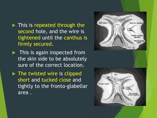
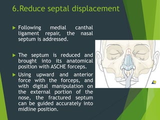
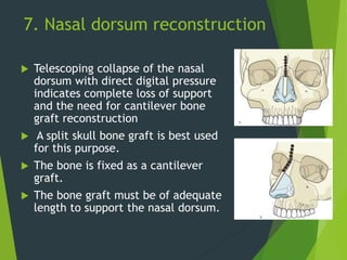
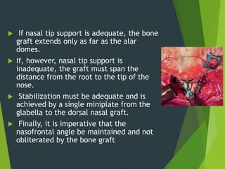
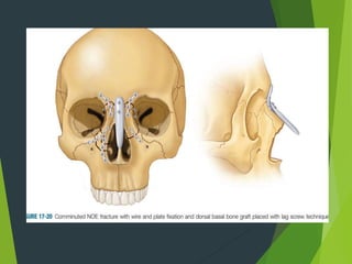
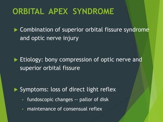
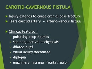
SlideShare a Scribd company logo
NOE FRACTURES
PRESENTED BY,
DR. BHAVIK MIYANI,
OMFS PG II,
NPDCH, VISNAGAR.
GUIDED BY,
DR. ANIL MANAGUTTI,
DR. SHAILESH MENAT,
DR. RUSHIT PATEL,
DR. JIGAR PATEL.
CONTENTS
 Introduction
 Anatomy
 Classification
 Diagnosis :
Clinical examination
Radiological examination
 Treatment
 Complications
 References
INTRODUCTION
 NOE region is an important area of face – not
only for cosmesis but also in determining facial
projection.
 NOE region relies on a complex inter-
relationship between uniquely specialized soft
tissues and bones formed into buttresses and
thin plates.
Peter Wardbooth
 NOE fracture refers to injuries involving the area of
confluence of the nose, orbit, ethmoids, the base
of the frontal sinus, and the floor of the anterior
cranial base.
 NOE fractures represent a spectrum of injury , from
simple nasal fractures with minimal ethmoid
involvement to grossly comminuted fractures with
displacement.
Peter Wardbooth
NOMENCLATURE
‘fracture of the ethmoids’ Dawson and Fordyce,1953
‘naso-orbital fractures’ Converse and Smith ,1963
‘naso-ethmoid injuries’ Stranc ,1970
‘naso-ethmoido-maxillo-fronto-
orbitaire’complex injuries
Vaillant and Perry,1971
‘orbito-ethmoid’ Jackson,1989
‘upper central face injury’ Bowerman,1992
INCIDENCE
 Infrequent : 2-15% of all facial fractures
 According to Fonseca (Cruse et al) , in 182
major facial fractures approx. 18% were NOE
fractures.
 60% patients with NOE fractures have
associated nonfacial injuries
 CNS injury - 51% patients
CSF leak - 42 % cases
 In male – 66-91% cases
 In young – Average : 31 years
ANATOMY AND APPLIED ASPECTS
 The naso-orbito-ethmoidal region is situated in
the central upper mid face.
 It consists of a strong triangular frame.
BONY ANATOMY
 4 cavities involved:
 Cranium
 Orbits
 Nasal
 Maxilla
 4 bones involved:
 Paired nasal
 Frontal process of maxilla
 Ethmoids
 Lacrimal bones(Medial orbital rim and wall)
Other bones involved:
‒ Perpendicular and cribriform
plate of ethmoid
‒ Nasal process of frontal bone
‒ Sphenoid bone
 Strength :
• Vertical buttress : frontal
process of maxilla
• Horizontal buttress :
superior/inferior orbital rims
 Additional strength:
• Lattice network of bones
• Articulation at various angles.
Nasal bone
Ethmoid bone
Significance of ethmoidal foramina :
 Structure traversing
 Ant. ethmoid art – 24mm
 Post. ethmoid art – 34mm
o Can contribute to Retrobulbar
Hemorrhage
 The safe exploration in superomedial
wall can extend till 30-35 mm.
 A high medial wall exploration places
orbital apex and optic canal at risk.
 The foraminae are at the same level
as optic canal and hence the
exploration should not exceed post
foramina.
Medial orbital wall
 Paper thin  lamina papyracea of ethmoid bone
 Traversed by:
• Ant. ethmoid artery and Post. ethmoid artery
 Entrapment of orbital fat medially  horizontal
diplopia – restriction of abduction-retraction of globe
Lacrimal fossa :
• Formed by maxillary and lacrimal bones
• Bound by anterior and posterior lacrimal crests.
• 16mm high x 4-9mm wide x 2mm deep
• Mean thickness of lacrimal bone : 106micron  easy perforation
Sutura notha/ Sutura longitudinalis imperfecta of Weber:
• Fine groove on frontal process of maxilla
• Anterior to anterior lacrimal crest
• Contains small branches of infraorbital artery.
• Anticipate their presence during dissection
SOFT TISSUE ANATOMY
Medial canthal ligament
Lacrimal drainage apparatus
Associated vessels
Medial canthal ligament
 MCL Intimately related to :
• Lacrimal drainage apparatus
• Lacrimal bone
• Frontal process of maxilla – reinforces.
ANTERIOR LIMB:
 11.7mm length/4.9mm width – longer and more
prominent.
Medial attachment :
 Frontal process of maxilla just lateral to suture with
nasal bone.
 Superior aspect of Ant lacrimal crest and beyond.
Superior branch – periosteum of frontal bone(corrugator
super cilli)
POSTERIOR LIMB :
 Small and poorly defined.
 Attaches to posterior lacrimal
crest.
 Periosteum in this region is thicker
and extends till anterior lacrimal
crest in a triangular fashion
 Applied :
• Makes post attachment strong.
• Strengthens the whole structure.
• Hence important to reconstruct.
Both anterior and posterior limbs envelope the lacrimal sac.
Other Relations:
 MCL TO ANT. CRANIAL FOSSA :
• Mean vertical dimension between MCL and level of
cribriform plate : 17mm +/- 4mm McCann’1998 Invest Opthal
• Distance between common internal punctum and most
ant. part of cribriform plate is 25mm botek ’93 Opthal Surgery
 MCL AND ANGULAR ART AND VEIN :
• Superficial to MCL
• 5-8mm anteromedial to ant lacrimal crest
• Anticipate bleeding.
Lacrimal System
BLOOD SUPPLY
 Branches of internal and the external carotid
arteries.
 The anterior and posterior ethmoid arteries descend
from the internal carotid artery.
 The maxillary artery from the external carotid artery
NERVE SUPPLY
 Ophthalmic and maxillary nerves, which are
derived from the Trigeminal nerve.
CAUSES
 RTA (44-85%)
 Sport injuries
 Fights
 Work related accidents
CLASSIFICATION
MARKOWITZ BL , MANSON PN , SARGENT L , et al
(1991) :
 These can be unilateral or bilateral injuries.
TYPE I :
There is a single large NOE fragment bearing the medial
canthal tendon.
 The nasal bone may also be involved and, in cases of
comminution,may not provide adequate dorsal support to
the nasal bridge.
Plast Reconstr Surg. 87(5):843-53
UNILATERAL TYPE II :
There is often
comminution of the NOE
area, but the canthal
tendon remains attached to
a fragment of bone,
allowing the canthus to be
stabilized with wires or a
small plate on the
fractured segment.
UNILATERAL TYPE II +
INVOLVEMENT OF NASAL
BONE :
The nasal bone may also
be involved and, in cases of
comminution,may not
provide adequate dorsal
support to the nasal bridge.
BILATERAL TYPE II
FRACTURE WITH NASAL
BONE INVOLVEMENT :
Bone grafting of the nasal
dorsum may be necessary.
TYPE III :
There is often comminution
of the NOE area (as in type
II fractures) and a
detachment of the medial
canthal tendon from the
bone.
 TYPE III WITH
INVOLVEMENT OF NASAL
BONE
 BILATERAL TYPE III WITH
NASAL BONE
INVOLVEMENT
AYLIFF’S CLASSIFICATION :
 Type I – en bloc minimum displaced fractures
of the entire NOE complex
 Type II– en bloc displaced fractures, usually
associated with large pneumatized sinus and
minimal fragmentation
 Type III – comminuted fracture but canthal
ligament firmly attached with bone fragments
which are big enough to plate.
TYPE I
TYPE II
TYPE III
 Type IV– comminuted fracture with
free canthal ligament not large
enough to be plate.
 Type V – gross comminution needing
bone grafting
TYPE IV
TYPE V
STRANC AND ROBERTSON (1979) :
 Plane I:
Injuries do not extend beyond a line joining the lower
end of the nasal bones to the anterior nasal spine.
 Plane II:
Injuries are limited to the external nose and do not
transgress the orbital rim.
 Plane III:
Injuries are more serious involve orbital and possibly
intracranial structures.
ROWE AND WILLIAM’S / BOWERMAN ET AL (1985) :
 Isolated naso-ethmoid and frontal region injury without
other fractures of the mid-face
a) Bilateral
b) Unilateral
 Combined naso-ethmoid and frontal region injury with other
fractures of the mid-face
a) Bilateral
b) Unilateral
 This is essentially the same as Manson’s classification in content.
YAREMCHUK CLASSIFICATION : /
J.S. GRUSS (1993) :
 Type I: Isolated bony NOE
 Type II: Bony NOE and central maxilla
II A: Central maxilla only
II B: Central and unilateral maxilla.
II C:Central and bilateral maxilla.
 Type III: extended NOE
III A :with craniofacial injuries
IIIB: with LF II and LF III
 Type IV: NOE with orbital displacement
IV A: with occulo-orbital displacement
IV B: with orbital dystopia
 Type V: NOE with bone loss
DIAGNOSIS
a) Clinical examination
b) Radiological examination
c) Examination of lacrimal system
d) CSF examination
INITIAL EVALUATION
 Establish ABCs.
 Diagnose any associated injuries.
 Direct examination of NOE complex.
 A thorough head and neck examination to reveal
injuries to the brain,spine, orbit and facial skeleton
is required.
 Team approach involving the otolaryngologist, plastic
surgeon, neurosurgeon and ophthalmologic
consultation is mandatory.
a) Clinical examination :
1) Clinical features
2) Clinical assessment
3) Distinguishing nasal # from NOE #
1) Clinical features
 Associated with the location and severity of the NOE fracture.
GENERAL SIGNS AND SYMPTOMS :
 Gross facial edema may show firstly in the early stage of
fracture, which will result in distortion of soft tissue landmarks
 Laceration in the nose and forehead
 Intracranial involvement
 Eye, forehead, and nose pain
 Forehead paraesthesias
 Traumatic hypertelorism
 Mongoloid slant.
Nasal injuries :
 Reduced Dorsal nasal projection , Upturned nasal tip
 Accentuation of Naso-Frontal angle PIG SNOUT
 Inward Telescoping medial wall into ethmoid
 Flattened nasal bridge with splaying of nasal complex
 Saddle shaped deformity of nose from side
 Epistaxis
 Tenderness ,crepitus and mobility of nasal complex
 Reduced nasal projection and height
 Septal deviation or dislocation
 Anosmia caused by damage to the cribiform plate
 Nasal congestion secondary to septal hematoma or bony/cartilaginous deformity
 Nasolacrimal duct obstruction/severage  epiphora
 Nasal airway obstruction
Associated ocular injuries :
 Enopthalmos
 Diplopia
 Vertical dystopia
 Loss of globe integrity
 Almond shaped palpebral fissure
 Rounding of medial canthal angle
 Traumatic telecanthus
 Circumorbital oedema and ecchymosis
 Subconjuctival haemorrhage.
 Possible supraorbital/supra trochlear
nerve parasthesia.
 Mongoloid slant
 Cerebrospinal fluid leakage
• Subconjuctival
haemorrhage.
• Traumatic
telecanthus
• Circumorbital
oedema and
ecchymosis
• Epistaxis
2) Clinical assessment
 According to Leipsiger and Manson
(1992), when properly performed,
bimanual palpation is the ‘gold
standard’ in evaluating NOE
fractures.
 Firm palpation of ant. Lacrimal
crest and frontal process of maxilla
 Firm compression of MC region to
displace the edema with thumb and
forefinger while displacing lateral
canthus laterally allows palpation of
 Fractured fragment,
 Mobility of MCL attachment
 Mobility of adjacent bone
Principles of management of complex craniofacial trauma; Marciani et al, JOMS ‘93
3) Distinguishing Nasal from NOE fractures
 BIMANUAL EXAMINATION
 Place thumb and index finger over
Medial Canthus bilaterally
 Any Movement implies instability and
requires open reduction and stabilization
 Place instrument (Kelly’s Clamp) high
into the nose , with its tip directly
beneath the MCT
 Gentle lifting with the contralateral
finger palpates the canthal tendons &
allows – assessment of instability of
tendon attachment and necessity for
open reduction
 BOWSTRING TEST /
FURNAS TEST / EYELID
TRACTION TEST
 Pull the lids laterally while
palpating the tendon area
to detect movement of
fractured segments.
b) Radiographic examination :
 Plain films(conventional – standard PNS view) are of almost
no use in diagnosing NOE fractures because most of # will go
undetected.
 CT :
Is of greatest value.
HRCT adds to the existing value.
Ask for :
1-2 mm axial and coronal slices with 3D recon.
 3D CT :
Help in assessing the severity of the injury but the final
diagnosis requires clinical findings and is usually made
intraoperatively.
Tests for Secretory
system
Tests for
Drainage/Excretory system
• Schirmer’s test(basal
tear secretion test)
• Primary dye test
c) Examination of lacrimal system :
• Syringing and probing
• Dye disappearance test
• Jones 1 and Jones 2
• DCG
• HRCT
• Tc 99 scan
Tests for Secretory system
 Schirmer’s test (Basal tear secretion test)
 Primary dye test (Jones 1)
Tests for Drainage/Excretory system
SYRINGING AND PROBING :
DYE DISAPPEARANCE TEST :
Simplest of tests
 2% Fluorescein dye placed on
conjunctival fornix
 Dye disappears.
 System patent
JONE’S TEST :
Jone’s 1 :
• 1 drop 2% fluorescein dye placed into conjunctival sac.
• Cotton bud soaked in LA placed in inferior meatus.
• Wait for 5 min and remove the bud.
• If bud stained with dye  test +ve
If Negative , then proceed to JONE’S 2 :
Clear saline irrigated through cannula inserted into inf. canaliculus
Patient bends forward
Nothing from nostril
Complete obstr.
distal to tip
Fluid regurgutates –
opp. punctum
Patency of both canaliculi till
int canaliculus
Clear fluid from nose
Dye not entered canaliculi
Blocked
punctum/canaliculi
Stained fluid
+ve test
DACROCYSTOGRAPHY :
 Radioactive oilbased dye injected into lacrimal drainage.
 Radiographed to know the course of duct
 CT used for imaging  CT dacrocystography.
DACROSCINTIGRAPHY :
• Dacroscintigraphy is done
using radionuclide (Tc-99)
• Mainly used to confirm a
diagnosis of functional
blockage when there is
delayed or no out- flow of
radioactive media in the
presence of a normal DCG.
d) CSF examination :
CLINICAL DIAGNOSIS
Reservoir sign
Salty taste
Handkerchief sign
Halo sign/double ring sign
Tramline effect
LABORATORY DIAGNOSIS
Glucose test
Protein analysis
Beta transferrin test
Beta-trace Protein
Electronic nose
High resolution CT
CT cisternography
Radionuclide cisternography
Intrathecal flourescein
MRI cisternography
RADIOGRAPHIC EVALUATION
RESERVOIR SIGN
• Simple bed side procedure 
nonspecific.
• Performed upon patient arising in
the morning.
• Place patients chin to chest for
1min.
• Copious leakage through nose like
an open faucet.
CLINICAL DIAGNOSIS :
 SALTY TASTE
 HANDKERCHIEF SIGN : mucous stiffens linen on drying but
CSF keeps it soft  distinguishes from allergic rhinitis.
 HALO SIGN / DOUBLE RING SIGN :
• Blood CSF mixture spreads on linen.
• Dark ring of blood encircles more lightly stained CSF
 TRAMLINE EFFECT :
• Occurs when CSF mixed with
blood.
• CSF appears later as yellowish
discharge  mixes with blood.
• CSF  higher protein content.
• More viscous CSF forms central
track with blood on either side
which diffuses to edge.
LABORATORY DIAGNOSIS :
 Glucose test - >30mg/dl
 Protein analysis
 Beta transferrin test
 Beta-trace Protein
 Electronic nose
RADIOGRAPHIC EVALUATION:
 High resolution CT : bone defect is filled with CSF
density fluid extracranially.
 CT cisternography
 Radionuclide cisternography
 Intrathecal flourescein
 MRI cisternography
TREATMENT
 Better overtreated than undertreated .
 Why overtreat?
Inadequate treatment
Secondary deformities
Missing or displaced bony fragments, soft
tissue retraction, scarring, malposition
Difficult to correct
Ellis E. Sequencing treatment for naso-orbito-ethmoid fractures. Journal of Oral
Early Versus Late Management??
• Treatment should begin as soon as the edema
from the initial traumatic event has resolved,
but waiting no later than 10 to 14 days, as long
as the patient is stable enough to undergo the
procedure.
Papadopoulos H . Management of Naso-Orbital-EthmoidalFractures
Oral Maxillofacial SurgClinN Am 21 (2009) 221–225
Closed Versus Open Reduction??
 Open techniques are recognized as the
best way to manage NOE fractures.
(given by Mustarde & Dingman in 1964)
Papadopoulos H . Management of Naso-Orbital-EthmoidalFractures
Oral Maxillofacial SurgClinN Am 21 (2009) 221–225
GOALS
a) Management of CSF leaks.
b) Management of damage to nasolacrimal
drainage system – dacrocystorhinostomy.
c) Sequencing treatment for naso-orbito
ethmoidal fractures.
a) Management of CSF leaks
 Tailored to individual : fracture reduction often stops the leak
 Conservative approach
 Antibiotic prophylaxis
 Surgical repair of dura
o Extracranial - Endoscopic
o Intracranial
• Extradural
• Intradural
• Combination
o Grafts: pericranium, fascia lata, muscle etc.
 Obliteration / Cranialization of frontal sinus
b) Management of lacrimal drainage
DACROCYSTORHINOSTOMY (8-18%) :
 Is the repair of the lacrimal drainage system through the
creation of a new “ostomy” or tract from the lacrimal
canaliculi to the nasal cavity.
 Principle: Large nasal osteotomy can allow greater lacrimal
drainage in upright position than will a lacrimal sac with an
interrupted lacrimal pump.
 Techniques:-
 Open (external)
 Endonasal
 Soft tissue conjuctivorhinostomy.
1. Exposure
2. Identify the MCL or the MCL bearing bone.
3. Reduce / reconstruct medial orbital rims
4. Reconstruct medial orbital walls
5. Transnasal canthopexy
6. Reduce septal fracture/displacement
7. Nasal dorsum reconstruction/augmentation
8. Soft tissue re-adaptation/nasal splint
c)
1. Exposure (surgical approaches)
 Existing lacerations
 Bicoronal / coronal incision
 Eyelid incisions
 Vertical/horizontal radix incisions (Stranc ‘70)
 Open sky approach - H shape incision (Converse & Smith ‘80)
 W shape incision (Bowermann ‘75)
 Bilateral Z approach (Dingman ‘60)
 Lynch incision.
 Transcaruncular incision No external scars
 Pre caruncular incision
 Transoral - degloving incision
 Midfacial degloving incision - great access - No scar.
Bicoronal /coronal approach
 This approach provides excellent exposure of the
nasal bones with the frontal bone down to the
upper lateral cartilages, with sufficient exposure
for plate and screw application.
 Rarely indicated for isolated fractures limited to
the nasal skeleton.
 Commonly used for fractures extending into the
nasoorbitoethmoidal (NOE), nasofrontal, or frontal
sinus regions.
Incision placement with patients with
male patterns of hair recession .the
incision is stripped posteriorly just
above the attachment of helix of ear
.the incision is moved more posteriorly
as required
Incision placement in most females and
male with no family history baldness .
Incision is kept 4cm behind the hair
line
VIDEO (Bicoronal flap)
Eyelid incisions
D - Transcaruncular approach
E - Lynch approach
Vertical incision over the nasal radix
Horizontal radix incision
Open sky
approach –
H shape
Two gull
wings
Butterfly - a
combination of
the open sky and
gull wing
Sewall -
Single side
medial orbital
incision
Transbuccal/transoral
Miniplate or wire osteosynthesis :
 Type I fracture – 3 point stabilization.
 Frontal process of maxilla to Frontal bone
 Nasofrontal junction to nasal complex.
 Nasal complex to maxillary buttress.
 Type II fracture –
 small bony fragments – interosseous wiring
 stabilize them to frontal process of maxilla – this to frontal bone.
 Type III fracture – Interosseous wiring of small fragments – anatomic
reduction, rigid fixation – canthopexy, bone grafting if required.
2. Identify MCL
Capturing/tagging MCL:
 Canthal ligament grasped
with forceps and pierced
with braided 2.0 / 3.0
Mersilene/ethibond.
 MCL pierced again but at 90
degree to previous first pass
 This completely encircles
and secures the tendon.
A.Wire support of avulsed medial canthal tendon
B.Transnasal reduction of medial canthal tendon
with transnasal wire fixation to raise tendon
superiorly and posteriorly
3. Reconstruction Of Medial Orbital Rim
 Most important step in preserving intercanthal
distance.
 Done by : Transnasal wiring of this bone fragment
with 25- or 26-gauge wire.
 Fragment bearing the MCL identified. Loose nasal bones
may be removed temporarily for better access.
 If fragment is large enough, reduce and fix it to
adjacent bone with miniplates
 Transnasal wiring for type II & III:
 Drill one hole posterior to lacrimal fossa
to prevent lateral splaying posteriorly
and telecanthus
 Other wire passed superior and posterior
to lacrimal fossa on other side.
 Wires tightened as much as possible to
“overreduce” and narrow the base to
gain the projection.
 Once the canthal-bearing bone has been
reduced with a transnasal wire, other
large fragments are positioned and either
wired or plated in place
4. Reconstruction of medial orbital wall
Importance :
 To regain anatomic morphology
 To regain lost orbital volume
 To achieve normal eye position after injury
 Bone graft of choice for reconstruction : calvarial
graft/rib graft
 Long pieces of bone used and should extend just behind
the medial orbital rim
 Fixed with lag screws or miniplates or wires.
5. Medial Canthal Reconstruction / Medial
canthopexy
A. Transnasal wiring
B. Ipsilateral/homolateral techniques :
 Nylon anchor suture,
 Stainless steel screw,
 Cantilevered miniplate (Y-shaped, five holes),
 Bone anchor systems.
 Holes:
 Medial orbital rim , posterior and superior to
posterior lacrimal crest.
 2-4mm diameter.
 Direction of transnasal wire is from high to low
 The essential biomechanical principle is that
although the tightening produces a vertical force,
the MCT moves medially in its prepared area of
attachment.
 A contouring burr is used to create a depression in
the frontal process of the maxilla just superior and
posterior to the anterior lacrimal crest to insert the
MCT.
 On the contralateral fronto-glabellar area, a 1.5-mm
hole is drilled and taken through to the depression
created to receive the MCT. A second drill hole is
made 5 mm below the first.
 18-gauge syringe needle is passed through the first
hole to the medial canthal area and the superior
wire is fed through .
 This is repeated through the
second hole, and the wire is
tightened until the canthus is
firmly secured.
 This is again inspected from
the skin side to be absolutely
sure of the correct location.
 The twisted wire is clipped
short and tucked close and
tightly to the fronto-glabellar
area .
6.Reduce septal displacement
 Following medial canthal
ligament repair, the nasal
septum is addressed.
 The septum is reduced and
brought into its anatomical
position with ASCHE forceps.
 Using upward and anterior
force with the forceps, and
with digital manipulation on
the external portion of the
nose, the fractured septum
can be guided accurately into
midline position.
7. Nasal dorsum reconstruction
 Telescoping collapse of the nasal
dorsum with direct digital pressure
indicates complete loss of support
and the need for cantilever bone
graft reconstruction
 A split skull bone graft is best used
for this purpose.
 The bone is fixed as a cantilever
graft.
 The bone graft must be of adequate
length to support the nasal dorsum.
 If nasal tip support is adequate, the bone
graft extends only as far as the alar
domes.
 If, however, nasal tip support is
inadequate, the graft must span the
distance from the root to the tip of the
nose.
 Stabilization must be adequate and is
achieved by a single miniplate from the
glabella to the dorsal nasal graft.
 Finally, it is imperative that the
nasofrontal angle be maintained and not
obliterated by the bone graft
8. Soft tissue re-adaptation
 Control of soft-tissue redraping is the single most
problematic issue in NOE fracture reconstruction
 Postoperative edema and formation of subperiosteal
seroma or hematoma result in a
• permanent thickening of the soft tissue
• loss of definition in the nasofrontal angle and
nasoorbital valley
• development of epicanthal fullness.
External Splint :
• Problem with NOE
fracture – even after
perfect bony reduction –
lack of definition in the
medial canthal area
(Epicanthal fold)
• Placing external nasal
splints at the end of
procedure
 PERCUTANEOUS
BOLSTERS – to
ensure adaptation of
the skin to the
underlying bone
Should be applied
with great caution
to avoid underlying
skin necrosis
COMPLICATIONS
 PRINCIPAL TYPES – those that occur :
- Directly at the time of injury
- Infectious nature
- Chronic problems
 Most devastating – Neuro problems :
- displacement/penetration of frontal bones – into brain
Can Result in : Concussion , Severe Brain injury , Death
 Displacement of frontal bone – Orbital damage
 Most common ocular complication – Diplopia
 Trauma to the floor of the frontal sinus or displacement of the medial
supraorbital rim may cause a CSF leak.
Generally, reduction of the fractures corrects this problem. If it is
persistent,however, neurosurgical repair is indicated.
 Retrobulbar haemorrhage
 Superior orbital fissure syndrome
 Orbital apex syndrome-blindness
 Carotid-cavernous fistula
RETROBULBAR HAEMORRHAGE :
 Signs-
-Proptosis
- Globe very hard on palpation
- Increased intraocular pressure
-Dilating pupil
-Loss of direct light reflex
-Preservation of consensual light reflex
-Constriction of the retinal arterioles
-Ophthalmoplegia
 Symptoms -
-Pain, diplopia and decreased visual acuity or blindness,
chemosis
Clinical features :
1. Loss of sensation over forehead
2. Edema of the periorbital regions
3. Proptosis
4. Dilation of pupils
5. Ptosis
6. Opthalmoplegia
7. Loss of corneal reflex
8. Loss of direct light reflex
9. Presence of consensual reflex
SUPERIOR ORBITAL FISSURE SYNDROME
ORBITAL APEX SYNDROME
 Combination of superior orbital fissure syndrome
and optic nerve injury
 Etiology: bony compression of optic nerve and
superior orbital fissure
 Symptoms: loss of direct light reflex
• fundoscopic changes -- pallor of disk
• maintenance of consensual reflex
CAROTID-CAVERNOUS FISTULA
 Injury extends to cause cranial base fracture
 Tears carotid artery -- arterio-venous fistula
 Clinical features :
• pulsating exopthalmos
• sub-conjunctival ecchymosis
• dilated pupil
• visual acuity decreased
• diplopia
• machinery murmur frontal region
References
 Fonseca – Trauma vol 2
 OMFS Fonseca – vol 3
 Trauma and Esthetic reconstruction – Peter Ward Booth
 Surgical approaches to facial skeleton – Edward Ellis
 Rowe and William’s Maxillofacial Injuries – Vol 2 -3rd edition
 Oral & Maxillofacial Surgery Clinics of North America – Orbital
Trauma
 Text book of oral and maxillofacial surgery R M BORLE
Naso-orbito-ethmoidal fracture

More Related Content

What's hot

Mandibular osteotomies
Mandibular osteotomiesMandibular osteotomies
Mandibular osteotomies
Ram Yadav
 
Pedicle flap in Maxillofacial Surgery
 Pedicle flap in Maxillofacial Surgery Pedicle flap in Maxillofacial Surgery
Pedicle flap in Maxillofacial Surgery
DrKamini Dadsena
 
NASO-ORBITO-ETHMOIDAL fracture and management
NASO-ORBITO-ETHMOIDAL fracture and managementNASO-ORBITO-ETHMOIDAL fracture and management
NASO-ORBITO-ETHMOIDAL fracture and management
Md Roohia
 
Management of nasoethmoidal fractures
Management of nasoethmoidal fracturesManagement of nasoethmoidal fractures
Management of nasoethmoidal fractures
Notre Dame De Chartres Hospital
 
Genioplasty
 Genioplasty Genioplasty
Genioplasty
Padmasree Patowary
 
Tmj arthroscopy
Tmj arthroscopyTmj arthroscopy
Tmj arthroscopy
Rince Mohammed
 
Condylar fractures
Condylar fracturesCondylar fractures
Condylar fractures
Zeeshan Arif
 
Sequencing in panfacial trauma
Sequencing in panfacial traumaSequencing in panfacial trauma
Sequencing in panfacial trauma
shivani gaba
 
Access osteotomy
Access osteotomyAccess osteotomy
Access osteotomy
Cathrine Diana
 
surgical approaches to the mandibular condyle
surgical approaches to the mandibular condylesurgical approaches to the mandibular condyle
surgical approaches to the mandibular condyle
Jamil Kifayatullah
 
3 approaches to the tmj
3 approaches to the tmj3 approaches to the tmj
3 approaches to the tmj
DrKamini Dadsena
 
Bsso
BssoBsso
Surgical approaches to tmj
Surgical approaches to tmjSurgical approaches to tmj
Surgical approaches to tmj
Aditi Rajvanshi
 
Lefort 1 osteotomy
Lefort 1 osteotomyLefort 1 osteotomy
Lefort 1 osteotomy
shalinisinghchauhan
 
Masticatory space infection
Masticatory space infectionMasticatory space infection
Masticatory space infection
Dr. swati sahu
 
Mandibular fractures
Mandibular fracturesMandibular fractures
Mandibular fractures
chaitanyeah
 
Frontal sinus fracture
Frontal sinus fractureFrontal sinus fracture
Frontal sinus fracture
Jamil Kifayatullah
 
Genioplasty
GenioplastyGenioplasty
Genioplasty
shalinisinghchauhan
 
Principles of fixation and osteosynthesis in trauma
Principles of fixation and osteosynthesis in traumaPrinciples of fixation and osteosynthesis in trauma
Principles of fixation and osteosynthesis in trauma
Dr Bhavik Miyani
 

What's hot (20)

Mandibular osteotomies
Mandibular osteotomiesMandibular osteotomies
Mandibular osteotomies
 
Pedicle flap in Maxillofacial Surgery
 Pedicle flap in Maxillofacial Surgery Pedicle flap in Maxillofacial Surgery
Pedicle flap in Maxillofacial Surgery
 
NASO-ORBITO-ETHMOIDAL fracture and management
NASO-ORBITO-ETHMOIDAL fracture and managementNASO-ORBITO-ETHMOIDAL fracture and management
NASO-ORBITO-ETHMOIDAL fracture and management
 
Management of nasoethmoidal fractures
Management of nasoethmoidal fracturesManagement of nasoethmoidal fractures
Management of nasoethmoidal fractures
 
Genioplasty
 Genioplasty Genioplasty
Genioplasty
 
Tmj arthroscopy
Tmj arthroscopyTmj arthroscopy
Tmj arthroscopy
 
Condylar fractures
Condylar fracturesCondylar fractures
Condylar fractures
 
Sequencing in panfacial trauma
Sequencing in panfacial traumaSequencing in panfacial trauma
Sequencing in panfacial trauma
 
Access osteotomy
Access osteotomyAccess osteotomy
Access osteotomy
 
surgical approaches to the mandibular condyle
surgical approaches to the mandibular condylesurgical approaches to the mandibular condyle
surgical approaches to the mandibular condyle
 
3 approaches to the tmj
3 approaches to the tmj3 approaches to the tmj
3 approaches to the tmj
 
Zygomatic fractures
Zygomatic fracturesZygomatic fractures
Zygomatic fractures
 
Bsso
BssoBsso
Bsso
 
Surgical approaches to tmj
Surgical approaches to tmjSurgical approaches to tmj
Surgical approaches to tmj
 
Lefort 1 osteotomy
Lefort 1 osteotomyLefort 1 osteotomy
Lefort 1 osteotomy
 
Masticatory space infection
Masticatory space infectionMasticatory space infection
Masticatory space infection
 
Mandibular fractures
Mandibular fracturesMandibular fractures
Mandibular fractures
 
Frontal sinus fracture
Frontal sinus fractureFrontal sinus fracture
Frontal sinus fracture
 
Genioplasty
GenioplastyGenioplasty
Genioplasty
 
Principles of fixation and osteosynthesis in trauma
Principles of fixation and osteosynthesis in traumaPrinciples of fixation and osteosynthesis in trauma
Principles of fixation and osteosynthesis in trauma
 

Similar to Naso-orbito-ethmoidal fracture

5 NOE FRACTURE seminar 5.pptx
5 NOE FRACTURE seminar 5.pptx5 NOE FRACTURE seminar 5.pptx
5 NOE FRACTURE seminar 5.pptx
sneha
 
Naso orbito-ethmoid fractures
Naso orbito-ethmoid fracturesNaso orbito-ethmoid fractures
Naso orbito-ethmoid fractures
Saarang Hansraj
 
Naso-Orbital-Ethmoidal Fractures
Naso-Orbital-Ethmoidal FracturesNaso-Orbital-Ethmoidal Fractures
Naso-Orbital-Ethmoidal Fractures
Mohammed Sayed
 
Naso orbito ethmoidal fracture
Naso orbito ethmoidal fractureNaso orbito ethmoidal fracture
Naso orbito ethmoidal fracture
Dr.Preethi Gopinath
 
Surgical anatomy of Noe complex in context of trauma
Surgical anatomy of Noe complex in context of traumaSurgical anatomy of Noe complex in context of trauma
Surgical anatomy of Noe complex in context of trauma
Dr. Hani Yousuf
 
Mid facial fracture
Mid facial fractureMid facial fracture
Mid facial fracture
Indian dental academy
 
Nasal and noe fractures
Nasal and noe fracturesNasal and noe fractures
Nasal and noe fractures
SANDEEP KASHYAP
 
Anatomy of human orbit
Anatomy of human orbitAnatomy of human orbit
Anatomy of human orbit
rakshyabasnet1
 
orbit anatomy.pptx
orbit anatomy.pptxorbit anatomy.pptx
orbit anatomy.pptx
SouvikMukherjee95
 
merin facial fractures a topic in ENT residency
merin facial fractures a topic in ENT residencymerin facial fractures a topic in ENT residency
merin facial fractures a topic in ENT residency
DrsiyaMedfriend
 
Le fort fracture by Dr. Amit T. Suryawanshi, Oral Surgeon, Pune
Le fort fracture by Dr. Amit T. Suryawanshi,  Oral Surgeon, Pune Le fort fracture by Dr. Amit T. Suryawanshi,  Oral Surgeon, Pune
Le fort fracture by Dr. Amit T. Suryawanshi, Oral Surgeon, Pune
All Good Things
 
Le fort fracture by Dr. Amit Suryawanshi .Dentist in Kolhapur (MDS). Oral &...
Le fort fracture  by  Dr. Amit Suryawanshi .Dentist in Kolhapur (MDS). Oral &...Le fort fracture  by  Dr. Amit Suryawanshi .Dentist in Kolhapur (MDS). Oral &...
Le fort fracture by Dr. Amit Suryawanshi .Dentist in Kolhapur (MDS). Oral &...
All Good Things
 
Nasal fracture
Nasal fractureNasal fracture
Nasal fracture
Padmasree Patowary
 
Nasal bone fracture
Nasal bone fractureNasal bone fracture
Nasal bone fracture
ahmedaan84
 
Mid face fractures 1 8
Mid face fractures  1  8Mid face fractures  1  8
Mid face fractures 1 8
Dhaval Trivedi
 
Facial Trauma Surgery Minimal Read
Facial Trauma Surgery Minimal ReadFacial Trauma Surgery Minimal Read
Facial Trauma Surgery Minimal Read
Arjun Shenoy
 
Temporal & infra temporal region
Temporal & infra temporal regionTemporal & infra temporal region
Temporal & infra temporal region
Dr. Anindya Chakrabarty
 
Maxillofacial injury
Maxillofacial injuryMaxillofacial injury
Maxillofacial injury
Dennis Lee
 
Mid facial fractures ( Mid facial trauma )
Mid facial fractures ( Mid facial trauma ) Mid facial fractures ( Mid facial trauma )
Mid facial fractures ( Mid facial trauma )
Ahmed Elgamal
 
Wiring of-mandible
Wiring of-mandibleWiring of-mandible
Wiring of-mandible
Zohaib Saleem
 

Similar to Naso-orbito-ethmoidal fracture (20)

5 NOE FRACTURE seminar 5.pptx
5 NOE FRACTURE seminar 5.pptx5 NOE FRACTURE seminar 5.pptx
5 NOE FRACTURE seminar 5.pptx
 
Naso orbito-ethmoid fractures
Naso orbito-ethmoid fracturesNaso orbito-ethmoid fractures
Naso orbito-ethmoid fractures
 
Naso-Orbital-Ethmoidal Fractures
Naso-Orbital-Ethmoidal FracturesNaso-Orbital-Ethmoidal Fractures
Naso-Orbital-Ethmoidal Fractures
 
Naso orbito ethmoidal fracture
Naso orbito ethmoidal fractureNaso orbito ethmoidal fracture
Naso orbito ethmoidal fracture
 
Surgical anatomy of Noe complex in context of trauma
Surgical anatomy of Noe complex in context of traumaSurgical anatomy of Noe complex in context of trauma
Surgical anatomy of Noe complex in context of trauma
 
Mid facial fracture
Mid facial fractureMid facial fracture
Mid facial fracture
 
Nasal and noe fractures
Nasal and noe fracturesNasal and noe fractures
Nasal and noe fractures
 
Anatomy of human orbit
Anatomy of human orbitAnatomy of human orbit
Anatomy of human orbit
 
orbit anatomy.pptx
orbit anatomy.pptxorbit anatomy.pptx
orbit anatomy.pptx
 
merin facial fractures a topic in ENT residency
merin facial fractures a topic in ENT residencymerin facial fractures a topic in ENT residency
merin facial fractures a topic in ENT residency
 
Le fort fracture by Dr. Amit T. Suryawanshi, Oral Surgeon, Pune
Le fort fracture by Dr. Amit T. Suryawanshi,  Oral Surgeon, Pune Le fort fracture by Dr. Amit T. Suryawanshi,  Oral Surgeon, Pune
Le fort fracture by Dr. Amit T. Suryawanshi, Oral Surgeon, Pune
 
Le fort fracture by Dr. Amit Suryawanshi .Dentist in Kolhapur (MDS). Oral &...
Le fort fracture  by  Dr. Amit Suryawanshi .Dentist in Kolhapur (MDS). Oral &...Le fort fracture  by  Dr. Amit Suryawanshi .Dentist in Kolhapur (MDS). Oral &...
Le fort fracture by Dr. Amit Suryawanshi .Dentist in Kolhapur (MDS). Oral &...
 
Nasal fracture
Nasal fractureNasal fracture
Nasal fracture
 
Nasal bone fracture
Nasal bone fractureNasal bone fracture
Nasal bone fracture
 
Mid face fractures 1 8
Mid face fractures  1  8Mid face fractures  1  8
Mid face fractures 1 8
 
Facial Trauma Surgery Minimal Read
Facial Trauma Surgery Minimal ReadFacial Trauma Surgery Minimal Read
Facial Trauma Surgery Minimal Read
 
Temporal & infra temporal region
Temporal & infra temporal regionTemporal & infra temporal region
Temporal & infra temporal region
 
Maxillofacial injury
Maxillofacial injuryMaxillofacial injury
Maxillofacial injury
 
Mid facial fractures ( Mid facial trauma )
Mid facial fractures ( Mid facial trauma ) Mid facial fractures ( Mid facial trauma )
Mid facial fractures ( Mid facial trauma )
 
Wiring of-mandible
Wiring of-mandibleWiring of-mandible
Wiring of-mandible
 

More from Dr Bhavik Miyani

Cleft Palate & It's Management
Cleft Palate & It's ManagementCleft Palate & It's Management
Cleft Palate & It's Management
Dr Bhavik Miyani
 
Case of Trauma- Bilateral body of mandible fracture and Lefort I fracture
Case of Trauma- Bilateral body of mandible fracture and Lefort I fractureCase of Trauma- Bilateral body of mandible fracture and Lefort I fracture
Case of Trauma- Bilateral body of mandible fracture and Lefort I fracture
Dr Bhavik Miyani
 
Case of odontogeic fibromyxoma of maxilla case report: a rare entity.
Case of odontogeic fibromyxoma of maxilla case report: a rare entity.Case of odontogeic fibromyxoma of maxilla case report: a rare entity.
Case of odontogeic fibromyxoma of maxilla case report: a rare entity.
Dr Bhavik Miyani
 
Case of epidermoid cyst in mandible a rare entity and review.
Case of epidermoid cyst in mandible  a rare entity and review.Case of epidermoid cyst in mandible  a rare entity and review.
Case of epidermoid cyst in mandible a rare entity and review.
Dr Bhavik Miyani
 
Pre-Prosthetic Surgery
Pre-Prosthetic SurgeryPre-Prosthetic Surgery
Pre-Prosthetic Surgery
Dr Bhavik Miyani
 
Journal Club Use of two lag screws for ORIF of mandibular condylar sagittal s...
Journal Club Use of two lag screws for ORIF of mandibular condylar sagittal s...Journal Club Use of two lag screws for ORIF of mandibular condylar sagittal s...
Journal Club Use of two lag screws for ORIF of mandibular condylar sagittal s...
Dr Bhavik Miyani
 
Case of Radicular Cyst
Case of Radicular CystCase of Radicular Cyst
Case of Radicular Cyst
Dr Bhavik Miyani
 
Journal Club Bad splits in bilateral sagittal split osteotomy: systematic rev...
Journal Club Bad splits in bilateral sagittal split osteotomy: systematic rev...Journal Club Bad splits in bilateral sagittal split osteotomy: systematic rev...
Journal Club Bad splits in bilateral sagittal split osteotomy: systematic rev...
Dr Bhavik Miyani
 
Case of TMJ Subluxation
Case of TMJ SubluxationCase of TMJ Subluxation
Case of TMJ Subluxation
Dr Bhavik Miyani
 
Case of Trauma- Bilateral Condylar and Parasymphysis Fracture
Case of Trauma- Bilateral Condylar and Parasymphysis FractureCase of Trauma- Bilateral Condylar and Parasymphysis Fracture
Case of Trauma- Bilateral Condylar and Parasymphysis Fracture
Dr Bhavik Miyani
 
Journal Club Percutaneous sclerotherapy of sialoceles after parotidectomy wit...
Journal Club Percutaneous sclerotherapy of sialoceles after parotidectomy wit...Journal Club Percutaneous sclerotherapy of sialoceles after parotidectomy wit...
Journal Club Percutaneous sclerotherapy of sialoceles after parotidectomy wit...
Dr Bhavik Miyani
 
Journal Club Impact of Ultra Sound Therapy on Myofascial Pain Dysfunction Syn...
Journal Club Impact of Ultra Sound Therapy on Myofascial Pain Dysfunction Syn...Journal Club Impact of Ultra Sound Therapy on Myofascial Pain Dysfunction Syn...
Journal Club Impact of Ultra Sound Therapy on Myofascial Pain Dysfunction Syn...
Dr Bhavik Miyani
 
Case of mandibular parasymphysis and angle fracture
Case of mandibular parasymphysis and angle fractureCase of mandibular parasymphysis and angle fracture
Case of mandibular parasymphysis and angle fracture
Dr Bhavik Miyani
 
Journal Club on The clinical and radiographical characteristics of zygomatic ...
Journal Club on The clinical and radiographical characteristics of zygomatic ...Journal Club on The clinical and radiographical characteristics of zygomatic ...
Journal Club on The clinical and radiographical characteristics of zygomatic ...
Dr Bhavik Miyani
 
Journal Club on Clinical comparison of ultrasonic surgery and conventional su...
Journal Club on Clinical comparison of ultrasonic surgery and conventional su...Journal Club on Clinical comparison of ultrasonic surgery and conventional su...
Journal Club on Clinical comparison of ultrasonic surgery and conventional su...
Dr Bhavik Miyani
 
Journal Club on Tooth in the line of fracture its prognosis and its effects o...
Journal Club on Tooth in the line of fracture its prognosis and its effects o...Journal Club on Tooth in the line of fracture its prognosis and its effects o...
Journal Club on Tooth in the line of fracture its prognosis and its effects o...
Dr Bhavik Miyani
 
Journal Club on A novel approach to the management of a central giant cell gr...
Journal Club on A novel approach to the management of a central giant cell gr...Journal Club on A novel approach to the management of a central giant cell gr...
Journal Club on A novel approach to the management of a central giant cell gr...
Dr Bhavik Miyani
 
Journal Club on Autologous blood injection for the treatment of recurrent tmj...
Journal Club on Autologous blood injection for the treatment of recurrent tmj...Journal Club on Autologous blood injection for the treatment of recurrent tmj...
Journal Club on Autologous blood injection for the treatment of recurrent tmj...
Dr Bhavik Miyani
 
Journal Club New protocol to prevent TMJ reankylosis and potentially life thr...
Journal Club New protocol to prevent TMJ reankylosis and potentially life thr...Journal Club New protocol to prevent TMJ reankylosis and potentially life thr...
Journal Club New protocol to prevent TMJ reankylosis and potentially life thr...
Dr Bhavik Miyani
 
"Sinus Lift in Implant Dentistry"
"Sinus Lift in Implant Dentistry""Sinus Lift in Implant Dentistry"
"Sinus Lift in Implant Dentistry"
Dr Bhavik Miyani
 

More from Dr Bhavik Miyani (20)

Cleft Palate & It's Management
Cleft Palate & It's ManagementCleft Palate & It's Management
Cleft Palate & It's Management
 
Case of Trauma- Bilateral body of mandible fracture and Lefort I fracture
Case of Trauma- Bilateral body of mandible fracture and Lefort I fractureCase of Trauma- Bilateral body of mandible fracture and Lefort I fracture
Case of Trauma- Bilateral body of mandible fracture and Lefort I fracture
 
Case of odontogeic fibromyxoma of maxilla case report: a rare entity.
Case of odontogeic fibromyxoma of maxilla case report: a rare entity.Case of odontogeic fibromyxoma of maxilla case report: a rare entity.
Case of odontogeic fibromyxoma of maxilla case report: a rare entity.
 
Case of epidermoid cyst in mandible a rare entity and review.
Case of epidermoid cyst in mandible  a rare entity and review.Case of epidermoid cyst in mandible  a rare entity and review.
Case of epidermoid cyst in mandible a rare entity and review.
 
Pre-Prosthetic Surgery
Pre-Prosthetic SurgeryPre-Prosthetic Surgery
Pre-Prosthetic Surgery
 
Journal Club Use of two lag screws for ORIF of mandibular condylar sagittal s...
Journal Club Use of two lag screws for ORIF of mandibular condylar sagittal s...Journal Club Use of two lag screws for ORIF of mandibular condylar sagittal s...
Journal Club Use of two lag screws for ORIF of mandibular condylar sagittal s...
 
Case of Radicular Cyst
Case of Radicular CystCase of Radicular Cyst
Case of Radicular Cyst
 
Journal Club Bad splits in bilateral sagittal split osteotomy: systematic rev...
Journal Club Bad splits in bilateral sagittal split osteotomy: systematic rev...Journal Club Bad splits in bilateral sagittal split osteotomy: systematic rev...
Journal Club Bad splits in bilateral sagittal split osteotomy: systematic rev...
 
Case of TMJ Subluxation
Case of TMJ SubluxationCase of TMJ Subluxation
Case of TMJ Subluxation
 
Case of Trauma- Bilateral Condylar and Parasymphysis Fracture
Case of Trauma- Bilateral Condylar and Parasymphysis FractureCase of Trauma- Bilateral Condylar and Parasymphysis Fracture
Case of Trauma- Bilateral Condylar and Parasymphysis Fracture
 
Journal Club Percutaneous sclerotherapy of sialoceles after parotidectomy wit...
Journal Club Percutaneous sclerotherapy of sialoceles after parotidectomy wit...Journal Club Percutaneous sclerotherapy of sialoceles after parotidectomy wit...
Journal Club Percutaneous sclerotherapy of sialoceles after parotidectomy wit...
 
Journal Club Impact of Ultra Sound Therapy on Myofascial Pain Dysfunction Syn...
Journal Club Impact of Ultra Sound Therapy on Myofascial Pain Dysfunction Syn...Journal Club Impact of Ultra Sound Therapy on Myofascial Pain Dysfunction Syn...
Journal Club Impact of Ultra Sound Therapy on Myofascial Pain Dysfunction Syn...
 
Case of mandibular parasymphysis and angle fracture
Case of mandibular parasymphysis and angle fractureCase of mandibular parasymphysis and angle fracture
Case of mandibular parasymphysis and angle fracture
 
Journal Club on The clinical and radiographical characteristics of zygomatic ...
Journal Club on The clinical and radiographical characteristics of zygomatic ...Journal Club on The clinical and radiographical characteristics of zygomatic ...
Journal Club on The clinical and radiographical characteristics of zygomatic ...
 
Journal Club on Clinical comparison of ultrasonic surgery and conventional su...
Journal Club on Clinical comparison of ultrasonic surgery and conventional su...Journal Club on Clinical comparison of ultrasonic surgery and conventional su...
Journal Club on Clinical comparison of ultrasonic surgery and conventional su...
 
Journal Club on Tooth in the line of fracture its prognosis and its effects o...
Journal Club on Tooth in the line of fracture its prognosis and its effects o...Journal Club on Tooth in the line of fracture its prognosis and its effects o...
Journal Club on Tooth in the line of fracture its prognosis and its effects o...
 
Journal Club on A novel approach to the management of a central giant cell gr...
Journal Club on A novel approach to the management of a central giant cell gr...Journal Club on A novel approach to the management of a central giant cell gr...
Journal Club on A novel approach to the management of a central giant cell gr...
 
Journal Club on Autologous blood injection for the treatment of recurrent tmj...
Journal Club on Autologous blood injection for the treatment of recurrent tmj...Journal Club on Autologous blood injection for the treatment of recurrent tmj...
Journal Club on Autologous blood injection for the treatment of recurrent tmj...
 
Journal Club New protocol to prevent TMJ reankylosis and potentially life thr...
Journal Club New protocol to prevent TMJ reankylosis and potentially life thr...Journal Club New protocol to prevent TMJ reankylosis and potentially life thr...
Journal Club New protocol to prevent TMJ reankylosis and potentially life thr...
 
"Sinus Lift in Implant Dentistry"
"Sinus Lift in Implant Dentistry""Sinus Lift in Implant Dentistry"
"Sinus Lift in Implant Dentistry"
 

Recently uploaded

678020731-Sumas-y-Restas-Para-Colorear.pdf
678020731-Sumas-y-Restas-Para-Colorear.pdf678020731-Sumas-y-Restas-Para-Colorear.pdf
678020731-Sumas-y-Restas-Para-Colorear.pdf
CarlosHernanMontoyab2
 
Palestine last event orientationfvgnh .pptx
Palestine last event orientationfvgnh .pptxPalestine last event orientationfvgnh .pptx
Palestine last event orientationfvgnh .pptx
RaedMohamed3
 
Embracing GenAI - A Strategic Imperative
Embracing GenAI - A Strategic ImperativeEmbracing GenAI - A Strategic Imperative
Embracing GenAI - A Strategic Imperative
Peter Windle
 
Digital Tools and AI for Teaching Learning and Research
Digital Tools and AI for Teaching Learning and ResearchDigital Tools and AI for Teaching Learning and Research
Digital Tools and AI for Teaching Learning and Research
Vikramjit Singh
 
Welcome to TechSoup New Member Orientation and Q&A (May 2024).pdf
Welcome to TechSoup   New Member Orientation and Q&A (May 2024).pdfWelcome to TechSoup   New Member Orientation and Q&A (May 2024).pdf
Welcome to TechSoup New Member Orientation and Q&A (May 2024).pdf
TechSoup
 
Sha'Carri Richardson Presentation 202345
Sha'Carri Richardson Presentation 202345Sha'Carri Richardson Presentation 202345
Sha'Carri Richardson Presentation 202345
beazzy04
 
The Challenger.pdf DNHS Official Publication
The Challenger.pdf DNHS Official PublicationThe Challenger.pdf DNHS Official Publication
The Challenger.pdf DNHS Official Publication
Delapenabediema
 
2024.06.01 Introducing a competency framework for languag learning materials ...
2024.06.01 Introducing a competency framework for languag learning materials ...2024.06.01 Introducing a competency framework for languag learning materials ...
2024.06.01 Introducing a competency framework for languag learning materials ...
Sandy Millin
 
Adversarial Attention Modeling for Multi-dimensional Emotion Regression.pdf
Adversarial Attention Modeling for Multi-dimensional Emotion Regression.pdfAdversarial Attention Modeling for Multi-dimensional Emotion Regression.pdf
Adversarial Attention Modeling for Multi-dimensional Emotion Regression.pdf
Po-Chuan Chen
 
The basics of sentences session 5pptx.pptx
The basics of sentences session 5pptx.pptxThe basics of sentences session 5pptx.pptx
The basics of sentences session 5pptx.pptx
heathfieldcps1
 
How to Make a Field invisible in Odoo 17
How to Make a Field invisible in Odoo 17How to Make a Field invisible in Odoo 17
How to Make a Field invisible in Odoo 17
Celine George
 
"Protectable subject matters, Protection in biotechnology, Protection of othe...
"Protectable subject matters, Protection in biotechnology, Protection of othe..."Protectable subject matters, Protection in biotechnology, Protection of othe...
"Protectable subject matters, Protection in biotechnology, Protection of othe...
SACHIN R KONDAGURI
 
Thesis Statement for students diagnonsed withADHD.ppt
Thesis Statement for students diagnonsed withADHD.pptThesis Statement for students diagnonsed withADHD.ppt
Thesis Statement for students diagnonsed withADHD.ppt
EverAndrsGuerraGuerr
 
Acetabularia Information For Class 9 .docx
Acetabularia Information For Class 9  .docxAcetabularia Information For Class 9  .docx
Acetabularia Information For Class 9 .docx
vaibhavrinwa19
 
special B.ed 2nd year old paper_20240531.pdf
special B.ed 2nd year old paper_20240531.pdfspecial B.ed 2nd year old paper_20240531.pdf
special B.ed 2nd year old paper_20240531.pdf
Special education needs
 
Introduction to AI for Nonprofits with Tapp Network
Introduction to AI for Nonprofits with Tapp NetworkIntroduction to AI for Nonprofits with Tapp Network
Introduction to AI for Nonprofits with Tapp Network
TechSoup
 
The Accursed House by Émile Gaboriau.pptx
The Accursed House by Émile Gaboriau.pptxThe Accursed House by Émile Gaboriau.pptx
The Accursed House by Émile Gaboriau.pptx
DhatriParmar
 
Lapbook sobre os Regimes Totalitários.pdf
Lapbook sobre os Regimes Totalitários.pdfLapbook sobre os Regimes Totalitários.pdf
Lapbook sobre os Regimes Totalitários.pdf
Jean Carlos Nunes Paixão
 
Synthetic Fiber Construction in lab .pptx
Synthetic Fiber Construction in lab .pptxSynthetic Fiber Construction in lab .pptx
Synthetic Fiber Construction in lab .pptx
Pavel ( NSTU)
 
BÀI TẬP BỔ TRỢ TIẾNG ANH GLOBAL SUCCESS LỚP 3 - CẢ NĂM (CÓ FILE NGHE VÀ ĐÁP Á...
BÀI TẬP BỔ TRỢ TIẾNG ANH GLOBAL SUCCESS LỚP 3 - CẢ NĂM (CÓ FILE NGHE VÀ ĐÁP Á...BÀI TẬP BỔ TRỢ TIẾNG ANH GLOBAL SUCCESS LỚP 3 - CẢ NĂM (CÓ FILE NGHE VÀ ĐÁP Á...
BÀI TẬP BỔ TRỢ TIẾNG ANH GLOBAL SUCCESS LỚP 3 - CẢ NĂM (CÓ FILE NGHE VÀ ĐÁP Á...
Nguyen Thanh Tu Collection
 

Recently uploaded (20)

678020731-Sumas-y-Restas-Para-Colorear.pdf
678020731-Sumas-y-Restas-Para-Colorear.pdf678020731-Sumas-y-Restas-Para-Colorear.pdf
678020731-Sumas-y-Restas-Para-Colorear.pdf
 
Palestine last event orientationfvgnh .pptx
Palestine last event orientationfvgnh .pptxPalestine last event orientationfvgnh .pptx
Palestine last event orientationfvgnh .pptx
 
Embracing GenAI - A Strategic Imperative
Embracing GenAI - A Strategic ImperativeEmbracing GenAI - A Strategic Imperative
Embracing GenAI - A Strategic Imperative
 
Digital Tools and AI for Teaching Learning and Research
Digital Tools and AI for Teaching Learning and ResearchDigital Tools and AI for Teaching Learning and Research
Digital Tools and AI for Teaching Learning and Research
 
Welcome to TechSoup New Member Orientation and Q&A (May 2024).pdf
Welcome to TechSoup   New Member Orientation and Q&A (May 2024).pdfWelcome to TechSoup   New Member Orientation and Q&A (May 2024).pdf
Welcome to TechSoup New Member Orientation and Q&A (May 2024).pdf
 
Sha'Carri Richardson Presentation 202345
Sha'Carri Richardson Presentation 202345Sha'Carri Richardson Presentation 202345
Sha'Carri Richardson Presentation 202345
 
The Challenger.pdf DNHS Official Publication
The Challenger.pdf DNHS Official PublicationThe Challenger.pdf DNHS Official Publication
The Challenger.pdf DNHS Official Publication
 
2024.06.01 Introducing a competency framework for languag learning materials ...
2024.06.01 Introducing a competency framework for languag learning materials ...2024.06.01 Introducing a competency framework for languag learning materials ...
2024.06.01 Introducing a competency framework for languag learning materials ...
 
Adversarial Attention Modeling for Multi-dimensional Emotion Regression.pdf
Adversarial Attention Modeling for Multi-dimensional Emotion Regression.pdfAdversarial Attention Modeling for Multi-dimensional Emotion Regression.pdf
Adversarial Attention Modeling for Multi-dimensional Emotion Regression.pdf
 
The basics of sentences session 5pptx.pptx
The basics of sentences session 5pptx.pptxThe basics of sentences session 5pptx.pptx
The basics of sentences session 5pptx.pptx
 
How to Make a Field invisible in Odoo 17
How to Make a Field invisible in Odoo 17How to Make a Field invisible in Odoo 17
How to Make a Field invisible in Odoo 17
 
"Protectable subject matters, Protection in biotechnology, Protection of othe...
"Protectable subject matters, Protection in biotechnology, Protection of othe..."Protectable subject matters, Protection in biotechnology, Protection of othe...
"Protectable subject matters, Protection in biotechnology, Protection of othe...
 
Thesis Statement for students diagnonsed withADHD.ppt
Thesis Statement for students diagnonsed withADHD.pptThesis Statement for students diagnonsed withADHD.ppt
Thesis Statement for students diagnonsed withADHD.ppt
 
Acetabularia Information For Class 9 .docx
Acetabularia Information For Class 9  .docxAcetabularia Information For Class 9  .docx
Acetabularia Information For Class 9 .docx
 
special B.ed 2nd year old paper_20240531.pdf
special B.ed 2nd year old paper_20240531.pdfspecial B.ed 2nd year old paper_20240531.pdf
special B.ed 2nd year old paper_20240531.pdf
 
Introduction to AI for Nonprofits with Tapp Network
Introduction to AI for Nonprofits with Tapp NetworkIntroduction to AI for Nonprofits with Tapp Network
Introduction to AI for Nonprofits with Tapp Network
 
The Accursed House by Émile Gaboriau.pptx
The Accursed House by Émile Gaboriau.pptxThe Accursed House by Émile Gaboriau.pptx
The Accursed House by Émile Gaboriau.pptx
 
Lapbook sobre os Regimes Totalitários.pdf
Lapbook sobre os Regimes Totalitários.pdfLapbook sobre os Regimes Totalitários.pdf
Lapbook sobre os Regimes Totalitários.pdf
 
Synthetic Fiber Construction in lab .pptx
Synthetic Fiber Construction in lab .pptxSynthetic Fiber Construction in lab .pptx
Synthetic Fiber Construction in lab .pptx
 
BÀI TẬP BỔ TRỢ TIẾNG ANH GLOBAL SUCCESS LỚP 3 - CẢ NĂM (CÓ FILE NGHE VÀ ĐÁP Á...
BÀI TẬP BỔ TRỢ TIẾNG ANH GLOBAL SUCCESS LỚP 3 - CẢ NĂM (CÓ FILE NGHE VÀ ĐÁP Á...BÀI TẬP BỔ TRỢ TIẾNG ANH GLOBAL SUCCESS LỚP 3 - CẢ NĂM (CÓ FILE NGHE VÀ ĐÁP Á...
BÀI TẬP BỔ TRỢ TIẾNG ANH GLOBAL SUCCESS LỚP 3 - CẢ NĂM (CÓ FILE NGHE VÀ ĐÁP Á...
 

Naso-orbito-ethmoidal fracture

  • 1. NOE FRACTURES PRESENTED BY, DR. BHAVIK MIYANI, OMFS PG II, NPDCH, VISNAGAR. GUIDED BY, DR. ANIL MANAGUTTI, DR. SHAILESH MENAT, DR. RUSHIT PATEL, DR. JIGAR PATEL.
  • 2. CONTENTS  Introduction  Anatomy  Classification  Diagnosis : Clinical examination Radiological examination  Treatment  Complications  References
  • 4.  NOE region is an important area of face – not only for cosmesis but also in determining facial projection.  NOE region relies on a complex inter- relationship between uniquely specialized soft tissues and bones formed into buttresses and thin plates. Peter Wardbooth
  • 5.  NOE fracture refers to injuries involving the area of confluence of the nose, orbit, ethmoids, the base of the frontal sinus, and the floor of the anterior cranial base.  NOE fractures represent a spectrum of injury , from simple nasal fractures with minimal ethmoid involvement to grossly comminuted fractures with displacement. Peter Wardbooth
  • 6. NOMENCLATURE ‘fracture of the ethmoids’ Dawson and Fordyce,1953 ‘naso-orbital fractures’ Converse and Smith ,1963 ‘naso-ethmoid injuries’ Stranc ,1970 ‘naso-ethmoido-maxillo-fronto- orbitaire’complex injuries Vaillant and Perry,1971 ‘orbito-ethmoid’ Jackson,1989 ‘upper central face injury’ Bowerman,1992
  • 7. INCIDENCE  Infrequent : 2-15% of all facial fractures  According to Fonseca (Cruse et al) , in 182 major facial fractures approx. 18% were NOE fractures.  60% patients with NOE fractures have associated nonfacial injuries  CNS injury - 51% patients CSF leak - 42 % cases  In male – 66-91% cases  In young – Average : 31 years
  • 9.  The naso-orbito-ethmoidal region is situated in the central upper mid face.  It consists of a strong triangular frame.
  • 11.  4 cavities involved:  Cranium  Orbits  Nasal  Maxilla  4 bones involved:  Paired nasal  Frontal process of maxilla  Ethmoids  Lacrimal bones(Medial orbital rim and wall)
  • 12. Other bones involved: ‒ Perpendicular and cribriform plate of ethmoid ‒ Nasal process of frontal bone ‒ Sphenoid bone  Strength : • Vertical buttress : frontal process of maxilla • Horizontal buttress : superior/inferior orbital rims  Additional strength: • Lattice network of bones • Articulation at various angles.
  • 15. Significance of ethmoidal foramina :  Structure traversing  Ant. ethmoid art – 24mm  Post. ethmoid art – 34mm o Can contribute to Retrobulbar Hemorrhage  The safe exploration in superomedial wall can extend till 30-35 mm.  A high medial wall exploration places orbital apex and optic canal at risk.  The foraminae are at the same level as optic canal and hence the exploration should not exceed post foramina.
  • 16. Medial orbital wall  Paper thin  lamina papyracea of ethmoid bone  Traversed by: • Ant. ethmoid artery and Post. ethmoid artery  Entrapment of orbital fat medially  horizontal diplopia – restriction of abduction-retraction of globe
  • 17. Lacrimal fossa : • Formed by maxillary and lacrimal bones • Bound by anterior and posterior lacrimal crests. • 16mm high x 4-9mm wide x 2mm deep • Mean thickness of lacrimal bone : 106micron  easy perforation Sutura notha/ Sutura longitudinalis imperfecta of Weber: • Fine groove on frontal process of maxilla • Anterior to anterior lacrimal crest • Contains small branches of infraorbital artery. • Anticipate their presence during dissection
  • 19. Medial canthal ligament Lacrimal drainage apparatus Associated vessels
  • 20. Medial canthal ligament  MCL Intimately related to : • Lacrimal drainage apparatus • Lacrimal bone • Frontal process of maxilla – reinforces.
  • 21. ANTERIOR LIMB:  11.7mm length/4.9mm width – longer and more prominent. Medial attachment :  Frontal process of maxilla just lateral to suture with nasal bone.  Superior aspect of Ant lacrimal crest and beyond. Superior branch – periosteum of frontal bone(corrugator super cilli)
  • 22. POSTERIOR LIMB :  Small and poorly defined.  Attaches to posterior lacrimal crest.  Periosteum in this region is thicker and extends till anterior lacrimal crest in a triangular fashion  Applied : • Makes post attachment strong. • Strengthens the whole structure. • Hence important to reconstruct. Both anterior and posterior limbs envelope the lacrimal sac.
  • 23. Other Relations:  MCL TO ANT. CRANIAL FOSSA : • Mean vertical dimension between MCL and level of cribriform plate : 17mm +/- 4mm McCann’1998 Invest Opthal • Distance between common internal punctum and most ant. part of cribriform plate is 25mm botek ’93 Opthal Surgery  MCL AND ANGULAR ART AND VEIN : • Superficial to MCL • 5-8mm anteromedial to ant lacrimal crest • Anticipate bleeding.
  • 25. BLOOD SUPPLY  Branches of internal and the external carotid arteries.  The anterior and posterior ethmoid arteries descend from the internal carotid artery.  The maxillary artery from the external carotid artery
  • 26. NERVE SUPPLY  Ophthalmic and maxillary nerves, which are derived from the Trigeminal nerve.
  • 27. CAUSES  RTA (44-85%)  Sport injuries  Fights  Work related accidents
  • 29. MARKOWITZ BL , MANSON PN , SARGENT L , et al (1991) :  These can be unilateral or bilateral injuries. TYPE I : There is a single large NOE fragment bearing the medial canthal tendon.  The nasal bone may also be involved and, in cases of comminution,may not provide adequate dorsal support to the nasal bridge. Plast Reconstr Surg. 87(5):843-53
  • 30. UNILATERAL TYPE II : There is often comminution of the NOE area, but the canthal tendon remains attached to a fragment of bone, allowing the canthus to be stabilized with wires or a small plate on the fractured segment. UNILATERAL TYPE II + INVOLVEMENT OF NASAL BONE : The nasal bone may also be involved and, in cases of comminution,may not provide adequate dorsal support to the nasal bridge.
  • 31. BILATERAL TYPE II FRACTURE WITH NASAL BONE INVOLVEMENT : Bone grafting of the nasal dorsum may be necessary. TYPE III : There is often comminution of the NOE area (as in type II fractures) and a detachment of the medial canthal tendon from the bone.
  • 32.  TYPE III WITH INVOLVEMENT OF NASAL BONE  BILATERAL TYPE III WITH NASAL BONE INVOLVEMENT
  • 33. AYLIFF’S CLASSIFICATION :  Type I – en bloc minimum displaced fractures of the entire NOE complex  Type II– en bloc displaced fractures, usually associated with large pneumatized sinus and minimal fragmentation  Type III – comminuted fracture but canthal ligament firmly attached with bone fragments which are big enough to plate. TYPE I TYPE II TYPE III
  • 34.  Type IV– comminuted fracture with free canthal ligament not large enough to be plate.  Type V – gross comminution needing bone grafting TYPE IV TYPE V
  • 35. STRANC AND ROBERTSON (1979) :  Plane I: Injuries do not extend beyond a line joining the lower end of the nasal bones to the anterior nasal spine.  Plane II: Injuries are limited to the external nose and do not transgress the orbital rim.  Plane III: Injuries are more serious involve orbital and possibly intracranial structures.
  • 36. ROWE AND WILLIAM’S / BOWERMAN ET AL (1985) :  Isolated naso-ethmoid and frontal region injury without other fractures of the mid-face a) Bilateral b) Unilateral  Combined naso-ethmoid and frontal region injury with other fractures of the mid-face a) Bilateral b) Unilateral  This is essentially the same as Manson’s classification in content.
  • 37. YAREMCHUK CLASSIFICATION : / J.S. GRUSS (1993) :  Type I: Isolated bony NOE  Type II: Bony NOE and central maxilla II A: Central maxilla only II B: Central and unilateral maxilla. II C:Central and bilateral maxilla.  Type III: extended NOE III A :with craniofacial injuries IIIB: with LF II and LF III  Type IV: NOE with orbital displacement IV A: with occulo-orbital displacement IV B: with orbital dystopia  Type V: NOE with bone loss
  • 39. a) Clinical examination b) Radiological examination c) Examination of lacrimal system d) CSF examination
  • 40. INITIAL EVALUATION  Establish ABCs.  Diagnose any associated injuries.  Direct examination of NOE complex.  A thorough head and neck examination to reveal injuries to the brain,spine, orbit and facial skeleton is required.  Team approach involving the otolaryngologist, plastic surgeon, neurosurgeon and ophthalmologic consultation is mandatory.
  • 41. a) Clinical examination : 1) Clinical features 2) Clinical assessment 3) Distinguishing nasal # from NOE #
  • 42. 1) Clinical features  Associated with the location and severity of the NOE fracture. GENERAL SIGNS AND SYMPTOMS :  Gross facial edema may show firstly in the early stage of fracture, which will result in distortion of soft tissue landmarks  Laceration in the nose and forehead  Intracranial involvement  Eye, forehead, and nose pain  Forehead paraesthesias  Traumatic hypertelorism  Mongoloid slant.
  • 43. Nasal injuries :  Reduced Dorsal nasal projection , Upturned nasal tip  Accentuation of Naso-Frontal angle PIG SNOUT  Inward Telescoping medial wall into ethmoid  Flattened nasal bridge with splaying of nasal complex  Saddle shaped deformity of nose from side  Epistaxis  Tenderness ,crepitus and mobility of nasal complex  Reduced nasal projection and height  Septal deviation or dislocation  Anosmia caused by damage to the cribiform plate  Nasal congestion secondary to septal hematoma or bony/cartilaginous deformity  Nasolacrimal duct obstruction/severage  epiphora  Nasal airway obstruction
  • 44. Associated ocular injuries :  Enopthalmos  Diplopia  Vertical dystopia  Loss of globe integrity  Almond shaped palpebral fissure  Rounding of medial canthal angle  Traumatic telecanthus  Circumorbital oedema and ecchymosis  Subconjuctival haemorrhage.  Possible supraorbital/supra trochlear nerve parasthesia.  Mongoloid slant
  • 45.  Cerebrospinal fluid leakage • Subconjuctival haemorrhage. • Traumatic telecanthus • Circumorbital oedema and ecchymosis • Epistaxis
  • 46. 2) Clinical assessment  According to Leipsiger and Manson (1992), when properly performed, bimanual palpation is the ‘gold standard’ in evaluating NOE fractures.  Firm palpation of ant. Lacrimal crest and frontal process of maxilla  Firm compression of MC region to displace the edema with thumb and forefinger while displacing lateral canthus laterally allows palpation of  Fractured fragment,  Mobility of MCL attachment  Mobility of adjacent bone Principles of management of complex craniofacial trauma; Marciani et al, JOMS ‘93
  • 47. 3) Distinguishing Nasal from NOE fractures  BIMANUAL EXAMINATION  Place thumb and index finger over Medial Canthus bilaterally  Any Movement implies instability and requires open reduction and stabilization  Place instrument (Kelly’s Clamp) high into the nose , with its tip directly beneath the MCT  Gentle lifting with the contralateral finger palpates the canthal tendons & allows – assessment of instability of tendon attachment and necessity for open reduction
  • 48.  BOWSTRING TEST / FURNAS TEST / EYELID TRACTION TEST  Pull the lids laterally while palpating the tendon area to detect movement of fractured segments.
  • 49. b) Radiographic examination :  Plain films(conventional – standard PNS view) are of almost no use in diagnosing NOE fractures because most of # will go undetected.  CT : Is of greatest value. HRCT adds to the existing value. Ask for : 1-2 mm axial and coronal slices with 3D recon.  3D CT : Help in assessing the severity of the injury but the final diagnosis requires clinical findings and is usually made intraoperatively.
  • 50. Tests for Secretory system Tests for Drainage/Excretory system • Schirmer’s test(basal tear secretion test) • Primary dye test c) Examination of lacrimal system : • Syringing and probing • Dye disappearance test • Jones 1 and Jones 2 • DCG • HRCT • Tc 99 scan
  • 51. Tests for Secretory system  Schirmer’s test (Basal tear secretion test)  Primary dye test (Jones 1)
  • 52. Tests for Drainage/Excretory system SYRINGING AND PROBING :
  • 53. DYE DISAPPEARANCE TEST : Simplest of tests  2% Fluorescein dye placed on conjunctival fornix  Dye disappears.  System patent
  • 54. JONE’S TEST : Jone’s 1 : • 1 drop 2% fluorescein dye placed into conjunctival sac. • Cotton bud soaked in LA placed in inferior meatus. • Wait for 5 min and remove the bud. • If bud stained with dye  test +ve
  • 55. If Negative , then proceed to JONE’S 2 : Clear saline irrigated through cannula inserted into inf. canaliculus Patient bends forward Nothing from nostril Complete obstr. distal to tip Fluid regurgutates – opp. punctum Patency of both canaliculi till int canaliculus Clear fluid from nose Dye not entered canaliculi Blocked punctum/canaliculi Stained fluid +ve test
  • 56. DACROCYSTOGRAPHY :  Radioactive oilbased dye injected into lacrimal drainage.  Radiographed to know the course of duct  CT used for imaging  CT dacrocystography.
  • 57. DACROSCINTIGRAPHY : • Dacroscintigraphy is done using radionuclide (Tc-99) • Mainly used to confirm a diagnosis of functional blockage when there is delayed or no out- flow of radioactive media in the presence of a normal DCG.
  • 58. d) CSF examination : CLINICAL DIAGNOSIS Reservoir sign Salty taste Handkerchief sign Halo sign/double ring sign Tramline effect LABORATORY DIAGNOSIS Glucose test Protein analysis Beta transferrin test Beta-trace Protein Electronic nose High resolution CT CT cisternography Radionuclide cisternography Intrathecal flourescein MRI cisternography RADIOGRAPHIC EVALUATION
  • 59. RESERVOIR SIGN • Simple bed side procedure  nonspecific. • Performed upon patient arising in the morning. • Place patients chin to chest for 1min. • Copious leakage through nose like an open faucet. CLINICAL DIAGNOSIS :
  • 60.  SALTY TASTE  HANDKERCHIEF SIGN : mucous stiffens linen on drying but CSF keeps it soft  distinguishes from allergic rhinitis.  HALO SIGN / DOUBLE RING SIGN : • Blood CSF mixture spreads on linen. • Dark ring of blood encircles more lightly stained CSF
  • 61.  TRAMLINE EFFECT : • Occurs when CSF mixed with blood. • CSF appears later as yellowish discharge  mixes with blood. • CSF  higher protein content. • More viscous CSF forms central track with blood on either side which diffuses to edge.
  • 62. LABORATORY DIAGNOSIS :  Glucose test - >30mg/dl  Protein analysis  Beta transferrin test  Beta-trace Protein  Electronic nose
  • 63. RADIOGRAPHIC EVALUATION:  High resolution CT : bone defect is filled with CSF density fluid extracranially.  CT cisternography  Radionuclide cisternography  Intrathecal flourescein  MRI cisternography
  • 65.  Better overtreated than undertreated .  Why overtreat? Inadequate treatment Secondary deformities Missing or displaced bony fragments, soft tissue retraction, scarring, malposition Difficult to correct Ellis E. Sequencing treatment for naso-orbito-ethmoid fractures. Journal of Oral
  • 66. Early Versus Late Management?? • Treatment should begin as soon as the edema from the initial traumatic event has resolved, but waiting no later than 10 to 14 days, as long as the patient is stable enough to undergo the procedure. Papadopoulos H . Management of Naso-Orbital-EthmoidalFractures Oral Maxillofacial SurgClinN Am 21 (2009) 221–225
  • 67. Closed Versus Open Reduction??  Open techniques are recognized as the best way to manage NOE fractures. (given by Mustarde & Dingman in 1964) Papadopoulos H . Management of Naso-Orbital-EthmoidalFractures Oral Maxillofacial SurgClinN Am 21 (2009) 221–225
  • 68. GOALS a) Management of CSF leaks. b) Management of damage to nasolacrimal drainage system – dacrocystorhinostomy. c) Sequencing treatment for naso-orbito ethmoidal fractures.
  • 69. a) Management of CSF leaks  Tailored to individual : fracture reduction often stops the leak  Conservative approach  Antibiotic prophylaxis  Surgical repair of dura o Extracranial - Endoscopic o Intracranial • Extradural • Intradural • Combination o Grafts: pericranium, fascia lata, muscle etc.  Obliteration / Cranialization of frontal sinus
  • 70. b) Management of lacrimal drainage DACROCYSTORHINOSTOMY (8-18%) :  Is the repair of the lacrimal drainage system through the creation of a new “ostomy” or tract from the lacrimal canaliculi to the nasal cavity.  Principle: Large nasal osteotomy can allow greater lacrimal drainage in upright position than will a lacrimal sac with an interrupted lacrimal pump.  Techniques:-  Open (external)  Endonasal  Soft tissue conjuctivorhinostomy.
  • 71. 1. Exposure 2. Identify the MCL or the MCL bearing bone. 3. Reduce / reconstruct medial orbital rims 4. Reconstruct medial orbital walls 5. Transnasal canthopexy 6. Reduce septal fracture/displacement 7. Nasal dorsum reconstruction/augmentation 8. Soft tissue re-adaptation/nasal splint c)
  • 72. 1. Exposure (surgical approaches)  Existing lacerations  Bicoronal / coronal incision  Eyelid incisions  Vertical/horizontal radix incisions (Stranc ‘70)  Open sky approach - H shape incision (Converse & Smith ‘80)  W shape incision (Bowermann ‘75)  Bilateral Z approach (Dingman ‘60)  Lynch incision.  Transcaruncular incision No external scars  Pre caruncular incision  Transoral - degloving incision  Midfacial degloving incision - great access - No scar.
  • 73. Bicoronal /coronal approach  This approach provides excellent exposure of the nasal bones with the frontal bone down to the upper lateral cartilages, with sufficient exposure for plate and screw application.  Rarely indicated for isolated fractures limited to the nasal skeleton.  Commonly used for fractures extending into the nasoorbitoethmoidal (NOE), nasofrontal, or frontal sinus regions.
  • 74. Incision placement with patients with male patterns of hair recession .the incision is stripped posteriorly just above the attachment of helix of ear .the incision is moved more posteriorly as required Incision placement in most females and male with no family history baldness . Incision is kept 4cm behind the hair line
  • 75.
  • 77. Eyelid incisions D - Transcaruncular approach E - Lynch approach
  • 78. Vertical incision over the nasal radix Horizontal radix incision
  • 79. Open sky approach – H shape Two gull wings Butterfly - a combination of the open sky and gull wing Sewall - Single side medial orbital incision
  • 81.
  • 82. Miniplate or wire osteosynthesis :  Type I fracture – 3 point stabilization.  Frontal process of maxilla to Frontal bone  Nasofrontal junction to nasal complex.  Nasal complex to maxillary buttress.  Type II fracture –  small bony fragments – interosseous wiring  stabilize them to frontal process of maxilla – this to frontal bone.  Type III fracture – Interosseous wiring of small fragments – anatomic reduction, rigid fixation – canthopexy, bone grafting if required.
  • 83. 2. Identify MCL Capturing/tagging MCL:  Canthal ligament grasped with forceps and pierced with braided 2.0 / 3.0 Mersilene/ethibond.  MCL pierced again but at 90 degree to previous first pass  This completely encircles and secures the tendon. A.Wire support of avulsed medial canthal tendon B.Transnasal reduction of medial canthal tendon with transnasal wire fixation to raise tendon superiorly and posteriorly
  • 84. 3. Reconstruction Of Medial Orbital Rim  Most important step in preserving intercanthal distance.  Done by : Transnasal wiring of this bone fragment with 25- or 26-gauge wire.  Fragment bearing the MCL identified. Loose nasal bones may be removed temporarily for better access.  If fragment is large enough, reduce and fix it to adjacent bone with miniplates
  • 85.  Transnasal wiring for type II & III:  Drill one hole posterior to lacrimal fossa to prevent lateral splaying posteriorly and telecanthus  Other wire passed superior and posterior to lacrimal fossa on other side.  Wires tightened as much as possible to “overreduce” and narrow the base to gain the projection.  Once the canthal-bearing bone has been reduced with a transnasal wire, other large fragments are positioned and either wired or plated in place
  • 86. 4. Reconstruction of medial orbital wall Importance :  To regain anatomic morphology  To regain lost orbital volume  To achieve normal eye position after injury  Bone graft of choice for reconstruction : calvarial graft/rib graft  Long pieces of bone used and should extend just behind the medial orbital rim  Fixed with lag screws or miniplates or wires.
  • 87. 5. Medial Canthal Reconstruction / Medial canthopexy A. Transnasal wiring B. Ipsilateral/homolateral techniques :  Nylon anchor suture,  Stainless steel screw,  Cantilevered miniplate (Y-shaped, five holes),  Bone anchor systems.
  • 88.  Holes:  Medial orbital rim , posterior and superior to posterior lacrimal crest.  2-4mm diameter.  Direction of transnasal wire is from high to low  The essential biomechanical principle is that although the tightening produces a vertical force, the MCT moves medially in its prepared area of attachment.
  • 89.  A contouring burr is used to create a depression in the frontal process of the maxilla just superior and posterior to the anterior lacrimal crest to insert the MCT.  On the contralateral fronto-glabellar area, a 1.5-mm hole is drilled and taken through to the depression created to receive the MCT. A second drill hole is made 5 mm below the first.  18-gauge syringe needle is passed through the first hole to the medial canthal area and the superior wire is fed through .
  • 90.  This is repeated through the second hole, and the wire is tightened until the canthus is firmly secured.  This is again inspected from the skin side to be absolutely sure of the correct location.  The twisted wire is clipped short and tucked close and tightly to the fronto-glabellar area .
  • 91. 6.Reduce septal displacement  Following medial canthal ligament repair, the nasal septum is addressed.  The septum is reduced and brought into its anatomical position with ASCHE forceps.  Using upward and anterior force with the forceps, and with digital manipulation on the external portion of the nose, the fractured septum can be guided accurately into midline position.
  • 92. 7. Nasal dorsum reconstruction  Telescoping collapse of the nasal dorsum with direct digital pressure indicates complete loss of support and the need for cantilever bone graft reconstruction  A split skull bone graft is best used for this purpose.  The bone is fixed as a cantilever graft.  The bone graft must be of adequate length to support the nasal dorsum.
  • 93.  If nasal tip support is adequate, the bone graft extends only as far as the alar domes.  If, however, nasal tip support is inadequate, the graft must span the distance from the root to the tip of the nose.  Stabilization must be adequate and is achieved by a single miniplate from the glabella to the dorsal nasal graft.  Finally, it is imperative that the nasofrontal angle be maintained and not obliterated by the bone graft
  • 94.
  • 95. 8. Soft tissue re-adaptation  Control of soft-tissue redraping is the single most problematic issue in NOE fracture reconstruction  Postoperative edema and formation of subperiosteal seroma or hematoma result in a • permanent thickening of the soft tissue • loss of definition in the nasofrontal angle and nasoorbital valley • development of epicanthal fullness.
  • 96. External Splint : • Problem with NOE fracture – even after perfect bony reduction – lack of definition in the medial canthal area (Epicanthal fold) • Placing external nasal splints at the end of procedure
  • 97.  PERCUTANEOUS BOLSTERS – to ensure adaptation of the skin to the underlying bone Should be applied with great caution to avoid underlying skin necrosis
  • 99.  PRINCIPAL TYPES – those that occur : - Directly at the time of injury - Infectious nature - Chronic problems  Most devastating – Neuro problems : - displacement/penetration of frontal bones – into brain Can Result in : Concussion , Severe Brain injury , Death  Displacement of frontal bone – Orbital damage  Most common ocular complication – Diplopia  Trauma to the floor of the frontal sinus or displacement of the medial supraorbital rim may cause a CSF leak. Generally, reduction of the fractures corrects this problem. If it is persistent,however, neurosurgical repair is indicated.
  • 100.  Retrobulbar haemorrhage  Superior orbital fissure syndrome  Orbital apex syndrome-blindness  Carotid-cavernous fistula
  • 101. RETROBULBAR HAEMORRHAGE :  Signs- -Proptosis - Globe very hard on palpation - Increased intraocular pressure -Dilating pupil -Loss of direct light reflex -Preservation of consensual light reflex -Constriction of the retinal arterioles -Ophthalmoplegia  Symptoms - -Pain, diplopia and decreased visual acuity or blindness, chemosis
  • 102. Clinical features : 1. Loss of sensation over forehead 2. Edema of the periorbital regions 3. Proptosis 4. Dilation of pupils 5. Ptosis 6. Opthalmoplegia 7. Loss of corneal reflex 8. Loss of direct light reflex 9. Presence of consensual reflex SUPERIOR ORBITAL FISSURE SYNDROME
  • 103. ORBITAL APEX SYNDROME  Combination of superior orbital fissure syndrome and optic nerve injury  Etiology: bony compression of optic nerve and superior orbital fissure  Symptoms: loss of direct light reflex • fundoscopic changes -- pallor of disk • maintenance of consensual reflex
  • 104. CAROTID-CAVERNOUS FISTULA  Injury extends to cause cranial base fracture  Tears carotid artery -- arterio-venous fistula  Clinical features : • pulsating exopthalmos • sub-conjunctival ecchymosis • dilated pupil • visual acuity decreased • diplopia • machinery murmur frontal region
  • 105. References  Fonseca – Trauma vol 2  OMFS Fonseca – vol 3  Trauma and Esthetic reconstruction – Peter Ward Booth  Surgical approaches to facial skeleton – Edward Ellis  Rowe and William’s Maxillofacial Injuries – Vol 2 -3rd edition  Oral & Maxillofacial Surgery Clinics of North America – Orbital Trauma  Text book of oral and maxillofacial surgery R M BORLE