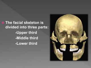
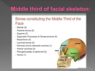
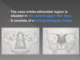
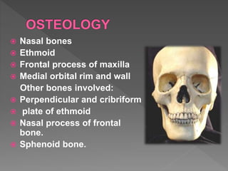
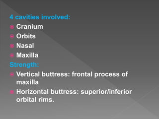
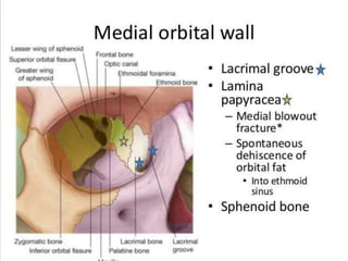
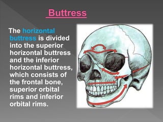
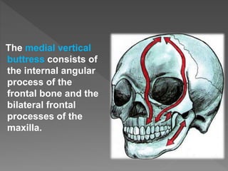
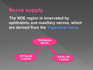
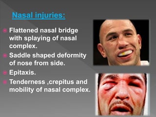
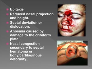
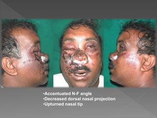
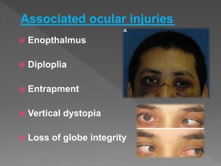
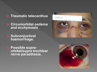
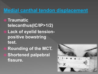
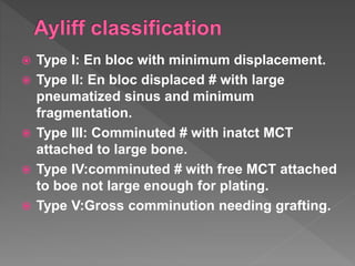
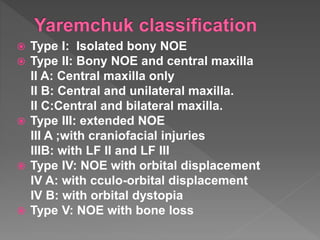
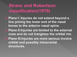
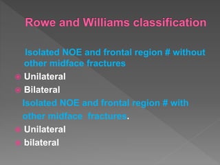
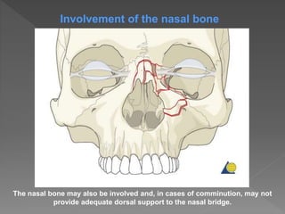
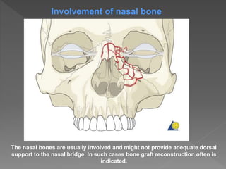
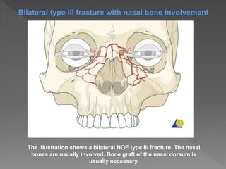
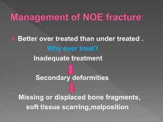
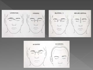
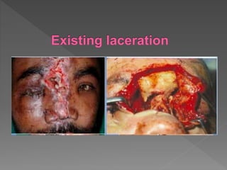
The naso-orbitoethmoid complex (NOE) fracture represents the most wearisome and challenging of all facial fractures due to the complexity and intricacy of its surgical & anatomic components. A good working knowledge with regards its surgical anatomy, clinical features, sequence of treatment & surgical approaches, potential pitfalls in its treatment & postoperative consideration,. Appropriate diagnosis and timely treatment is crucial to avoid unfavorable & difficult to treat sequelae.
Clinical significance of submental artery island flap. department of oral and maxillofacial surgery. presentation from international science conference 2016-17
Naso-orbital-ethmoid (NOE) fractures: Management principles, options and rec...Dibya Falgoon Sarkar
Comprehensive discussion on diagnosis and management of NOE fractures. Surgical anatomy and approaches to NOE region is also discussed. Reconstruction of NOE complex is discussed. Recent advances in management of NOE fractures are also highlighted in this presentation
Frontal sinus fractures are currently managed by various medical specialists, including otolaryngologists/head and neck surgeons, maxillofacial surgeons, plastic surgeons, and neurosurgeons. As a result, consensus does not exist regarding the timing, indications, and treatment modality of these injuries.
Naso orbital ethmoid fractures- part 2 /certified fixed orthodontic courses ...Indian dental academy
The Indian Dental Academy is the Leader in continuing dental education , training dentists in all aspects of dentistry and offering a wide range of dental certified courses in different formats.
Indian dental academy provides dental crown & Bridge,rotary endodontics,fixed orthodontics,
Dental implants courses.for details pls visit www.indiandentalacademy.com ,or call
0091-9248678078
surgical anatomy of nose is a humble attempt to make the anatomy of nose simpler and easy for medical students and fellow physicians. at the end of the presentation the students will be able to identify all the structures.
Orbital anatomy and orbital fracture/oral surgery courses by indian dental ac...Indian dental academy
Indian Dental Academy: will be one of the most relevant and exciting training center with best faculty and flexible training programs for dental professionals who wish to advance in their dental practice,Offers certified courses in Dental implants,Orthodontics,Endodontics,Cosmetic Dentistry, Prosthetic Dentistry, Periodontics and General Dentistry.
This PowerPoint presentation provides a concise and technical exploration of NOE fractures, encompassing fracture classifications, diagnostic modalities, and treatment approaches. Delve into the intricacies of fracture pathology, radiological assessments, and surgical interventions
Couples presenting to the infertility clinic- Do they really have infertility...Sujoy Dasgupta
Dr Sujoy Dasgupta presented the study on "Couples presenting to the infertility clinic- Do they really have infertility? – The unexplored stories of non-consummation" in the 13th Congress of the Asia Pacific Initiative on Reproduction (ASPIRE 2024) at Manila on 24 May, 2024.
Prix Galien International 2024 Forum ProgramLevi Shapiro
June 20, 2024, Prix Galien International and Jerusalem Ethics Forum in ROME. Detailed agenda including panels:
- ADVANCES IN CARDIOLOGY: A NEW PARADIGM IS COMING
- WOMEN’S HEALTH: FERTILITY PRESERVATION
- WHAT’S NEW IN THE TREATMENT OF INFECTIOUS,
ONCOLOGICAL AND INFLAMMATORY SKIN DISEASES?
- ARTIFICIAL INTELLIGENCE AND ETHICS
- GENE THERAPY
- BEYOND BORDERS: GLOBAL INITIATIVES FOR DEMOCRATIZING LIFE SCIENCE TECHNOLOGIES AND PROMOTING ACCESS TO HEALTHCARE
- ETHICAL CHALLENGES IN LIFE SCIENCES
- Prix Galien International Awards Ceremony
Pulmonary Thromboembolism - etilogy, types, medical- Surgical and nursing man...VarunMahajani
Disruption of blood supply to lung alveoli due to blockage of one or more pulmonary blood vessels is called as Pulmonary thromboembolism. In this presentation we will discuss its causes, types and its management in depth.
Tom Selleck Health: A Comprehensive Look at the Iconic Actor’s Wellness Journeygreendigital
Tom Selleck, an enduring figure in Hollywood. has captivated audiences for decades with his rugged charm, iconic moustache. and memorable roles in television and film. From his breakout role as Thomas Magnum in Magnum P.I. to his current portrayal of Frank Reagan in Blue Bloods. Selleck's career has spanned over 50 years. But beyond his professional achievements. fans have often been curious about Tom Selleck Health. especially as he has aged in the public eye.
Follow us on: Pinterest
Introduction
Many have been interested in Tom Selleck health. not only because of his enduring presence on screen but also because of the challenges. and lifestyle choices he has faced and made over the years. This article delves into the various aspects of Tom Selleck health. exploring his fitness regimen, diet, mental health. and the challenges he has encountered as he ages. We'll look at how he maintains his well-being. the health issues he has faced, and his approach to ageing .
Early Life and Career
Childhood and Athletic Beginnings
Tom Selleck was born on January 29, 1945, in Detroit, Michigan, and grew up in Sherman Oaks, California. From an early age, he was involved in sports, particularly basketball. which played a significant role in his physical development. His athletic pursuits continued into college. where he attended the University of Southern California (USC) on a basketball scholarship. This early involvement in sports laid a strong foundation for his physical health and disciplined lifestyle.
Transition to Acting
Selleck's transition from an athlete to an actor came with its physical demands. His first significant role in "Magnum P.I." required him to perform various stunts and maintain a fit appearance. This role, which he played from 1980 to 1988. necessitated a rigorous fitness routine to meet the show's demands. setting the stage for his long-term commitment to health and wellness.
Fitness Regimen
Workout Routine
Tom Selleck health and fitness regimen has evolved. adapting to his changing roles and age. During his "Magnum, P.I." days. Selleck's workouts were intense and focused on building and maintaining muscle mass. His routine included weightlifting, cardiovascular exercises. and specific training for the stunts he performed on the show.
Selleck adjusted his fitness routine as he aged to suit his body's needs. Today, his workouts focus on maintaining flexibility, strength, and cardiovascular health. He incorporates low-impact exercises such as swimming, walking, and light weightlifting. This balanced approach helps him stay fit without putting undue strain on his joints and muscles.
Importance of Flexibility and Mobility
In recent years, Selleck has emphasized the importance of flexibility and mobility in his fitness regimen. Understanding the natural decline in muscle mass and joint flexibility with age. he includes stretching and yoga in his routine. These practices help prevent injuries, improve posture, and maintain mobilit
Ethanol (CH3CH2OH), or beverage alcohol, is a two-carbon alcohol
that is rapidly distributed in the body and brain. Ethanol alters many
neurochemical systems and has rewarding and addictive properties. It
is the oldest recreational drug and likely contributes to more morbidity,
mortality, and public health costs than all illicit drugs combined. The
5th edition of the Diagnostic and Statistical Manual of Mental Disorders
(DSM-5) integrates alcohol abuse and alcohol dependence into a single
disorder called alcohol use disorder (AUD), with mild, moderate,
and severe subclassifications (American Psychiatric Association, 2013).
In the DSM-5, all types of substance abuse and dependence have been
combined into a single substance use disorder (SUD) on a continuum
from mild to severe. A diagnosis of AUD requires that at least two of
the 11 DSM-5 behaviors be present within a 12-month period (mild
AUD: 2–3 criteria; moderate AUD: 4–5 criteria; severe AUD: 6–11 criteria).
The four main behavioral effects of AUD are impaired control over
drinking, negative social consequences, risky use, and altered physiological
effects (tolerance, withdrawal). This chapter presents an overview
of the prevalence and harmful consequences of AUD in the U.S.,
the systemic nature of the disease, neurocircuitry and stages of AUD,
comorbidities, fetal alcohol spectrum disorders, genetic risk factors, and
pharmacotherapies for AUD.
These simplified slides by Dr. Sidra Arshad present an overview of the non-respiratory functions of the respiratory tract.
Learning objectives:
1. Enlist the non-respiratory functions of the respiratory tract
2. Briefly explain how these functions are carried out
3. Discuss the significance of dead space
4. Differentiate between minute ventilation and alveolar ventilation
5. Describe the cough and sneeze reflexes
Study Resources:
1. Chapter 39, Guyton and Hall Textbook of Medical Physiology, 14th edition
2. Chapter 34, Ganong’s Review of Medical Physiology, 26th edition
3. Chapter 17, Human Physiology by Lauralee Sherwood, 9th edition
4. Non-respiratory functions of the lungs https://academic.oup.com/bjaed/article/13/3/98/278874
These lecture slides, by Dr Sidra Arshad, offer a quick overview of physiological basis of a normal electrocardiogram.
Learning objectives:
1. Define an electrocardiogram (ECG) and electrocardiography
2. Describe how dipoles generated by the heart produce the waveforms of the ECG
3. Describe the components of a normal electrocardiogram of a typical bipolar leads (limb II)
4. Differentiate between intervals and segments
5. Enlist some common indications for obtaining an ECG
Study Resources:
1. Chapter 11, Guyton and Hall Textbook of Medical Physiology, 14th edition
2. Chapter 9, Human Physiology - From Cells to Systems, Lauralee Sherwood, 9th edition
3. Chapter 29, Ganong’s Review of Medical Physiology, 26th edition
4. Electrocardiogram, StatPearls - https://www.ncbi.nlm.nih.gov/books/NBK549803/
5. ECG in Medical Practice by ABM Abdullah, 4th edition
6. ECG Basics, http://www.nataliescasebook.com/tag/e-c-g-basics
New Directions in Targeted Therapeutic Approaches for Older Adults With Mantl...i3 Health
i3 Health is pleased to make the speaker slides from this activity available for use as a non-accredited self-study or teaching resource.
This slide deck presented by Dr. Kami Maddocks, Professor-Clinical in the Division of Hematology and
Associate Division Director for Ambulatory Operations
The Ohio State University Comprehensive Cancer Center, will provide insight into new directions in targeted therapeutic approaches for older adults with mantle cell lymphoma.
STATEMENT OF NEED
Mantle cell lymphoma (MCL) is a rare, aggressive B-cell non-Hodgkin lymphoma (NHL) accounting for 5% to 7% of all lymphomas. Its prognosis ranges from indolent disease that does not require treatment for years to very aggressive disease, which is associated with poor survival (Silkenstedt et al, 2021). Typically, MCL is diagnosed at advanced stage and in older patients who cannot tolerate intensive therapy (NCCN, 2022). Although recent advances have slightly increased remission rates, recurrence and relapse remain very common, leading to a median overall survival between 3 and 6 years (LLS, 2021). Though there are several effective options, progress is still needed towards establishing an accepted frontline approach for MCL (Castellino et al, 2022). Treatment selection and management of MCL are complicated by the heterogeneity of prognosis, advanced age and comorbidities of patients, and lack of an established standard approach for treatment, making it vital that clinicians be familiar with the latest research and advances in this area. In this activity chaired by Michael Wang, MD, Professor in the Department of Lymphoma & Myeloma at MD Anderson Cancer Center, expert faculty will discuss prognostic factors informing treatment, the promising results of recent trials in new therapeutic approaches, and the implications of treatment resistance in therapeutic selection for MCL.
Target Audience
Hematology/oncology fellows, attending faculty, and other health care professionals involved in the treatment of patients with mantle cell lymphoma (MCL).
Learning Objectives
1.) Identify clinical and biological prognostic factors that can guide treatment decision making for older adults with MCL
2.) Evaluate emerging data on targeted therapeutic approaches for treatment-naive and relapsed/refractory MCL and their applicability to older adults
3.) Assess mechanisms of resistance to targeted therapies for MCL and their implications for treatment selection
Report Back from SGO 2024: What’s the Latest in Cervical Cancer?bkling
Are you curious about what’s new in cervical cancer research or unsure what the findings mean? Join Dr. Emily Ko, a gynecologic oncologist at Penn Medicine, to learn about the latest updates from the Society of Gynecologic Oncology (SGO) 2024 Annual Meeting on Women’s Cancer. Dr. Ko will discuss what the research presented at the conference means for you and answer your questions about the new developments.
The prostate is an exocrine gland of the male mammalian reproductive system
It is a walnut-sized gland that forms part of the male reproductive system and is located in front of the rectum and just below the urinary bladder
Function is to store and secrete a clear, slightly alkaline fluid that constitutes 10-30% of the volume of the seminal fluid that along with the spermatozoa, constitutes semen
A healthy human prostate measures (4cm-vertical, by 3cm-horizontal, 2cm ant-post ).
It surrounds the urethra just below the urinary bladder. It has anterior, median, posterior and two lateral lobes
It’s work is regulated by androgens which are responsible for male sex characteristics
Generalised disease of the prostate due to hormonal derangement which leads to non malignant enlargement of the gland (increase in the number of epithelial cells and stromal tissue)to cause compression of the urethra leading to symptoms (LUTS
2. Fractures of the middle third of the facial
skeleton are known as maxillofacial
skeletal injuries and they are associated
with varying degrees of injuries of soft
tissue.
3. The facial skeleton is
divided into three parts:
-Upper third
-Middle third
-Lower third
4.
5. The naso-orbito-ethmoidal region is
situated in the central upper mid face.
It consists of a strong triangular frame.
6. Due to the complexity and the density of
the anatomic components of the area,
the fracture in this region has been a
great challenge in maxillofacial trauma.
Successful results depend upon
knowledge of the complex anatomy and
ability to provide correct early
management.
8. Nasal bones
Ethmoid
Frontal process of maxilla
Medial orbital rim and wall
Other bones involved:
Perpendicular and cribriform
plate of ethmoid
Nasal process of frontal
bone.
Sphenoid bone.
9. 4 cavities involved:
Cranium
Orbits
Nasal
Maxilla
Strength:
Vertical buttress: frontal process of
maxilla
Horizontal buttress: superior/inferior
orbital rims.
10. The main structural buttress is the frontal
process of the maxilla and internal
angular process of the frontal bone,
supplemented by the thick proximal
portion of the nasal bone.
This reinforced pillar provides a central
skeletal framework to which more
delicate bones are joined.
11. The medial orbital wall is made up of
lacrimal bone and the delicate lamina
papyracea of the ethmoid bones posteriorly.
These structures are susceptible to
communition,allowing for a medial
displacement of the orbital contents after
blunt trauma(medial blowout).
12.
13. Superiorly, thin ethmoid
bones form part of the
floor of the anterior skull
base, in this region dural
injury and resultant CSF
leakage is possible.
The ethmoid bone is
located posterior to the
nasal bones,the ethmoid
air cells are present at
birth and enlarge to adult
size by the age 12 years.
15. The overall growth and size of ethmoid
complex is highly variable among
individuals.
The ethmoid labyrinth separates the
orbits from the nasal cavity ,while the
fovea ethmoidalis forms the roof of the
ethmoid sinuses laterally.
17. The medial canthal
ligament (MCT) is a
crucial soft tissue
component of NOE
complex.
It arises from the
anterior and posterior
lacrimal crest and
frontal process of
maxilla.
18. MCT surrounds the lacrimal sac and diverges to
become the orbicularis occuli muscle.,
tarsal plate, and suspensory ligaments of eyelids.
19. The tendon splits
around the lacrimal
sac and attaches to
the anterior and
posterior lacrimal
crests, as well as to
the frontal process
of maxilla.
The canthal tendon
diverges to become
the pretarsal
,preseptal, and
orbital orbicularis
oculi muscle.
20. The action of the muscles and tendon
thus allow for pumping action of the
lacrimal sac and ducts allowing for
propagation of tears through
nasolacrimal system.
In addition, the MCT acts as a
suspensory sling for the globe,
maintaining its support along with the
lateral canthal tendon.
21. A normal intercanthal distance is 30-35
mm ,which is approximately half of the
interpupilary distance and is equivalent
to the width of the nasal base.
22.
23. Has the potential to be disrupted on a
NOE fracture especially a comminuted
one.
The system consists of a lacrimal gland
situated in the superolateral anterior
portion of the orbit and two lacrmial
canaliculi that drain the eye via puncta
that are situated in the medial aspect of
each eye.
24.
25. The sac drains into the inferior meatus
via the nasolacrimal duct.
The duct is around 20 mm in length half
of which is bony.
The portion of the nasolacrimal system
that is most prone to damage is the bony
nasolactimal duct.
26. The cribriform plate is loacted
approximately 1 cm inferior to the fovea
ethmoidalis and it formss the roof of the
nasal cavity medially.
27.
28. The horizontal
buttress is divided
into the superior
horizontal buttress
and the inferior
horizontal buttress,
which consists of
the frontal bone,
superior orbital
rims and inferior
orbital rims.
29. The medial vertical
buttress consists of
the internal angular
process of the
frontal bone and the
bilateral frontal
processes of the
maxilla.
30. The blood supplying for the midface and
nasal region comes from the branches of
internal and the external carotid arteries.
The anterior and posterior ethmoid
arteries descend from the internal carotid
artery.
The maxillary artery from the external
carotid artery and subsequent branches
play a mainstay role for supporting the
midface.
31. The NOE region is innervated by
ophthalmic and maxillary nerves, which
are derived from the Trigeminal nerve.
TRIGEMINAL
NERVE
OPTHALMI
C NERVE
MAXILLAR
Y NERVE
32. The nasal bones and underlying cartilage are
susceptible to fractures because the nose
maintains a prominent position and central
location on the face and because it has a low
breaking strength.
Patterns of fractures are known to vary with
momentum of the striking object and the
density of the underlying bone.(Murray,1994)
33. As with other facial bones,younger
patients tend to have larger nasoseptal
fracture segments,whereas older patients
are more likely to present with more-
comminuted fracture patterns.
(Cummings,1998)
34. RTA
Sport injuries
Fights
Work related accidents
Falls are the most common cause of
nasal injury in children.
35. Violation of the primary
buttresses of the NOE
complex
Comminution of the
entire complex may
occur
Telecanthus,enophthalmos,diplopia,
midface retrusion
36. The clinical symptoms associate wit the
location and severity of the NOE fracture.
Patients with naso-orbito-ethmoidal
(NOE) fractures often have associated
facial injuries or panfacial fractures.
38. Gross facial edema may
show firstly in the early
stage of fracture, which
will result in distortion
of soft tissue landmarks.
39. Laceration in the nose and
forehead.
Intracranial involvement.
Eye, forehead, and nose pain
Forehead paraesthesias
Traumatic hypertelorism
Mongoloid slant.
40. Flattened nasal bridge
with splaying of nasal
complex.
Saddle shaped deformity
of nose from side.
Epitaxis.
Tenderness ,crepitus and
mobility of nasal complex.
Nasal injuries:
41. Epitaxis
Reduced nasal projection
and height.
Septal deviation or
dislocation.
Anosmia caused by
damage to the cribiform
plate.
Nasal congestion
secondary to septal
hematoma or
bony/cartilaginous
deformity.
46. Traumatic
telecanthus(IC/IP>1/2)
Lack of eyelid tension-
positive bowstring
test.
Rounding of the MCT.
Shortened palpebral
fissure.
Medial canthal tendon displacement
47. For telecanthus to occur ,the fracture
must involve at least 4 sites:
Medial orbital wall
Nasomaxillary butress/inferior orbital rim
Nrontomaxillary junction
Lateral nasal bone
49. Fracture of floor of anterior cranial
fossa/base of skull.
Escape of CSF through
Ethmoidal sinus
Sphenoidal sinus
Cribriform plate
Frontal sinus
50. Communication between
Meninges
Nose
Paranasal sinuses
Dural laceration
Later becomes
epithelised to Fistula
Blood clot of brain tissue
may obstruct fluid
passage.
51. After lysis of clot or increased
intracranial pressure leakage is seen.
Mobile midface fractures often creates
pumping action –because of increased
CSF leakage.
52. How is NOE complex fracture classified?
The status of the resulting central segment of
bone left by an NOE fracture is the basis of
classification of fracture patterns for this type
of injury.
Each fracture type is sub classified as either
unilateral or bilateral.
Among many classifications of NOE fractures,
the most widely accepted classification system
was established by Markowitz et al.
.
53. Type I: En bloc with minimum displacement.
Type II: En bloc displaced # with large
pneumatized sinus and minimum
fragmentation.
Type III: Comminuted # with inatct MCT
attached to large bone.
Type IV:comminuted # with free MCT attached
to boe not large enough for plating.
Type V:Gross comminution needing grafting.
54. Type I: Isolated bony NOE
Type II: Bony NOE and central maxilla
II A: Central maxilla only
II B: Central and unilateral maxilla.
II C:Central and bilateral maxilla.
Type III: extended NOE
III A ;with craniofacial injuries
IIIB: with LF II and LF III
Type IV: NOE with orbital displacement
IV A: with cculo-orbital displacement
IV B: with orbital dystopia
Type V: NOE with bone loss
55. Plane I: Injuries do not extend beyond a
line joining the lower end of the nasal
bones to the anterior nasal spine.
Plane II:Injuries are limited to the external
nose and do not trangress the orbital rim.
Plane III:Injuries are more serious involve
orbital and possibly intracranial
structures.
56. Isolated NOE and frontal region # without
other midface fractures
Unilateral
Bilateral
Isolated NOE and frontal region # with
other midface fractures.
Unilateral
bilateral
57. TYPE I: Involves single segment central
fragment fractures.
TYPE II: Comminuted central fragment
with fracture lines remaining peripheral
to the MCT insertion.
TYPE III: Comminuted central fragment
with fracture lines extending beneath the
MCT insertion.
59. In this simplest form,NOE fractures are
isolated involving only the portion of the
medial orbital rim that contains medial
canthal tendon.
Type I pattern consists of single central
fragment bearing the medial canthus.
60. These fractures maybe bilateral ,complete
or displaced.
Uncommonly ,the medial canthal tendon
is torn or avulsed completely from an
intact medial bony wall.
61. In unilateral Markowitz type I fractures, there is a single large NOE
fragment bearing the medial canthal tendon.
62. Involvement of the nasal bone: the nasal bone may also be involved
and, in cases of comminution, may not provide adequate dorsal
support to the nasal bridge.
63. Type II fractures are complete and maybe
unilateral or bilateral.
They may be single segment or
communited external to the medial
canthal insertion in the central segment.
MCT maintains continuity with large
fractured segment of bone,which maybe
used in the surgical reduction.
64. In unilateral type II fractures, there is often comminution of the NOE area, but the
canthal tendon remains attached to a fragment of bone, allowing the canthus to
be stabilized with wires or a small plate on the fractured segment.
65. The nasal bone may also be involved and, in cases of comminution, may not
provide adequate dorsal support to the nasal bridge.
Involvement of the nasal bone
66. The illustration shows a bilateral NOE type II fracture. In bilateral fractures the
nasal bones are commonly involved. In some instances, bone grafting of the
nasal dorsum may be necessary.
Bilateral type II fracture with nasal bone involvement
67. Communition within the central fragment
allows fracture to extend beneath the
canthal insertion characterising the type
III fracture pattern.
The canthus is rarely avulsed but it is to
bone fragments that are too small to
utilize in reconstruction.
68. In type III fractures, there is often comminution of the NOE area (as in type II
fractures) and a detachment of the medial canthal tendon from the bone.
69. The nasal bones are usually involved and might not provide adequate dorsal
support to the nasal bridge. In such cases bone graft reconstruction often is
indicated.
Involvement of nasal bone
70. The illustration shows a bilateral NOE type III fracture. The nasal
bones are usually involved. Bone graft of the nasal dorsum is
usually necessary.
Bilateral type III fracture with nasal bone involvement
71. Establish ABCs.
Diagnose any associated injuries.
Direct examination of NOE complex.
A thorough head and neck examination to
reveal injuries to the brain,spine, orbit
and facial skeleton is required.
A team approach involving the
otolaryngologist,plastic surgeon,
neurosurgeon and pohthalmologic
consultation is mandatory.
73. Plain radiographs have limited
usefulness in aiding in diagnosis of
NOE #s.
Thin cut axial and coronal CT scans
are the criterion standard for the
diagnosis of NOE fractures.
74. Plain films are of almost no use in diagnosing
NOE fractures because most will be undetected.
75. Is of greatest value.
HRCT adds to the existing value.
What to ask for?
-1-2 mm axial and coronal slices with 3D
recon.
-top of skull –frontal-sinus –orbits-maxilla
- Bone window NOE bony complex
-soft tissue window brain/ocular
adnexia.
76. Axial cuts:
Position and status of
frontal process of
maxilla central
fragment
Medial walls of orbit if
they are “blown in”
nasally.
Anterior and posterior
tables of frontal bone.
Nasolacrimal system.
77. Coronal cuts:
Cuts taken from nasal
bridge to orbital apex.
Junction of floor to
medial wall assessed.
Disruption of ant.
cranial fossa around
cribriform plate.
CSF leak CT value.
Localisation of CSF
leaks.
78. A surgeon can often distinguish between
a type I NOE fracture versus a type II / III
fracture by the degree of comminution
noted on the CT.
It is generally very difficult to distinguish
between a type II and a type III NOE
fracture based on radiographic
examination
79.
80. A 3D CT, as shown, can help assessing
the severity of the injury but the final
diagnosis requires clinical findings and is
usually made intraoperatively.
This 3D CT shows a bilateral NOE type II
with involvement of the nasal bones.
Be aware that a 3D CT often
underestimates the severity of the injury.
81. Examine the nasal cavity for the presence
of CSF.
Query all conscious patients about the
presence of watery rhinorrhea or salty
postnasal drainage.
Test bloody fluid that is suspicious for CSF
rhinorrhea (see Lab Studies).
Examine facial lacerations under sterile
conditions to assess depth of penetration
or intracranial violation.
To evaluate the integrity of the medial
canthal tendon.
82. Measure and document telecanthus and
enophthalmos.
Assess and document pupil responses
and extraocular muscle mobility.
Palpate the nasal bones for crepitus and
comminution.
Evaluate the septum for septal
hematoma.
Evaluate the degree of nasal or midface
retrusion. Preinjury photographs may be
helpful.
83. Examine facial lacerations under sterile conditions
to assess depth of penetration or intracranial
violation.
85. An intercanthal distance of > 35 mm is suggestive of
traumatic telecanthus ,measurement approaching
40mm are almost diagnostic.
86. The patient often has swelling in the medial canthal
area and pain and crepitation with palpation.
87. Examine the nasal cavity for
the presence of CSF.
Query all the patients about
the presence of watery
rhinnorhea or salty postnasal
drainage.
Test bloody fluid that is
suspicious of CSF rhinnorhea.
With NOE fractures a CSF leak should be assumed to be present even
if it is not clinically demonstrable and appropriate chemoprophylaxis
should be commenced.
88. The following diagnostic
procedures can be
performed if there is a
suspected CSF leak (clinical
sign: straw-colored or clear
nasal drainage):
Tilt test with positive halo
sign (as illustrated)
Comparison of the
concentration of glucose
between fluid and patient’s
serum.
89. CT scan with thin coronal cuts (0.5 mm)
of the cribriform plate.
90. Bloody rhinorrhea
suspicious for CSF can be
placed on filter paper and
observed for a halo sign.
If CSF is present ,it diffuses
faster than blood and
results in a clear halo
around the central stain.
Routine chemistry analysis
of the rhinorrhea may reveal
an elevated glucose content
consistent with CSF.
91. Beta 2 transferrin is the definitive test for
CSF rhinorrhoea.collect 1 ml of the
suspected fluid in a red top tube.
Watery rhinorrhea that is positive for beta
2-transferrin is diagnostic for a CSF leak.
93. These two tests aids in the diagnosis of
instability of the Medial canthus tendon.
Bowstring test
Bimanual palpation by placing an
instrument into the nose to determine
canthal bearing bone fragment
displaced and mobile.
.
94. In the bow string test ,the eyelid is pulled
laterally while the tendon area is palpated
to detect movement of fracture segments.
A lack of resistance or movement of the
underlying bone is indicative of a
fracture.
The surgeon may be able to grab the
eyelid or use a forceps to grab the skin in
the medial canthal area and pull it
laterally (“bow-string” test).
95.
96. It requires placing an instrument (kelly
clamp) high into the nose,with its tip
directly beneath the MCT .
Gentle lifting with the contralateral finger
palpates the canthal tendons and allows
an assessment of instability of the tendon
attachement and necessity for open
reduction.
97.
98. The lacrimal drainage system is intimately
related to the NOE region and can be
damaged during the trauma.
The surgeon should assess the
patency/continuity of the nasolacrimal
system at the time of surgical treatment.
If there is a discontinuity in the
nasolacrimal system repair should be
considered at the time of fracture
treatment.
99. Dye disappearance test
Jones test(primary and secondary tests)
Lacrimal irrigation
Scintigraphy
Contrast dacryocystography
CT scan
100. DDT is useful for assessing the presence or
absence of adequate lacrimal outflow.
2% fluorescein dye solution or a moistened
fluorescein strip instilled in conjuctival
fornix.
Persistence of significant dye indicates an
obstruction.
If the DDT result is normal, severe lacrimal
drainage dysfunction is highly unlikely.
103. The Jones tests have been used in the
evaluation of epiphora.
JONES I JONES II
JONES TEST
104. Like DDT,this test investigates lacrimal
outflow under normal physiologic conditions.
1 drop of 2% fluorescein dye placed into
conjuctival sac.
After about 5 mins ,cotton tipped applicator
inserted under the inferior turbinate.
If bud stained with dye ,test is positive.
Test is negative If no dye is detected ,means
there is partial or absolute obstruction or
failure of lacrimal pump.
105.
106.
107. Radiographic visualization of
the lacrimal sacs and
associated structures after
injection of a contrast
medium.
Contrast dacryocystography
provides anatomical
information with dye injection
into the lacrimal system
followed by computerized
digital subtraction imaging.
108. Better over treated than under treated .
Why over treat?
Inadequate treatment
Secondary deformities
Missing or displaced bone fragments,
soft tissue scarring,malposition
109. Reconstitution of the skeletal framework
of NOE region.
Stabilization of the intercanthal width and
MCT..
Orbital reconstruction.
Establishment of nasal support.
Reconstruction of other craniofacial
injuries including frontal sinus.
Soft tissue repair.
110. Early one stage repair
Exposure of all fracture fragments
Precise anatomic rigid fixation
Immediate bone grafting as indicated for
bony loss.
Definitive soft tissue management.
111. Exposure
Identify the MCL or the MCL bearing
bone.
Reduce/reconstruct medial orbital rims.
Reconstruct medial orbital walls.
Transnasal conthopexy
Reduce septal displacement
Soft tissue readaptation.
112. Unobstructed visualization of the
articulations of all the bones in the region.
One of the main reasons for treating NOE
#s is esthetics ,hence incisions are made
keeping in mind the esthetics.
Remote incisions preferred.
116. Advantages:
Correction of associated frontal sinus fracture.
Harvesting of calvarial bone graft or primary
reconstruction
Harvesting of pericranial flap of sufficient
length for sealing of defects in the ant.cranial
fossa.
Disadvantage:
Cannot be used when the skull has been
opened up previously for craniotomies by the
neurosurgeons.
117.
118.
119.
120.
121.
122.
123. Curved incision over
lateral nasal bones
anterior to MCL
attachment.
Skin here is thin-allows
easy exposure.
Sufficient or limited
reconstruction.
Cannot be used in
bilateral
canthopexies,bone
grafting.
124. Skin incision approx. 3 cm in length
made along the superior medial
orbital rim from 1 cm medial to medial
canthus to the lower border of the
medial eyebrow.
Angles of limbs of the W-110 to 120
degree
Four limbs of the W can be placed
parallel or oblique to the RTSL
The lateral limb of the .W can be
extended laterally long the lower
border of the medial eyebrow,
depending on the desired exposure.
125. Muscledissection,supratrochlea
r nerve located and preserved.
Periosteum is incised from
upper half of the MCT to the
medial portion of superior
orbital rim-periorbita is laterally
reflected.
126. Advantages:
W has small segmented limbs parallel or
oblique to the relaxed skin tenion lines.
W limbs break up the scar into smaller
components- minimal external scar.
Pulling both ends of the W along its
longitudinal axis provides the increase of
its longitudinal length - allows implant
up to 3 cm to be inserted.
Superior access to medial orbital wall.
127. Incisions utilized:
Transoral degloving from 2nd molar.
Intercartilaginous incision.
Transfixion incision.
PROCEDURE:
Mucoperiosteal flap till piriform aperture
raised.
Both intercartilaginous and tranfixion
incisions connected across the septal angle.
128.
129. The osseocartilagenous nose is
degloved over the upper lateral
cartilage as for a septorhinoplasty.
The intranasal incisions connected with
the oral incision by a nasal sill incison.
Midface can now be degloved.
130. Advantages:
No external visible scars.
Excellent visibility-as good as coronal
incision.
Minimal risk to vital structures.
No aesthetic sagging of tissues.
Provides concurrent access to zygoma on
both sides.
Disadvantages:
Suturing is vital-?stenosis of nasal aperture.
?damage to infraorbital nerve.
131.
132. 1. Both MCL remain attached and the
laterization of the complex is counteracted
by the orbicularis oculi. Type I : b/l single
segment NOE #
2. Tendon is still attached to the bone but
the bone fragment is separate from complex
: U/l single segment type I injury.
3. Avulsion of tendon from bony connection
type III.
4. Bone into which the tendon inserts is
missing
133.
134.
135.
136. Biomechanics of midface made
complicated by:
– Nonuniform geometry of bones
– Number and orientation of various attached
ligaments and soft tissues.
Treatment aimed to restrict three types of
movements of a fractures segment in 6
directions
Translatory movement essentially 2D
restricted by wires as well as plates
137. Rotatory movements : 3-D need
restrictions at 3 separate points plates
more effective.
Farther apart the fixation points better
the stability wider plates thus preferred.
3 wires or several small plates oriented
at different angles increase stability.
138. Adjunct to primary bone
grafting.
Avoids supplemental
maxillomandibular or
extraskeletal fixation .
Better rigid support and
immobilization.
Prevents overriding of the
fractured fragments.
139. Transnasal reduction of canthal bearing
fragment most important step in
preserving intercanthal distance.
Loose nasal bones may be removed
temporarily for better access.
Fragment bearing the MCL identified.
If fragment is large enough reduce and
fix it to adjacent bone with miniplates
140. Imperative to drill one hole posterior to
lacrimal fossa to prevent lateral splaying
coronal section : horizontal mattress
posteriorly and telecanthus.
Other wire passed superior and posterior
to lacrimal fossa on Proper placement of
transnasal wires posteriorly other side.
Wires tightened as much as possible to
“overreduce” and narrow the base to
gain the projection.
141. Importance:
To regain anatomic morphology
To regain lost orbital volume in blow out #
To achieve normal eye position after injury.
142. Bone material of choice for
reconstruction calvarial graft/rib graft.
Long pieces of bone used should
extend just behind the medial orbital rim.
Fixed with lag screws or miniplates.
If Bone pieces extend too posteriorly
poor access. loss of stability
143.
144. Canthal ligament was identified and
tagged earlier.
Followed by orbital wall and rim
reconstruction.
Steps demanded greatest traction.
If canthopexy performed earlier :
– Vigorous traction could pull through the
MCL and further damage the ligament.
146. Holes:
– medial orbital rim posterior and
superior to posterior lacrimal crest.
– 2-4mm diameter.
Direction of transnasal wire high to low
The essential biomechanical principle
is that although the tightening produces
a vertical force, the MCT moves medially
in its prepared area of attachment.
147.
148. A contouring burr is used to create a
depression in the frontal process of the
maxilla just superior and posterior to the
anterior lacrimal crest to inset the MCT.
On the contralateral fronto-glabellar area,
a 1.5-mm hole is drilled and taken
through to the depression created to
receive the MCT. A second drill hole is
made 5 mm below the first.
149. 18-gauge syringe needle is passed
through the first hole to the medial
canthal area and the superior wire is fed
through .
This is repeated through the second hole,
and the wire is tightened until the
canthus is firmly secured.
150.
151.
152.
153. Nasal bone forming medial orbital wall and
the bridge of the nose fragile ?
Withstand wire tightening.
Glabellar portion of the frontal bone is solid
and can withstand wire tightening. • The
fixation is secure.
Due to the relatively large amount of soft
tissue covering the twisted wire, extrusion
of the wire through the skin does not occur.
No injury to delicate structures of the
contralateral medial orbit such as the
lacrimal sac or lacrimal duct.
154. Technically difficult.
Necessitates wide exposure sufficient to
allow transverse passage of a wire
through a bony fenestration deep within
the orbit.
Weakening of the bones ( when central
fragment is drilled twice),
Dissection of the contralateral orbit.
155. A Kirschner wire with one
of the tips hammered and
shaped into a simple drill
is passed from the left
orbit toward the right thru
central fragment.
Plastic catheter is pushed
forward over the Kirschner
wire guide and through the
transnasal hole.
156. A bent, looped wire is
introduced from left to
right through the plastic
tube left in the
transnasal hole after the
Kirschner wire is
removed.
157. A titanium microplate is placed
in the loop at the second
penetration site.
Second microplate is placed
between the exiting wires at the
first penetration site,
Ends of the wires are twisted
together
The free tips of the wire at the
site of first penetration can be
used for canthopexy without
microplate placement, if desired.
158.
159. After passing thru ligament;The 30G wire is
passed through the posterior hole of the
miniplate and loosely twisted.
The plate is positioned, with the medial canthal
tendon pushed deep, near the posterior lacrimal
crest. The drill hole is made in the area of the
anterior hole of the plate and fixed with a
stainless steel screw (2 × 6 mm).
The stainless steel wire is then tightened.
The frontal process of the maxilla in the region
of the lacrimal crest is utilized for fixing the two-
hole plate transversely .
160. A simple method for medical canthal
wiring reconstruction.
A homolaterally fixed osteosynthesis
plate and a metal wire is used.
Avoids transnasal wiring and gives
superior control when correcting the
position of the lacerated medial canthus.
161. 20 metal wire is fixated to the ligament by
a double stitch.
One end of the metal wire is brought
through the last hole of the plate and the
plate is then fixed at the nasal bone in
such a way that the end of the plate is at
least some millimetres posterior and
superior to the lacrymal fossa.
Reach the desired position the wire
can be twisted and the wound closed.
162.
163.
164. Have provided for effective longterm
biomechanical stability in extremity
tendon reattachment to bone in
orthopedics
Prethreaded bone anchor system Mitek
mini bone anchor system used.
165. The key to replicating the delicate
three dimensional contour of the
medial canthus lies in addressing all
three vectors of attachment”.
166. Optimal position for bone anchor placement
is determined.
The hole for screw placement is positioned
within the central portion of the lacrimal
fossa.
If bone loss present no lacrimal fossa,
the screw hole is placed within a rigidly
fixated medial orbital wall bone graft at a
point corresponding to the contralateral
central lacrimal fossa position.
167.
168.
169. NOE # are associated with fractures of
perpendicular plate of ethmoid, septal
deviation, septal hematomas.
Goal should be to
– assure midline positioning of septum to
prevent airway compromise.
– Reduce septal fractures
170.
171.
172. Collapse of the bony
architecture broadening
of base.
Weakening of nasal
septal structures.
Damage to upper lateral
cartilages.
Complete loss of dorsal
nasal projection and loss
of support.
173.
174. Reinforcement of thin bones
Prevention of overriding and
displacement of fragments
Maintenance of vertical dimension
Provides substrate for osseous union
Prevention of soft tissue scarring
175. Bone graft sites: calvarial excellent
choice.
Shape it like a surf board gently tapering it
at the end.
Length should extend from frontonasal
junction to nasal tip.
Colummelar strut if needed.
Fixation:
- Single lag screw into the nasal pyramid.
-Microplate to cantilever off the frontal bone.
176.
177. Post surgical soft tissue thickening can
hamper esthetics.
Soft tissue thickening appearance of
telecanthus.
Solution: Soft tissue thermoplastic
stents.
Splint is contoured and overextended
into nasorbital valley. into junction of
nose and medial orbit. reinforced with
elastic tapes.
179. Temporary or permanent parasthesia.
CSF leak.
Meningitis.
Sinus infection or mucocoele.
Anosmia.
Infection of implants.
Postoperative telecanthus is a relatively
common complication of
nasoorbitoethmoid (NOE) fracture repair.
180. Pseudotelecanthus.
Enophthalmos results from inadequate
repair of the medial orbital wall or orbital
floor.
Midface retrusion may occur.
Extraocular dysfunction.
Blindness.
Possible need for additional surgery.
181. Postoperative ophthalmologic examination is
recommended, as well as gross visual acuity
checks every 6 hours for a 24-hour period.
The Penrose drains are removed from the
scalp at 24 hours, and the pressure dressing
is discontinued after 3 days. The lead
bolsters and scalp sutures are removed at 10
days postoperatively.
The patient should be examined and queried
again, looking for any evidence of a CSF leak.
Patients should be asked to perform standard
nasal hygiene (nasal saline irrigations and no
nose blowing).
182. Disruption of the delicate ethmoid
complex and comminution of the nasal
bones can make the repair of
nasoorbitoethmoid (NOE) complex
fractures extremely difficult.
These injuries often test the capabilities
of even the most experienced surgeons.
183. To obtain an aesthetic surgical result, the
surgeon must meticulously identify,
accurately reduce, and rigidly fixate the
medial canthal tendon and central
fragment.
Special attention also must be focused
on the overlying soft tissue to avoid
hematoma, chronic induration, and
pseudotelecanthus.
184. Aesthetic reconstruction of the nasal root
and medial canthal region continues to
be a significant surgical challenge.
Future advances may address this issue
with the use of surgical navigation
systems and/or intraoperative imaging,
which returns the bony architecture to its
premorbid state more accurately.
185. NOE injuries can be difficult to manage.
Proper assessment and early surgical
management of the NOE and concomitant
injuries are key to optimal outcomes.
Overcorrection of the bony position and
compression of the soft tissue overlying
the MCT are critical. Residual telecanthus
tends to be recalcitrant despite the best
efforts.