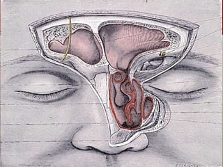
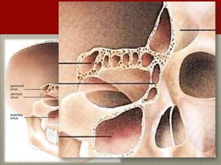
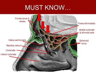
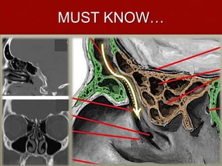
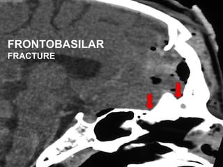
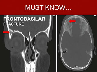
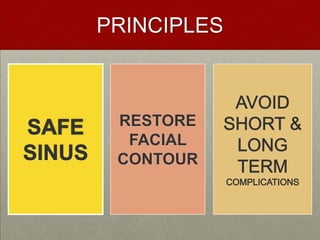
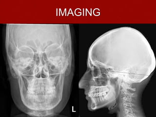
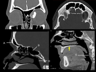
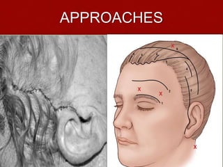
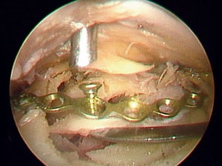
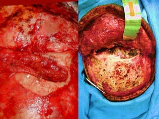
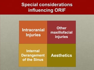
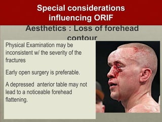
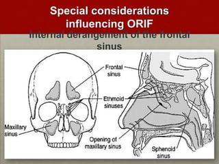
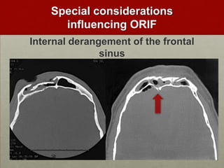
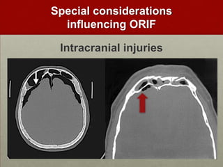
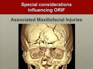
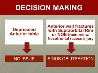
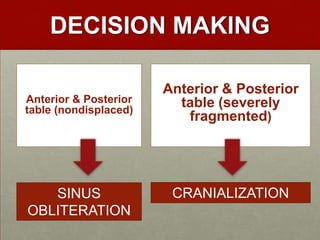
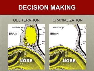
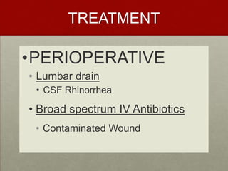
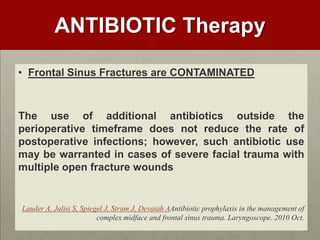
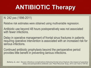
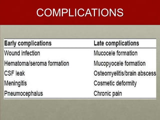
This document discusses frontal sinus fractures, including surgical anatomy, treatment approaches, considerations for open reduction and internal fixation (ORIF), and complications. It covers the anatomy of the olfactory bulb, cribriform plate, nasofrontal recess, and frontobasilar fractures that must be understood. Treatment goals are to restore facial contour and several surgical approaches are presented. Factors like intracranial injuries, other facial fractures, sinus derangement, and aesthetics can influence ORIF. Treatment may involve sinus obliteration or cranialization depending on fracture pattern. Perioperative care includes lumbar drains and antibiotics, though prolonged post-op antibiotics provide little benefit in preventing infections. Potential complications are also reviewed
Naso-orbital-ethmoid (NOE) fractures: Management principles, options and rec...Dibya Falgoon Sarkar
Comprehensive discussion on diagnosis and management of NOE fractures. Surgical anatomy and approaches to NOE region is also discussed. Reconstruction of NOE complex is discussed. Recent advances in management of NOE fractures are also highlighted in this presentation
This powerpoint describes the types of maxillectomy & operative steps for total maxillectomy. It also enumerates various flaps used for reconstruction of maxillectomy defect.
The Middle Third Of The Facial Skeleton Is Defined As An Area Bounded,
Superiorly –Line Drawn Across The skull from the Zygomatico frontal Suture across the Frontonasal & Frontomaxillary sutures to the Zygomaticofrontal suture on the opposite side
Inferiorly –by the occlusal plane of the upper teeth./upper alveolar ridge.
Posteriorly-The region is demarcated by the sphenoethmoidal junction but includes the free margin of the pterygoid laminae of the sphenoid bone inferiorly.
Inferiorly –by the occlusal plane of the upper teeth./upper alveolar ridge.
Posteriorly-The region is demarcated by the sphenoethmoidal junction but includes the free margin of the pterygoid laminae of the sphenoid bone inferiorly.
It is made up of the following bones:
1. Two maxillae
2. Tw o palatine bones
3. Two zygomatic bones and their temporal processes
4. Two zygomatic processes of the temporal bone
5. Two nasal bones
this presentation describes about each bone individually and its applied anatomy
A protocol for the management of frontal sinus /prosthodontic coursesIndian dental academy
The Indian Dental Academy is the Leader in continuing dental education , training dentists in all aspects of dentistry and
offering a wide range of dental certified courses in different formats.
The 6 Thinking hats ensures that groups think together in a focused manner, staying on task, & ensures that they focus their efforts on the most important elements of any issue being discussed.
The literature review usually precedes a research proposal and results section. Its goals are to situate the current study within the body of literature and to provide context for the particular reader. Literature reviews are important for research in nearly every academic field.
Le fort fracture by Dr. Amit Suryawanshi .Dentist in Kolhapur (MDS). Oral &...All Good Things
Description:
Hi. This is Dr. Amit T. Suryawanshi. Dentist in Kolhapur (MDS) Oral & Maxillofacial surgeon from Kolhapur, India. I am here on slideshare.com to share some of my own presentations presented at various levels in the field of OMFS. Hope this would somehow be helpful to you making your presentations. All the best & your replies are welcomed!
UCLA Plastic Surgeon, Jason Roostaeian, MD, presents the benefits of alar contour grafts to achieve natural looking results in cosmetic and functional rhinoplasty
Vertigo is a subtype of dizziness in which a patient inappropriately experiences the perception of motion (usually a spinning motion) due to dysfunction of the vestibular system.
For every new medicine we discover & invent - antibiotic resistance develops to the older one. For every microorganism that we eradicate, another one emerges to take its place.
i) The Vision of this Residents-in-training Manual is to create a perfect guide which is appropriate, competent & adaptive to the changing regional needs and culture of specialty training & dynamic enough to meet the demands of technology change, innovations in management & medical advancement.
The Vision of this BGH MC Residents-in-training Manual is to create a perfect guide which is appropriate, competent & adaptive to the changing regional needs and culture of specialty training & dynamic enough to meet the demands of technology change, innovations in management & medical advancement.
Evidence-based medicine (EBM) or evidence-based practice (EBP) aims to apply the best available evidence gained from the scientific method to clinical decision making. It seeks to assess the strength of evidence of the risks and benefits of treatments (including lack of treatment) and diagnostic tests. Evidence quality can range from meta-analyses and systematic reviews of double-blind, placebo-controlled clinical trials at the top end, down to conventional wisdom at the bottom.
The naso-orbitoethmoid complex (NOE) fracture represents the most wearisome and challenging of all facial fractures due to the complexity and intricacy of its surgical & anatomic components. A good working knowledge with regards its surgical anatomy, clinical features, sequence of treatment & surgical approaches, potential pitfalls in its treatment & postoperative consideration,. Appropriate diagnosis and timely treatment is crucial to avoid unfavorable & difficult to treat sequelae.
Ozempic: Preoperative Management of Patients on GLP-1 Receptor Agonists Saeid Safari
Preoperative Management of Patients on GLP-1 Receptor Agonists like Ozempic and Semiglutide
ASA GUIDELINE
NYSORA Guideline
2 Case Reports of Gastric Ultrasound
The Gram stain is a fundamental technique in microbiology used to classify bacteria based on their cell wall structure. It provides a quick and simple method to distinguish between Gram-positive and Gram-negative bacteria, which have different susceptibilities to antibiotics
ARTIFICIAL INTELLIGENCE IN HEALTHCARE.pdfAnujkumaranit
Artificial intelligence (AI) refers to the simulation of human intelligence processes by machines, especially computer systems. It encompasses tasks such as learning, reasoning, problem-solving, perception, and language understanding. AI technologies are revolutionizing various fields, from healthcare to finance, by enabling machines to perform tasks that typically require human intelligence.
TEST BANK for Operations Management, 14th Edition by William J. Stevenson, Ve...kevinkariuki227
TEST BANK for Operations Management, 14th Edition by William J. Stevenson, Verified Chapters 1 - 19, Complete Newest Version.pdf
Title: Sense of Smell
Presenter: Dr. Faiza, Assistant Professor of Physiology
Qualifications:
MBBS (Best Graduate, AIMC Lahore)
FCPS Physiology
ICMT, CHPE, DHPE (STMU)
MPH (GC University, Faisalabad)
MBA (Virtual University of Pakistan)
Learning Objectives:
Describe the primary categories of smells and the concept of odor blindness.
Explain the structure and location of the olfactory membrane and mucosa, including the types and roles of cells involved in olfaction.
Describe the pathway and mechanisms of olfactory signal transmission from the olfactory receptors to the brain.
Illustrate the biochemical cascade triggered by odorant binding to olfactory receptors, including the role of G-proteins and second messengers in generating an action potential.
Identify different types of olfactory disorders such as anosmia, hyposmia, hyperosmia, and dysosmia, including their potential causes.
Key Topics:
Olfactory Genes:
3% of the human genome accounts for olfactory genes.
400 genes for odorant receptors.
Olfactory Membrane:
Located in the superior part of the nasal cavity.
Medially: Folds downward along the superior septum.
Laterally: Folds over the superior turbinate and upper surface of the middle turbinate.
Total surface area: 5-10 square centimeters.
Olfactory Mucosa:
Olfactory Cells: Bipolar nerve cells derived from the CNS (100 million), with 4-25 olfactory cilia per cell.
Sustentacular Cells: Produce mucus and maintain ionic and molecular environment.
Basal Cells: Replace worn-out olfactory cells with an average lifespan of 1-2 months.
Bowman’s Gland: Secretes mucus.
Stimulation of Olfactory Cells:
Odorant dissolves in mucus and attaches to receptors on olfactory cilia.
Involves a cascade effect through G-proteins and second messengers, leading to depolarization and action potential generation in the olfactory nerve.
Quality of a Good Odorant:
Small (3-20 Carbon atoms), volatile, water-soluble, and lipid-soluble.
Facilitated by odorant-binding proteins in mucus.
Membrane Potential and Action Potential:
Resting membrane potential: -55mV.
Action potential frequency in the olfactory nerve increases with odorant strength.
Adaptation Towards the Sense of Smell:
Rapid adaptation within the first second, with further slow adaptation.
Psychological adaptation greater than receptor adaptation, involving feedback inhibition from the central nervous system.
Primary Sensations of Smell:
Camphoraceous, Musky, Floral, Pepperminty, Ethereal, Pungent, Putrid.
Odor Detection Threshold:
Examples: Hydrogen sulfide (0.0005 ppm), Methyl-mercaptan (0.002 ppm).
Some toxic substances are odorless at lethal concentrations.
Characteristics of Smell:
Odor blindness for single substances due to lack of appropriate receptor protein.
Behavioral and emotional influences of smell.
Transmission of Olfactory Signals:
From olfactory cells to glomeruli in the olfactory bulb, involving lateral inhibition.
Primitive, less old, and new olfactory systems with different path
- Video recording of this lecture in English language: https://youtu.be/lK81BzxMqdo
- Video recording of this lecture in Arabic language: https://youtu.be/Ve4P0COk9OI
- Link to download the book free: https://nephrotube.blogspot.com/p/nephrotube-nephrology-books.html
- Link to NephroTube website: www.NephroTube.com
- Link to NephroTube social media accounts: https://nephrotube.blogspot.com/p/join-nephrotube-on-social-media.html
Title: Sense of Taste
Presenter: Dr. Faiza, Assistant Professor of Physiology
Qualifications:
MBBS (Best Graduate, AIMC Lahore)
FCPS Physiology
ICMT, CHPE, DHPE (STMU)
MPH (GC University, Faisalabad)
MBA (Virtual University of Pakistan)
Learning Objectives:
Describe the structure and function of taste buds.
Describe the relationship between the taste threshold and taste index of common substances.
Explain the chemical basis and signal transduction of taste perception for each type of primary taste sensation.
Recognize different abnormalities of taste perception and their causes.
Key Topics:
Significance of Taste Sensation:
Differentiation between pleasant and harmful food
Influence on behavior
Selection of food based on metabolic needs
Receptors of Taste:
Taste buds on the tongue
Influence of sense of smell, texture of food, and pain stimulation (e.g., by pepper)
Primary and Secondary Taste Sensations:
Primary taste sensations: Sweet, Sour, Salty, Bitter, Umami
Chemical basis and signal transduction mechanisms for each taste
Taste Threshold and Index:
Taste threshold values for Sweet (sucrose), Salty (NaCl), Sour (HCl), and Bitter (Quinine)
Taste index relationship: Inversely proportional to taste threshold
Taste Blindness:
Inability to taste certain substances, particularly thiourea compounds
Example: Phenylthiocarbamide
Structure and Function of Taste Buds:
Composition: Epithelial cells, Sustentacular/Supporting cells, Taste cells, Basal cells
Features: Taste pores, Taste hairs/microvilli, and Taste nerve fibers
Location of Taste Buds:
Found in papillae of the tongue (Fungiform, Circumvallate, Foliate)
Also present on the palate, tonsillar pillars, epiglottis, and proximal esophagus
Mechanism of Taste Stimulation:
Interaction of taste substances with receptors on microvilli
Signal transduction pathways for Umami, Sweet, Bitter, Sour, and Salty tastes
Taste Sensitivity and Adaptation:
Decrease in sensitivity with age
Rapid adaptation of taste sensation
Role of Saliva in Taste:
Dissolution of tastants to reach receptors
Washing away the stimulus
Taste Preferences and Aversions:
Mechanisms behind taste preference and aversion
Influence of receptors and neural pathways
Impact of Sensory Nerve Damage:
Degeneration of taste buds if the sensory nerve fiber is cut
Abnormalities of Taste Detection:
Conditions: Ageusia, Hypogeusia, Dysgeusia (parageusia)
Causes: Nerve damage, neurological disorders, infections, poor oral hygiene, adverse drug effects, deficiencies, aging, tobacco use, altered neurotransmitter levels
Neurotransmitters and Taste Threshold:
Effects of serotonin (5-HT) and norepinephrine (NE) on taste sensitivity
Supertasters:
25% of the population with heightened sensitivity to taste, especially bitterness
Increased number of fungiform papillae
NVBDCP.pptx Nation vector borne disease control programSapna Thakur
NVBDCP was launched in 2003-2004 . Vector-Borne Disease: Disease that results from an infection transmitted to humans and other animals by blood-feeding arthropods, such as mosquitoes, ticks, and fleas. Examples of vector-borne diseases include Dengue fever, West Nile Virus, Lyme disease, and malaria.
These simplified slides by Dr. Sidra Arshad present an overview of the non-respiratory functions of the respiratory tract.
Learning objectives:
1. Enlist the non-respiratory functions of the respiratory tract
2. Briefly explain how these functions are carried out
3. Discuss the significance of dead space
4. Differentiate between minute ventilation and alveolar ventilation
5. Describe the cough and sneeze reflexes
Study Resources:
1. Chapter 39, Guyton and Hall Textbook of Medical Physiology, 14th edition
2. Chapter 34, Ganong’s Review of Medical Physiology, 26th edition
3. Chapter 17, Human Physiology by Lauralee Sherwood, 9th edition
4. Non-respiratory functions of the lungs https://academic.oup.com/bjaed/article/13/3/98/278874
Tom Selleck Health: A Comprehensive Look at the Iconic Actor’s Wellness Journeygreendigital
Tom Selleck, an enduring figure in Hollywood. has captivated audiences for decades with his rugged charm, iconic moustache. and memorable roles in television and film. From his breakout role as Thomas Magnum in Magnum P.I. to his current portrayal of Frank Reagan in Blue Bloods. Selleck's career has spanned over 50 years. But beyond his professional achievements. fans have often been curious about Tom Selleck Health. especially as he has aged in the public eye.
Follow us on: Pinterest
Introduction
Many have been interested in Tom Selleck health. not only because of his enduring presence on screen but also because of the challenges. and lifestyle choices he has faced and made over the years. This article delves into the various aspects of Tom Selleck health. exploring his fitness regimen, diet, mental health. and the challenges he has encountered as he ages. We'll look at how he maintains his well-being. the health issues he has faced, and his approach to ageing .
Early Life and Career
Childhood and Athletic Beginnings
Tom Selleck was born on January 29, 1945, in Detroit, Michigan, and grew up in Sherman Oaks, California. From an early age, he was involved in sports, particularly basketball. which played a significant role in his physical development. His athletic pursuits continued into college. where he attended the University of Southern California (USC) on a basketball scholarship. This early involvement in sports laid a strong foundation for his physical health and disciplined lifestyle.
Transition to Acting
Selleck's transition from an athlete to an actor came with its physical demands. His first significant role in "Magnum P.I." required him to perform various stunts and maintain a fit appearance. This role, which he played from 1980 to 1988. necessitated a rigorous fitness routine to meet the show's demands. setting the stage for his long-term commitment to health and wellness.
Fitness Regimen
Workout Routine
Tom Selleck health and fitness regimen has evolved. adapting to his changing roles and age. During his "Magnum, P.I." days. Selleck's workouts were intense and focused on building and maintaining muscle mass. His routine included weightlifting, cardiovascular exercises. and specific training for the stunts he performed on the show.
Selleck adjusted his fitness routine as he aged to suit his body's needs. Today, his workouts focus on maintaining flexibility, strength, and cardiovascular health. He incorporates low-impact exercises such as swimming, walking, and light weightlifting. This balanced approach helps him stay fit without putting undue strain on his joints and muscles.
Importance of Flexibility and Mobility
In recent years, Selleck has emphasized the importance of flexibility and mobility in his fitness regimen. Understanding the natural decline in muscle mass and joint flexibility with age. he includes stretching and yoga in his routine. These practices help prevent injuries, improve posture, and maintain mobilit
18. Special considerations
influencing ORIF
Aesthetics : Loss of forehead
contour
Physical Examination may be
inconsistent w/ the severity of the
fractures
Early open surgery is preferable.
A depressed anterior table may not
lead to a noticeable forehead
flattening.
28. ANTIBIOTIC Therapy
• Frontal Sinus Fractures are CONTAMINATED
The use of additional antibiotics outside the
perioperative timeframe does not reduce the rate of
postoperative infections; however, such antibiotic use
may be warranted in cases of severe facial trauma with
multiple open fracture wounds
Lauder A, Jalisi S, Spiegel J, Stram J, Devaiah AAntibiotic prophylaxis in the management of
complex midface and frontal sinus trauma. Laryngoscope. 2010 Oct.
29. ANTIBIOTIC Therapy
N: 242 pxs (1996-2011)
Relative risk estimates were obtained using multivariable regression.
Antibiotic use beyond 48 hours postoperatively was not associated
with fewer infections.
Delay in operative management of frontal sinus fractures in patients
requiring operative intervention is associated with an increased risk for
serious infections.
Continued antibiotic prophylaxis beyond the perioperative period
provides little benefit in preventing serious infections.
Bellamy JL, eta l. Severe infectious complications following frontal sinus fracture: the impact of operative
delay and perioperative antibiotic use. Plast Reconstr Surg. 2013 Jul;132(1):154-62.
Frontal sinus fracture treatment strategies lack statistical power so studies need to have a statistically valid treatment protocols for frontal sinus fracture based on injury pattern, nasofrontal outflow tract injury, and complication(s).This lecture will go thru the anatomy, the apporaches, special things to consider during the sugical repair & a review of complications.
The frontal sinus provides the convex contour of the frontal bar. It is thickened & gives structure to the supraciliary & glabellar areas. It is an epithelial lined cavity
The frontal bar is the thickened bone that bridges the zygomaticofrontal sutures to form the superior horizontal or also known as the TRANSVERSALBUTTRESS. The overlying frontal bar is the cornerstone of the forehead & anterior skull base.
The olfactory bulbs are & tracts are in close contact to the cribriform plate, & the dura is tightly adherent to the to bone in the olfactory groove.Underlying the cribriform plate is the olfactory mucosa of the upper nasal cavity
Also we note of the SINUS DRAINAGE or the the Nasofrontal duct which is not a correct term. We know that it is large enough to maintain drainage function during the acute phase of trauma.There is no true tubular connection between the frontal sinus and the nose exists; it is most often a relatively large opening, directly into the frontal recess of the nose or anterior ethmoid.the frontal recess may take the appearance of a duct when narrowed by theethmoid bulla or a pneumatized agger nasi cell.Laryngoscope. 2001 Apr;111(4 Pt 1):603-8.Surgicalanatomy of the nasofrontalduct: anatomical and computedtomographicanalysis.Kim KS, Kim HU, Chung IH, Lee JG, Park IY, Yoon JHAbstractOBJECTIVES:Although complete anatomicalknowledge of the nasofrontalducthasbeen of greatimportance, littleisknownaboutit. The aim of thisstudyis to examine the drainage site of the nasofrontalduct and to investigate the anatomicalboundaries of the nasofrontalductaccording to the drainage site.STUDY DESIGN:Onehundredsagittallydividedadult head specimenswereanalyzed by computedtomography and dissection under the surgicalmicroscope.METHODS:Computedtomographyscans of 50 adultcadaver heads weretakensagittallyat 1-mm intervals and coronallyat 3-mm intervals to find the nasofrontalduct. Onehundredspecimens, made up of sagittallydividedadultcadaver heads, weredissected under the microscope to study the structure of the nasofrontalduct.RESULTS:Weidentified the anterior, posterior, medial, and lateralboundaries of the nasofrontalduct. In the most common type, the superiorportion of the uncinate processformed the anteriorborder and the superiorportion of the bulla ethmoidalisformed the posteriorborder of the nasofrontalduct. The conchalplateformed the medialborder and the suprainfundibularplateformed the lateralborder of the nasofrontalduct. Othervariations are described in detail.CONCLUSIONS:To widen the nasofrontalcommunication, removing the upperportion of the ground lamella of the ethmoid bulla, whichis the posteriorboundary of the nasofrontalduct, with cuttingforcepsseems to be a safe and easy method.
FRONTAL SINUS fracture is defined as fractures involving one or more sinus wall fracture however,
FRONTOBASILAR Fractures are fracture extending into or beyond the ethmoid sinuses & cribriform plate which is a distinct & completely different & more complex injury.
The following are the most important principles we need to bear in mind in Frontal sinus fracture managementThe goal of frontal sinus fracture management is to create a safe sinus, restore facial contour and avoid short and long term complications Early complications – occur within the first 6 months after injury: Frontal sinusitis Meningitis Intracranial abscess Empyema Cavernous sinus thrombosis Concomitant neurologic injuries second- ary to penetrating trauma or displace- ment of the frontal bone into the neuro- cranium. CSF leak and fistulae Diplopia to blindness Limitation of extraocular motions Damage of the supraorbital or supra- trochlear nerves.Late complications – occur 6 months or more after the initial injury: Mucocele/ mucopyocele formation Late frontal sinusitis Brain abscess secondary to frontal sinus infection Frontal contour defects
Plain radiographs may be of value in fracture screening or for air fluid levels but it provides only insufficient information for dx & tx planning
Thin section axial, coronal or sagittal CT are required for accurate documentation of frontal sinus fractures. However, CT scans may only suggest direct evidence of potential outflow tract obstruction that could lead to infections because of the presence of the ethmoid cells surrounding the drainage opening.
In the absence of a big laceration on the forehead, the coronal incision is the standard for access for anterior table to extensive posterior table fractures.This facilitates fracture manipulation as well a internal management of sinus trauma.Among men with receeding hairlines, an incision over a forehead crease or above or below the brows may be preferable.
Endoscopic brow-lifting instruments have also been adapted for those that which a coronal incision might seem be excessiveThe OR field is viewedendoscopically. ORIF is done percutaneously. This approach is suitable & limited to anterior table fractures.
Management of the internal frontal sinus requires removal of the anterior table, through elevation of fractured segments or though osteotomies of intact segments. Ideally, periosteal attachments are maintained but is not necessary for the survival of the bigger bone fragments. Smaller fragments can be replaced by bone grafts.
Now we revisit the special factors that need to be considered when doing ORIF of the frontal sinus which are the following.
At the ER, forehead may be swollen & may mask actual depressed fracturesImmediate surgery is preferable than delayed complex reconstructive proceduresDO not wait for the edema to subsideMild anterior defects may be repaired endoscopically or delayed recontoruing with a graft.
The nasofrontal recess, although it is large, may not assure drainage & its response to trauma is often unpredictable.This may result to complicated & disastrous neurological complications due to its proximity to the orbit & intracranial cavity
A fracture in the posterior table is not an absolute indication for surgery unless it is displaced or there are associated intracranial findings
For the Intracranial injuries, Pneumocephalus is often seen near the fracture linesAlthough it does raise dural injury, pneumocephalus adjacent to a non displaced posterior table fracture does not demand surgery unless CT fails to document resolution.
A properly aligned frontal bar preceeds ORIF of the zygoma, orbits or NOE & maxilla
It is generally agreed upon that anterior table fractures can be managed w/o concern.More controversies arise in the management of Anterior table fracture w/ assoc Orbital rim or NOE fracture w/ Injury to the nasofrontal recess.Instead of using sinus stents, the trend has been to eliminate the sinus w/ an obliteration procedure. All sinus mucosa are removed & orifices are occluded w/ muscles, fascia or bone grafts, may also use hydroxyappatite cement or just leave the sinus to obliterate itself thru osteogenesis
Fractures of both anterior & posterior tables w/ nasofrontal recess involvement would necessitate an obliteration procedure IF posterior table fragments are intact.However, If the posterior table is severly fragmented &/or with dural tear & CSF leak, the sinus is cranialized
As a review, diagram shows you a sagittal section of the frontal sinus. Obliteration involves occlusion of the frontal sinus w/ muscles, fascia or bone grafts, may also use hydroxyappatite cement or just leave the sinus to obliterate itself thru osteogenesis.Cranialization, the posterior table is removed & the frontal sinus becomes part of the intracranial cavity. The frontal lobe will expand into this space for several months. Occlusion of the drainage orifices shall be accomplished by rotation of a pedicledpericranial flap.
Since, frontal fractures are extension of the nasal cavity, they are considered contaminated fractures. Therefore, a broad spectrum antibiotics is emperically started & continued for 3-7 days post-operatively. The use of lumbar drains is not routine. However they may be used in extensive surgery involving profuse CSF rhinorrhea
Laryngoscope. 2010 Oct;120(10):1940-5. doi: 10.1002/lary.21081.Antibiotic prophylaxis in the management of complex midface and frontal sinus trauma.Lauder A, Jalisi S, Spiegel J, Stram J, Devaiah A.Author informationAbstractOBJECTIVES/HYPOTHESIS:Although mandible trauma has been studied extensively, there is no standard for use of pre- and postoperative antibiotics in other facial trauma. We sought to determine whether antibiotic strategies have an effect on infection rates.STUDY DESIGN:Retrospective chart review and cohort analysis.METHODS:Patients seen by the otolaryngology service for traumatic facial injuries between January 1, 2003 and January 1, 2009, were included in a retrospective cohort analysis (N = 223). All patients received perioperative antibiotic coverage. Isolated mandible fractures were excluded.RESULTS:Patient demographics were 73% male and 27% female, with an average age of 35 years (range, 8-81 years). The most common causes of trauma were assault (39%), motor vehicle accidents (28%), and falls (11%). The overall infection rate was 9%. There was no significant difference (P = .248) between infection rates for patients in each antibiotic group (preoperative, postoperative, pre- and postoperative, only perioperative). Infection rate was independently correlated with both number of fractures (P < .0001) and open fracture wounds (P = .034). There was no significant difference in infection rate between patients who received only perioperative antibiotics and those who received additional antibiotics (P = .997). However, the cohort with the most antibiotic use (pre-, peri-, and postoperative) had more severe facial injuries than the cohort that received only perioperative antibiotics.CONCLUSIONS:The use of additional antibiotics outside the perioperative timeframe does not reduce the rate of postoperative infections; however, such antibiotic use may be warranted in cases of severe facial trauma with multiple open fracture wounds. Laryngoscope, 2010.
The purpose of this study was to investigate whether a delay in operative management of frontal sinus fractures is associated with increased risk of serious infections. Retrospective chart review was performed of 242 consecutive patients with surgically managed frontal sinus fracturesThere were 14 serious infections (5.8 percent). All patients with serious infections had both involvement of the posterior table and nasofrontal outflow tract injury.CONCLUSIONS:Delay in operative management of frontal sinus fractures in patients requiring operative intervention is associated with an increased risk for serious infections. Continued antibiotic prophylaxis beyond the perioperative period provides little benefit in preventing serious infections.Plast Reconstr Surg. 2013 Jul;132(1):154-62. doi: 10.1097/PRS.0b013e3182910b9b.Severe infectious complications following frontal sinus fracture: the impact of operative delay and perioperative antibiotic use.Bellamy JL1, Molendijk J, Reddy SK, Flores JM, Mundinger GS, Manson PN, Rodriguez ED, Dorafshar AH.Author informationAbstractBACKGROUND:The purpose of this study was to investigate whether a delay in operative management of frontal sinus fractures is associated with increased risk of serious infections.METHODS:Retrospective chart review was performed of 242 consecutive patients with surgically managed frontal sinus fractures who presented to the R Adams Cowley Shock Trauma Center between 1996 and 2011. Collected patient characteristics included demographics, surgical management, hospital course, and complications. All computed tomographic imaging was reviewed to evaluate involvement of the posterior table and nasofrontal outflow tract. Serious infections included meningitis, encephalitis, brain abscess, frontal sinus abscess, and osteomyelitis. Delayed operative interventions were defined as procedures performed more than 48 hours after admission. Adjusted relative risk estimates were obtained using multivariable regression.RESULTS:There were 14 serious infections (5.8 percent). All patients with serious infections had both involvement of the posterior table and nasofrontal outflow tract injury. The cumulative incidence of serious infection in these patients was 10.8 percent. After adjustments for confounding, multivariable regression showed that operative delay beyond 48 hours was independently associated with a 4.03-fold (p < 0.05) increased risk for serious infection; external cerebrospinal fluid drainage catheter use and local soft-tissue infection conferred a 4.09-fold (p < 0.05) and 5.10-fold (p < 0.001) increased risk, respectively. Antibiotic use beyond 48 hours postoperatively was not associated with fewer infections.CONCLUSIONS:Delay in operative management of frontal sinus fractures in patients requiring operative intervention is associated with an increased risk for serious infections. Continued antibiotic prophylaxis beyond the perioperative period provides little benefit in preventing serious infections.
Frontal sinus fracture represents 5 to 12% of all maxillofacial fractures. Because of the anatomic position of the frontal sinus and the enormous amount of force required to create a fracture in this area, these injuries are often devastating and associated with other trauma. Associated injuries include skull base, intracranial, ophthalmologic, and maxillofacial. Management of frontal sinus fractures is so controversial that the indications, timing, method of repair, and surveillance remain disputable among several surgical specialties. The one universal truth that is agreed upon is that all patients undergoing reconstructive surgery of the frontal sinus have a lifelong risk for delayed complications.
Conclusion: The management of FSFs presents a unique and challenging problem for allcontemporary surgeons. A clear understanding of corrective techniques is essential when approaching these challenging injuries. Each treatment method has its advocates, and controversies still abound regarding indications, applications, and ultimate success in given situations.