Downloaded 1,364 times

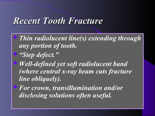
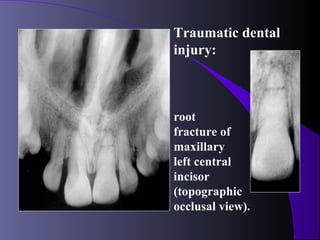
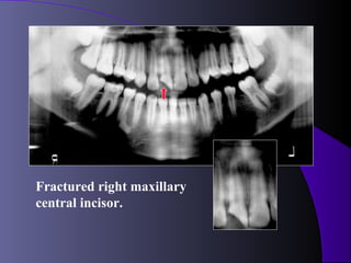


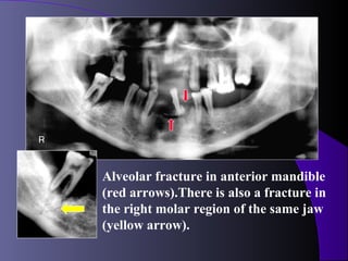
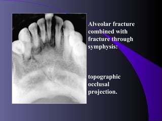
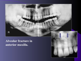

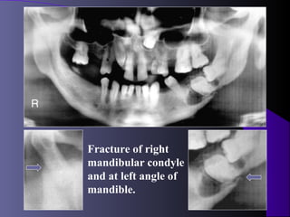
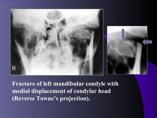
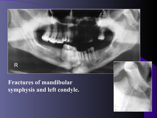
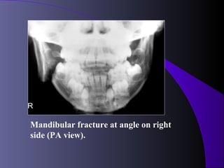
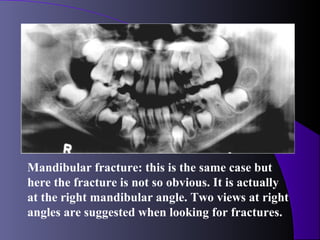
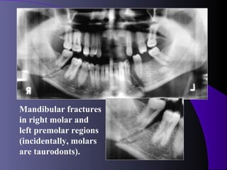
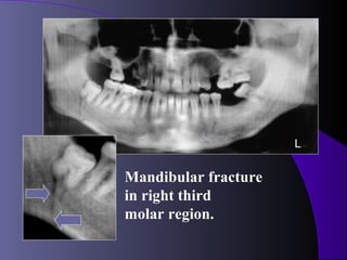
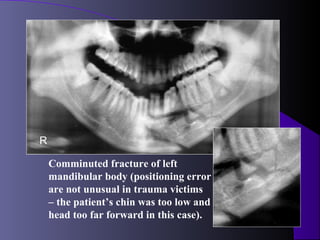
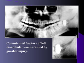
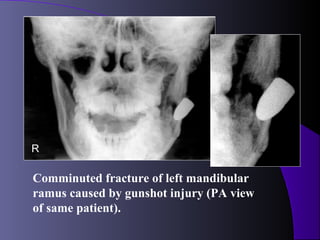
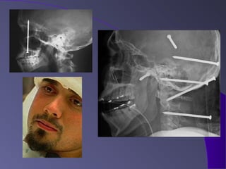
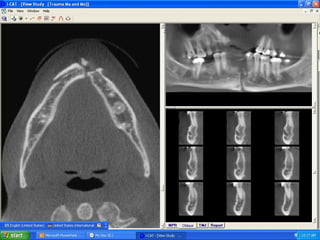
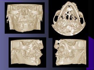
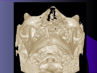
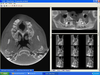


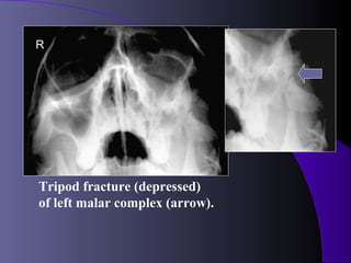
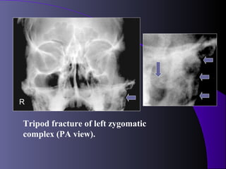
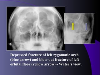
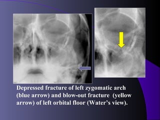


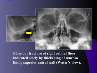
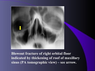
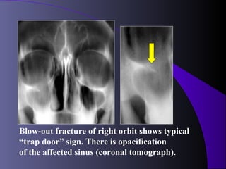
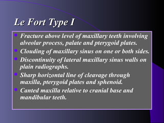
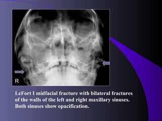
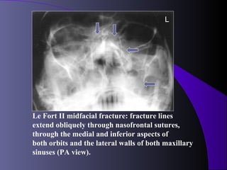
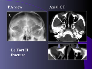
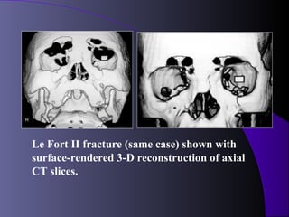
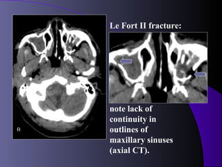
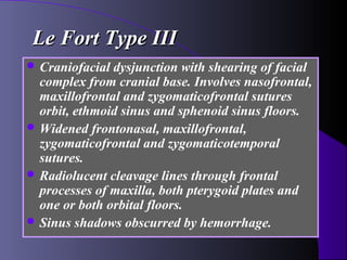
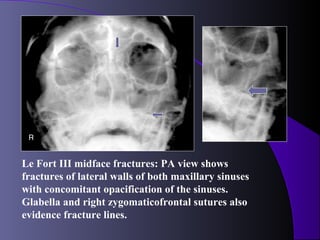
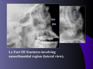
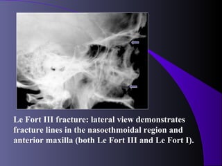

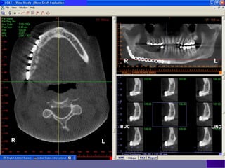
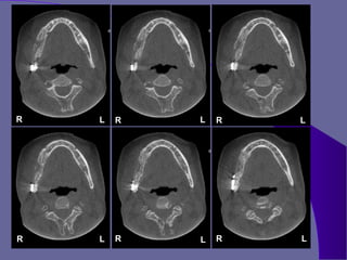
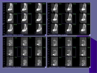

This document discusses various types of maxillofacial fractures seen on radiographs. It describes recent tooth fractures appearing as thin radiolucent lines through teeth. Alveolar fractures appear as sharply defined radiolucent lines in the alveolus. Mandibular condyle fractures involve the condylar head being "sheared off". Le Fort fractures are classified into types I, II, and III based on the anatomical structures involved. CT is the standard for evaluating maxillary fractures while panoramic radiography is best for the mandible.


