Cell junctions and the extracellular matrix
•
4 likes•1,799 views

Pharm. D - 1st Prof. Anatomy

Recommended
More Related Content
What's hot
What's hot (20)
Similar to Cell junctions and the extracellular matrix
Similar to Cell junctions and the extracellular matrix (20)
Recently uploaded
Recently uploaded (20)
Cell junctions and the extracellular matrix
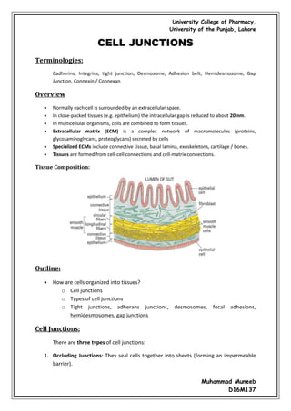
- 1. University College of Pharmacy, University of the Punjab, Lahore Muhammad Muneeb D16M137 CELL JUNCTIONS Terminologies: Cadherins, Integrins, tight junction, Desmosome, Adhesion belt, Hemidesmosome, Gap Junction, Connexin / Connexan Overview Normally each cell is surrounded by an extracellular space. In close-packed tissues (e.g. epithelium) the intracellular gap is reduced to about 20 nm. In multicellular organisms, cells are combined to form tissues. Extracellular matrix (ECM) is a complex network of macromolecules (proteins, glycosaminoglycans, proteoglycans) secreted by cells Specialized ECMs include connective tissue, basal lamina, exoskeletons, cartilage / bones. Tissues are formed from cell-cell connections and cell-matrix connections. Tissue Composition: Outline: How are cells organized into tissues? o Cell junctions o Types of cell junctions o Tight junctions, adherans junctions, desmosomes, focal adhesions, hemidesmosomes, gap junctions Cell Junctions: There are three types of cell junctions: 1. Occluding Junctions: They seal cells together into sheets (forming an impermeable barrier).
- 2. University College of Pharmacy, University of the Punjab, Lahore Muhammad Muneeb D16M137 2. Anchoring Junctions: They attach cells (and their cytoskeleton) to other cells or extracellular matrix (providing mechanical support). 3. Communicating Junctions: They allow exchange of chemical/electrical information between cells. Occluding Junctions: Tight junctions, also known as occluding junctions or zonulae occludentes (singular, zonula occludens), are the closely associated areas of two cells whose membranes join together forming a virtually impermeable barrier to fluid. It is a type of junctional complex present only in vertebrates. The corresponding junctions that occur in invertebrates are septate junctions. This variety of intracellular junctions is found in the epithelial tissues only. In tight junction, each cell possesses integral membrane proteins that bind to similar proteins in the adjacent, forming a continuous “weld”. Two types of occluding junctions are recognized: zonula occludens and fascia occludens. Structure: Tight junctions are composed of a branching network of sealing strands, each strand acting independently from the others. Therefore, the efficiency of the junction in preventing ion passage increases exponentially with the number of strands. Each strand is formed from a row of transmembrane proteins embedded in both plasma membranes, with extracellular domains joining one another directly. Although more proteins are present, the major types are the claudins and the occludins. These associate with different peripheral membrane proteins such as ZO-1 located on the intracellular side of plasma membrane, which anchor the
- 3. University College of Pharmacy, University of the Punjab, Lahore Muhammad Muneeb D16M137 strands to the actin component of the cytoskeleton. Thus, tight junctions join together the cytoskeletons of adjacent cells. Example: Tight junctions of intestinal epithelium Functions: They perform vital functions: They hold cells together. Barrier function, which can be further subdivided into protective barriers and functional barriers serving purposes such as material transport and maintenance of osmotic balance: o Tight Junctions help to maintain the polarity of cells by preventing the lateral diffusion of integral membrane proteins between the apical and lateral/basal surfaces. o Tight Junctions prevent the passage of molecules and ions through the space between plasma membranes of adjacent cells. In human physiology there are two main types of epithelia using distinct types of barrier mechanism. Dermal structures such as skin form a barrier from many layers of keratinised squamous cells. Internal epithelia on the other hand more often rely on tight junctions for their barrier function. This kind of barrier is mostly formed by only one or two layers of cells. Classification: Epithelia are classed as "tight" or "leaky", depending on the ability of the tight junctions to prevent water and solute movement: Tight epithelia have tight junctions that prevent most movement between cells. Examples of tight epithelia include the distal convoluted tubule, the collecting duct of the nephron in the kidney, and the bile ducts ramifying through liver tissue.
- 4. University College of Pharmacy, University of the Punjab, Lahore Muhammad Muneeb D16M137 Leaky epithelia do not have these tight junctions, or they have less complex tight junctions. For instance, the tight junction in the kidney proximal tubule, a very leaky epithelium, has only two to three junctional strands, and these strands exhibit infrequent large slit breaks. Anchoring Junctions: Cells have developed several types of junctional complexes to serve these functions, and in each case, anchoring proteins extend through the plasma membrane to link cytoskeletal proteins in one cell to cytoskeletal proteins in neighboring cells as well as to proteins in the extracellular matrix. Integral membrane proteins connect a cell’s cytoskeleton to another cell or extracellular matrix. Cytoskeletal fibers (Microfilament, intermediate filaments) connect to a Membrane protein receptor which attaches to another protein in either to: The extracellular matrix Another cell membrane Three types of anchoring junctions are observed, and differ from one another in the cytoskeletal protein anchor as well as the transmembrane linker protein that extends through the membrane: Junction Cytoskeletal anchor Transmembrane linker Ties cell to: Desmosomes Intermediate filaments Cadherin Other cells Hemidesmosomes Intermediate filaments Integrins EC matrix Adherens Junctions Actin filaments Cadherin / Integrins Other cells / EC matrix Anchoring-type junctions not only hold cells together but provide tissues with structural cohesion. These junctions are most abundant in tissues that are subject to constant mechanical stress such as skin and heart.
- 5. University College of Pharmacy, University of the Punjab, Lahore Muhammad Muneeb D16M137 Mainly, four types of adhering junctions are recognized: o Zonula adherens o Fascia adherens o Macula adherens o Hemidesmosomes Cadherins and Desmosomes: Epithelial cells adhere to each other due to the presence of a variety of adhesive glycoproteins in the intracellular gap; these glycoproteins are called cadherins. Cell to cell connections are mediated by cadherins. These receptors extend out from the cell, binding to other cadherins. Cadherins participate in adherans junctions. Under the cell membrane, contractile fibers of microfilaments connect to cell membrane proteins called cadherins. They surround the cell, forming a belt.
- 6. University College of Pharmacy, University of the Punjab, Lahore Muhammad Muneeb D16M137 Cadherins can also form localized spot connections Cadherins attach to intermediate filaments via anchoring proteins: a desmosome. Desmosomes, also termed as maculae adherents, can be visualized as rivets through the plasma membrane of adjacent cells. Intermediate filaments composed of keratin or desmin are attached to membrane-associated attachment proteins that form a dense plaque on the cytoplasmic face of the membrane. Cadherin molecules form the actual anchor by attaching to the cytoplasmic plaque, extending through the membrane and binding strongly to cadherins coming through the membrane of the adjacent cell. Focal Adhesions and Hemidesmosomes: Cytoskeletal fibers attach to transmembrane receptors (integrins) that are attached to extracellular matrix components Focal adhesions use MF Hemidesmosomes use IF Hemidesmosomes form rivet-like links between cytoskeleton and extracellular matrix components such as the basal lamina that underlie epithelia. Like desmosomes, they tie to intermediate filaments in the cytoplasm, but in contrast to desmosomes, their transmembrane anchors are integrins rather than cadherins.
- 7. University College of Pharmacy, University of the Punjab, Lahore Muhammad Muneeb D16M137 Gap Junctions: The gap junctions are also called as nexus, are communicating junctions which occur very frequently between the epithelial cells. Gap junctions allow cells to exchange electrical and / or chemical signals. They’re composed of proteins that form channels that allow small molecules to pass. Subunits of these channels are connexins that are assembled together to make connexons. The connexons from two cells join together to make a gap junction. Regulation of Connectivity: How do cells alter these connections? Alter the profile of cytoskeletal connections, receptors, and extracellular matrix Alter the binding affinity of receptors o many are Ca2+ dependent o many are affected by protein kinases Summary:
- 8. University College of Pharmacy, University of the Punjab, Lahore Muhammad Muneeb D16M137 Junctional Complex: In many locations (e.g. the small intestinal mucosa) the lateral surfaces of adjacent columnar epithelial cells are bound to each other by a series of cell junctions, which are collectively known as junctional complex. From the apical to the basal side a junctional complex is seen to consist of three components: Zonula Occludens Zonula Adherens Macula Adherans (desmosome)
- 9. University College of Pharmacy, University of the Punjab, Lahore Muhammad Muneeb D16M137 THE EXTRACELLULAR MATRIX Terminologies: Basal lamina, Connective tissue, Fibroblast, Proteoglycan, GAG (glycosaminoglycan), Hyaluronic, Aggrecan, Collagen, Fibronectin Overview In multicellular organisms, cells are combined to form tissues. Extracellular matrix (ECM) is a complex network of macromolecules (proteins, glycosaminoglycans, proteoglycans) secreted by cells Specialized ECMs include connective tissue, basal lamina, exoskeletons, cartilage / bones. Tissues are formed from cell-cell connections and cell-matrix connections. Outline: Extracellular matrix proteins o Types of ECM macromolecules o Synthesis and properties of hyaluronan, aggrecan, collagen, fibronectin Extracellular Space: A network of proteins, glycosaminoglycans (GAGs) and combinations of the two (proteoglycan) found in the extracellular space. All ECM components are secreted by cells. Most cells can secrete elements of the ECM but many ECMs are built by fibroblasts. Examples of ECM: Connective tissue, basal lamina, cartilage, bone, plant/fungi cell walls, myelin sheath Basal Lamina:
- 10. University College of Pharmacy, University of the Punjab, Lahore Muhammad Muneeb D16M137 Connective Tissue: Components of the Extracellular Matrix: Proteins / Glycoproteins: Proteins/glycoproteins (secreted by co-translational import) differ in physical properties (e.g. size, flexibility) and are able to bind to different combinations of macromolecules on cell membranes (e.g. integrins, cadherins) and other ECM elements. Examples include: collagen, elastin, fibronectin, laminin Proteins can provide elastic properties in many tissues (e.g. elastin). GAGs: GAGs differ in physical properties (e.g. size, flexibility, and hydration) They’re composed of repeating sugar + amino sugar units (e.g. N-acetylglucosamine, N-acetylgalactosamine) They occur in long strings, often attached to proteins
- 11. University College of Pharmacy, University of the Punjab, Lahore Muhammad Muneeb D16M137 Examples include hyaluronan, chondroitin sulfate, heparan sulfate, keratan sulfate GAGs attract a great deal of water. Hydroxyl groups form hydrogen bonds, and the many negative charges attract clouds of cations (Na+ ) that induce an osmotic movement of water. These hydrated gels resist compression (useful for joints). Proteoglycans: Proteoglycans (made of both proteins and GAGs) also differ in physical properties. They’re synthesized in Golgi prior to secretion. In addition to structural roles, proteoglycans can also bind hormones (e.g., inflammatory chemokines, FGF, TGFβ) to alter cell signaling pathways Examples include decoran, aggrecan (the main component of cartilage). Hyaluronan: Very simple GAG, consisting of 10,000+ repeats of glucuronic acid and N- acetylglucosamine. It spun directly from cell membranes by a surface enzyme complex It attracts water and fills spaces between cells with non-compressible gel (found around joints). Some cells secrete it to isolate themselves from other cells (e.g. myoblasts). These cells can secrete hyaluronidase to break it down, allowing contact
- 12. University College of Pharmacy, University of the Punjab, Lahore Muhammad Muneeb D16M137 (Very long macromolecule that hydrates and fills enormous volumes) Aggrecan: It is one of the largest macromolecules, consisting of a core protein with GAGs attached to form a feather-like appearance. An aggrecan core protein is very large but also binds many (different) GAGs (shown in red). Each aggrecan can be attached to a hyaluronan backbone to form an aggrecan aggregate.
- 13. University College of Pharmacy, University of the Punjab, Lahore Muhammad Muneeb D16M137 Collagen: All multicellular animals possess collagens, often with many different collagen genes It is synthesized as pro-collagen monomers (pro-a collagen) It is basically a peptide modified by hydroxylation and glycosylation. Prior to secretion they self- assemble into trimers. Upon secretion the trimers are processed by proteolytic enzymes then assemble into fibrils. Collagen Fibers: Collagen proteins (trimers) are then cross-linked to form collagen fibers (stiff, not very elastic)
- 14. University College of Pharmacy, University of the Punjab, Lahore Muhammad Muneeb D16M137 Fibronectin: Animals have a single fibronectin gene that can be alternatively spiced into 40+ forms. Different exons are able to bind different proteins/GAGs (e.g. integrins, collagen, etc) Fibronectin dimerizes using 2 similar (not identical) monomers Control of ECM: Cells control the synthesis of ECM by altering gene expression and co-translational import and secretion They also control the degradation of the ECM by secreting and activating/inactivating extracellular enzymes. Most important are a group of serine proteases called matrix metalloproteinases MMPs: Cells that need to migrate must first break down the connections to the ECM (e.g. tissue repair, metastasis of tumors) MMPs can be: secreted in active form (collagenase) secreted as inactive form (e.g. plasminogen is inactive until it is modified by “plasminogen activators) activated when cells stop secreting TIMPs (tissue inhibitors of metalloproteinases)