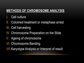
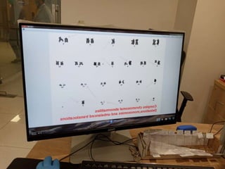
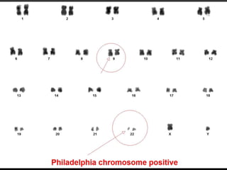
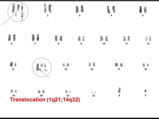
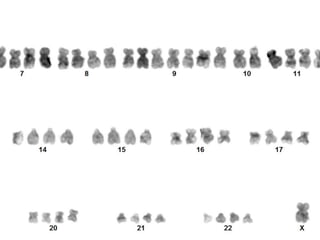
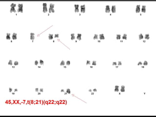
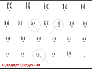
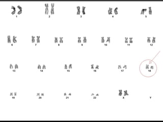
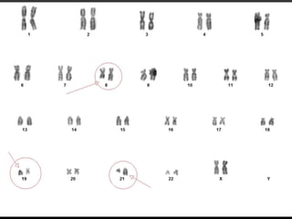
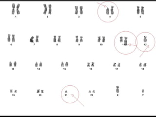
2. INTRODUCTION
1.Cytogenetic is a branch of pathology and genetics concerned with the study of normal
chromosomes and chromosome aberrations. Classical cytogenetic allows microscopic
visualization of whole chromosomes in order to assess their number and structure.
2.The study of chromosomes, which are long strands of DNA and protein that contain most of
the genetic information in a cell. Cytogenetic involves testing samples of tissue, blood, or
bone marrow in a laboratory to look for changes in chromosomes, including broken, missing,
rearranged, or extra chromosomes.
3. WHAT IS A CYTOGENETIC STUDY
• Cytogenetic involves testing samples of tissue, blood, or bone marrow in a laboratory to
look for changes in chromosomes, including.
• Broken. (Deletion)
• Missing. (Monosomy)
• Rearranged. (Translocation)
• Extra. (Trisomy)
• Changes in certain chromosomes may be a sign of a genetic disease or condition or
some types of cancer.
• Cytogenetic plays a key role in the detection of chromosomal abnormalities associated
with malignancies, as well as the characterization of new alterations that allow more
research and increase knowledge about the genetic aspects of these diseases.
4. WHAT IS A CHROMOSOME
• Chromosomes are thread-like structures present in the nucleus, which carries genetic
information from one generation to another. They play a vital role in cell division, heredity,
variation, mutation, repair and regeneration.
• DNA present on the chromosome not only carries most of the genetic information but also
controls the hereditary transfer. Chromosomes are essential for the process
of cell division, replication, division, and creation of daughter cells.
5. TYPES OF CHROMOSOME
There are four main types of chromosomes: Metacentric.
Sub metacentric.
Acrocentric.
Telocentric.
Chromosomes are found within the nucleus of most living cells and consist of DNA that is
tightly wound into thread-like structures.
6.
7. THE CELL CYCLE
The Cell cycle has the following phases.
I. Interphase.
II. Gap phase(G1)
III. Synthesis phase (S)
IV. Mitotic phase (M)
9. MITOSIS
Mitosis is a continuous process that usually lasts
1 to 2 hours but for descriptive purposes it is
convenient to distinguish five distinct stages.
These are
I. Prophase
II. Prometaphase
III. Metaphase
IV. Anaphase and telophase
10. METAPHASE
• The chromosomes become aligned along the
equatorial plane or plate of the cell, where each
chromosome is attached to the centriole by a
microtubule forming the mature spindle.
• At this point the chromosomes are maximally
contracted and therefore , most easily visible. Each
chromosome resembles the letter X in shape ,as
the chromatids of each chromosome have
separated longitudinally but remain attached at the
centromere.
11. METHODS OF CHROMOSOME ANALYSIS
I. Cell culture
II. Colcimed treatment or metaphase arrest
III. Cell harvesting
IV. Chromosome Preparation on the Slide
V. Ageing of chromosome
VI. Chromosome Banding
VII.Karyotype Analysis or Interpret of result
12. 1.CHROMOSOME PREPARATION
• Most commonly circulating lymphocytes from peripheral
blood are used.
• Samples for chromosomal analysis can be prepared
relatively easily using.
• Blood.
• Skin.
• Bone marrow.
• Chorionic villi.
• Amniotic fluid.( Aminiocytes)
• Any tissue with living nucleated cells that undergo
division can be used for studying human chromosomes.
13. FETAL AND NEONATAL BLOOD SAMPLES IN
WHICH PROBLEM MAY BE ENCOUNTERED
• Chromosomal syndrome chromosome implicated Problem encountered
Di George syndrome Abnormalities of # 22 Lack of T-Lymphocytes
Non-immune Hydrops 45,X or trisomy 21 Etiology complex?
Mechanism according to basic
cause
Intrauterine growth retardation Trisomy 18,4p-,5p-,most
deletion/duplication
Poor response to PHA
Chromosomal instability
syndrome AT,FA,BS,XP
Chromosomal breakages Poor response to mitogen
Roberts syndrome Separation of centromeric
heterochromatin
Poor response to mitogen
Fetal leukemia Patterns of abnormality
according to type
Altered growth patterns
Severe combined No specific chromosome Lake of T/B lymphocytes
14. PATIENT HISTORY IS MANDATORY
• Types of test
• Cytogenetic study test technique different.
• Blood
• Bone marrow
• Cultures
• Correlation with patient
• Treatment of patient
•
15. WHAT IS CHROMOSOMAL BANDING?
• A part of chromosome which is clearly
distinguishable from its adjacent
segments by appearing darker or
lighter with various banding methods.
19. G-BANDING
• G-banding generally provides high quality
chromosomes analysis with approximately 400-500
bands per haploid set.
• Each of these bands corresponds on average to
approximately 6000-8000 kilobases(kb) of DNA.
• This involves first inhibiting cell division with an
agent such as methotrexate or thymidine.
• Folic acid or deoxycytidine is added to the culture
medium,releasing the cells into mitosis.
20. KARYOTYPE ANALYSIS OR RESULT INTERPRET
• The next stage in chromosome analysis involves
first counting the number of chromosomes
present in a specified number of cells,
sometimes referred to as “Metaphase spreads”.
• The banding pattern of each chromosome is
specific and can be shown in the form of a
stylized ideal karyotype known as an Idiogram.
36. WHAT IS HEMATOLOGICAL MALIGNANCES
• Hematologic malignancies are cancers that affect the blood, bone marrow, and lymph
nodes.
• This classification includes various types of leukemia.
• ALL
• CLL
• AML
• CML
• Hodgkin’s Lymphoma
• Non Hodgkin’s Lymphoma
37. ROLE OF CYTOGENETIC STUDY IN
HEMATOLOGICAL MALIGNANCES
• Chromosome analysis has become a critical aspect in the workup of hematopoietic
neoplasms. Information obtained from cytogenetic studies is used on a clinical basis for
diagnosis and prognosis as well as a research basis for gene identification and potential
treatment advances.
• Strikingly, cancer cytogenetic not only provides key information to improve the care of
patients with leukemia and various cancers but also acts as a guide to identify the genes
responsible for the development of these neoplastic states.