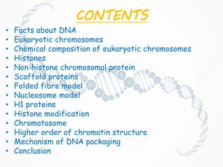
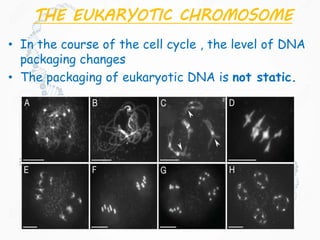
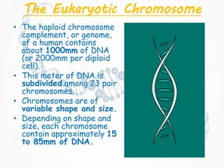
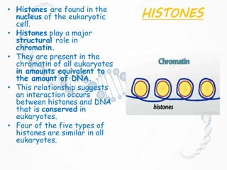
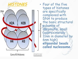
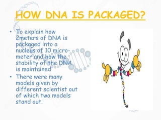
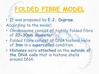
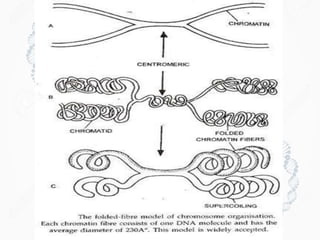
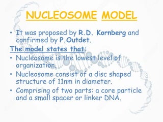
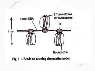
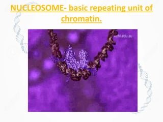
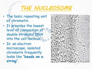
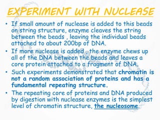
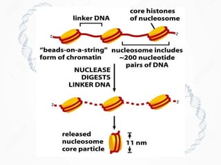
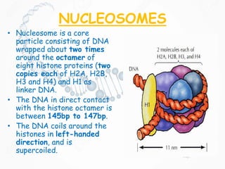
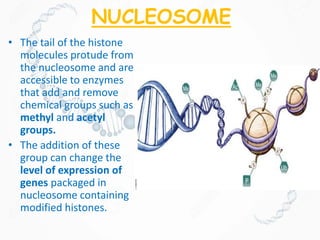
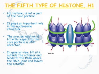
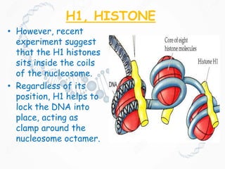
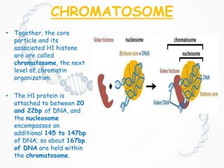
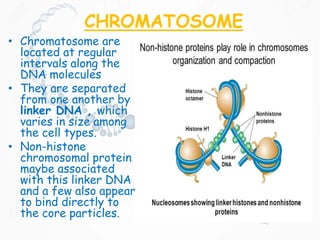
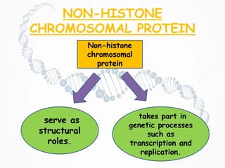
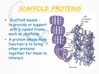
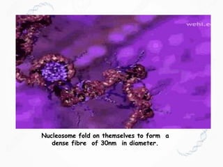
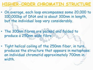
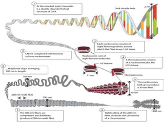
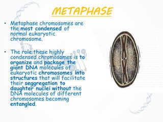
DNA ORGANIZATION IN
EUKARYOTIC CELLS
CONTENTS
• Facts about DNA
• Eukaryotic chromosomes
• Chemical composition of eukaryotic chromosomes
• Histones
• Non-histone chromosomal protein
• Scaffold proteins
• Folded fibre model
• Nucleosome model
• H1 proteins
• Histone modification
• Chromatosome
• Higher order of chromatin structure
• Mechanism of DNA packaging
• Conclusion
FACTS ABOUT DNA..!
• DNA stores all the information that makes up
an organism.
• DNA are made up of four building
blocks,which are- cytosine(C), Thymine(T),
Guanine (G) and adenine(A).
• Each DNA strand is about 1.8 meters long
but squeezed into a space of 0.09
micrometers.
• 99.9% of DNA is identical in all humans on this
Earth. The remaining 0.1% is what helps us to
differentiate between DNA sequences
allowing us to tell which DNA belongs to whom.
• Human DNA is 95% identical to DNA of
chimpanzees and interestingly it is 50%
identical to the DNA of the banana.
THE EUKARYOTIC
CHROMOSOME • Individual
eukaryotic
chromosome
contain enormous
amount of DNA.
• For all of these
DNA to fit into the
nucleus,
tremendous
packaging and
folding are
required.
• The chromosomes
are in an elongated,
relatively
uncondensed state
during interphase
of the cell cycle.
MEIOSIS
MITOSIS
THE EUKARYOTIC CHROMOSOME
• In the course of the cell cycle , the level of DNA
packaging changes
• The packaging of eukaryotic DNA is not static.
The Eukaryotic Chromosome
• The haploid chromosome
complement, or genome,
of a human contains
about 1000mm of DNA
(or 2000mm per diploid
cell).
• This meter of DNA is
subdivided among 23 pair
chromosomes.
• Chromosomes are of
variable shape and size.
• Depending on shape and
size, each chromosome
contain approximately 15
to 85mm of DNA.
CHEMICAL COMPOSITION OF
EUKARYOTIC CHROMOSOME
• DNA is closely associated with proteins,
creating chromatin.
• The two basic types of chromatins are:
-Euchromatin ,which undergoes the normal
process of condensation and decondensation
in the cell cycle.
-Heterochromatin , which remain in a highly
condensed state throughout the cell , even
during interphase.
CHEMICAL COMPOSITION OF
EUKARYOTIC CHROMOSOME
• Chemical analysis of isolated chromatin shows
that it consists primarily of DNA and proteins
with lesser amount of RNA.
• The proteins are of two major classes:
o Basic (positively charged at neutral pH) proteins
called histones.
o A heterogenous , largely acidic (negatively
charged at neutral pH) group of proteins
collectively referred to as non-histone
chromosomal proteins.
HISTONES
H1
H2A
H2B
H3
H4
HISTONES• Histones are found in the
nucleus of the eukaryotic
cell.
• Histones play a major
structural role in
chromatin.
• They are present in the
chromatin of all eukaryotes
in amounts equivalent to
the amount of DNA.
• This relationship suggests
an interaction occurs
between histones and DNA
that is conserved in
eukaryotes.
• Four of the five types of
histones are similar in all
eukaryotes.
HISTONES
• The histones of all plants and animals
consists of five classes of proteins.
• These five major histones types, called
H1, H2A, H2B, H3, H4, are present in
almost all cell types.
• The five histone type are present in molar
ratios of approximately
1:H1,2:H2A,2:H2B, 2:H3,2:H4.
HISTONES • Four of the five
types of histones
are specifically
complexed with
DNA to produce
the basic structural
subunits of
chromatin, small
(approximately
11nm in diameter by
6nm high)
ellipsoidal beads
called nucleosome.
• The histones are basic.
• Because they contain 20% to 30% arginine
and lysine, two positively charged amino
acids.
• The exposed –NH3
+ groups on histones are
important in their interaction with the DNA,
which is polyanionic because of the negatively
charged phosphate group.
HOW DNA IS PACKAGED?
• To explain how
2meters of DNA is
packaged into a
nucleus of 10 micro-
meter and how the
stability of the DNA
is maintained
• There were many
models given by
different scientist out
of which two models
stand out.
FOLDED FIBRE MODEL
• It was proposed by E.J. Dupraw.
According to the model:
• Chromosome consist of tightly folded fibre
of 20-30nm diameter.
• Folded fibre consist of DNA histone helix
of 3nm in a supercoiled condition.
• Histones were attached on the outside of
the DNA coils that is histone shells
around DNA.
NUCLEOSOME MODEL
• It was proposed by R.D. Kornberg and
confirmed by P.Outdet.
The model states that:
• Nucleosome is the lowest level of
organization.
• Nucleosome consist of a disc shaped
structure of 11nm in diameter.
• Comprising of two parts: a core particle
and a small spacer or linker DNA.
NUCLEOSOME- basic repeating unit of
chromatin.
THE NUCLEOSOME
• The basic repeating unit
of chromatin.
• It provides the lowest
level of compaction of
double-stranded DNA
into the cell nucleus.
• In an electron
microscope, isolated
chromatin frequently
looks like “beads on a
string”.
EXPERIMENT WITH NUCLEASE
• If small amount of nuclease is added to this beads
on string structure, enzyme cleaves the string
between the beads , leaving the individual beads
attached to about 200bp of DNA.
• If more nuclease is added , the enzyme chews up
all of the DNA between the beads and leaves a
core protein attached to a fragment of DNA.
• Such experiments demonstrated that chromatin is
not a random association of proteins and has a
fundamental repeating structure.
• The repeating core of proteins and DNA produced
by digestion with nuclease enzymes is the simplest
level of chromatin structure, the nucleosome.
NUCLEOSOMES
• Nucleosome is a core
particle consisting of DNA
wrapped about two times
around the octamer of
eight histone proteins (two
copies each of H2A, H2B,
H3 and H4) and H1 as
linker DNA.
• The DNA in direct contact
with the histone octamer is
between 145bp to 147bp.
• The DNA coils around the
histones in left-handed
direction, and is
supercoiled.
NUCLEOSOME
• The tail of the histone
molecules protude from
the nucleosome and are
accessible to enzymes
that add and remove
chemical groups such as
methyl and acetyl
groups.
• The addition of these
group can change the
level of expression of
genes packaged in
nucleosome containing
modified histones.
THE FIFTH TYPE OF HISTONE, H1
• H1, histone, is not a part
of the core particle.
• It plays an important role
in the nucleosome
structure.
• The precise location of
H1 with respect to the
core particle is still
uncertain.
• In general view, H1 sits
outside the octamer and
binds to the DNA where
the DNA joins and leaves
the octamer.
H1, HISTONE
• However, recent
experiment suggest
that the H1 histones
sits inside the coils
of the nucleosome.
• Regardless of its
position, H1 helps to
lock the DNA into
place, acting as
clamp around the
nucleosome octamer.
CHROMATOSOME
• Together, the core
particle and its
associated H1 histone
are are called
chromatosome, the next
level of chromatin
organization.
• The H1 protein is
attached to between 20
and 22bp of DNA, and
the nucleosome
encompasses an
additional 145 to 147bp
of DNA; so about 167bp
of DNA are held within
the chromatosome.
CHROMATOSOME
• Chromatosome are
located at regular
intervals along the
DNA molecules
• They are separated
from one another by
linker DNA , which
varies in size among
the cell types.
• Non-histone
chromosomal protein
maybe associated
with this linker DNA
and a few also appear
to bind directly to
the core particles.
NON-HISTONE
CHROMOSOMAL PROTEIN
Non-histone
chromosomal
protein
serve as
structural
roles.
takes part in
genetic processes
such as
transcription and
replication.
SCAFFOLD PROTEINS
• Scaffold means -
to provide or support
with a raised frame-
work or platform.
• A protein whose main
function is to bring
other proteins
together for them to
interact.
SCAFFOLD PROTEINS
• Chromatin is treated with a
concentrated salt solution.
• It removes histones and most of the
other chromosomal proteins,
• Leaving a “skeleton” to which DNA was
attached, known as scaffold proteins.
• These scaffold proteins play a role in
the folding and packaging of
chromosome.
SCAFFOLD PROTEINS
• Histone-depleted chromosomes
were studied in the electron
microscope.
• Results show that:
• the histone-depleted
chromosomes consist of a
scaffold or core, which has the
shape characteristic of a
metaphase chromosome,
surrounded by a halo of DNA;
• the halo consists of many loops
of DNA, each anchored in the
scaffold at its base;
• most of the DNA exists in
loops at least 10-30 µm long
(30-90 kilobases).
THREE LEVELS OF DNA
PACKAGING
HIGHER-ORDER CHROMATIN
STRUCTURE
• The first level of condensation involves packaging DNA
as a negative supercoil into nucleosomes , to produce
the 10nm interphase chromatin fiber. This clearly
involves an octamer of histone molecules, two each of
histones H2A, H2B, H3 and H4.
• In chromosomes, adjacent nucleosome are not
separated by space equal to the length of the linker
DNA.
• Rather nucleosomes fold on themselves to form a 30nm
dense, tightly packed structure.
• The next-higher level of chromatin structure is a series
of loop of 30nm fibres, each anchored at its base by
proteins in the nuclear scaffold.
Nucleosome fold on themselves to form a
dense fibre of 30nm in diameter.
HIGHER-ORDER CHROMATIN STRUCTURE
• On average, each loop encompasses some 20,000 to
100,000bp of DNA and is about 300nm in length,
but the individual loop vary considerably.
• The 300nm fibres are packed and folded to
produce a 250nm wide fibre.
• Tight helical coiling of the 250nm fiber, in turn,
produces the structure that appears in metaphase:
an individual chromatid approximately 700nm in
width.
METAPHASE
• Metaphase chromosomes are
the most condensed of
normal eukaryotic
chromosome.
• The role these highly
condensed chromosomes is to
organize and package the
giant DNA molecules of
eukaryotic chromosomes into
structures that will facilitate
their seggregation to
daughter nuclei without the
DNA molecules of different
chromosomes becoming
entangled.
METAPHASE
• There is evidence that
the gross structure of
metaphase chromosomes
is not dependent on
histones.
• Electron micrographs of
isolated metaphase
chromosome from which
the histones have been
removed revealed a
scaffold, which is
surrounded by huge pool
of DNA.
• This chromosome must
be composed of non-
histone chromosomal
protein.
CONCLUSION
• Each DNA strand is about 1.8 meters long but squeezed into a
space of 0.09 micrometers.
• Individual eukaryotic chromosome contain enormous amount of DNA.
• The packaging of eukaryotic DNA is not static.
• Eukaryotic chromosomes are made up two major types of proteins-
basic and largely acidic.
• Histones are of five types- H1, H2a, H2b, H3 and H4.
• Two units of each histone proteins wrapped by DNA, except H1,
forms the nucleosome.
• Nucleosome along with H1 forms the chromatosome.
• Each chromatosome is attached by linker DNA.
• They coil round to form a 30nm wide fibre, that forms loop
averaging from 300nm in length.
• The 300nm fibre are further compressed and folded to form
250nm wide fibre.
• Tight coiling of the 250nm fibre forms chromatids of
chromosomes.
REFERENCES
• https://www.youtube.com/watch?v=3wTAE
fjo20c&list=WL
• http://giphy.com/search/chromosome
• http://giphy.com/gifs/astronomy-
lOe630aMEzo76
• D.Peer Snustad, Michael J.
Simmons,Principle of Genetics,Fifth Edition.
• Eldon John Gardner, D.Peer Snustad,
Michael J. Simmons,Principle of
Genetics,Eight Edition.
• Peter J. Russell, iGenetics,Third Edition.
DNA organization in Eukaryotic cells

DNA organization in Eukaryotic cells

  • 1.
  • 2.
    CONTENTS • Facts aboutDNA • Eukaryotic chromosomes • Chemical composition of eukaryotic chromosomes • Histones • Non-histone chromosomal protein • Scaffold proteins • Folded fibre model • Nucleosome model • H1 proteins • Histone modification • Chromatosome • Higher order of chromatin structure • Mechanism of DNA packaging • Conclusion
  • 3.
    FACTS ABOUT DNA..! •DNA stores all the information that makes up an organism. • DNA are made up of four building blocks,which are- cytosine(C), Thymine(T), Guanine (G) and adenine(A). • Each DNA strand is about 1.8 meters long but squeezed into a space of 0.09 micrometers. • 99.9% of DNA is identical in all humans on this Earth. The remaining 0.1% is what helps us to differentiate between DNA sequences allowing us to tell which DNA belongs to whom. • Human DNA is 95% identical to DNA of chimpanzees and interestingly it is 50% identical to the DNA of the banana.
  • 4.
    THE EUKARYOTIC CHROMOSOME •Individual eukaryotic chromosome contain enormous amount of DNA. • For all of these DNA to fit into the nucleus, tremendous packaging and folding are required. • The chromosomes are in an elongated, relatively uncondensed state during interphase of the cell cycle. MEIOSIS MITOSIS
  • 5.
    THE EUKARYOTIC CHROMOSOME •In the course of the cell cycle , the level of DNA packaging changes • The packaging of eukaryotic DNA is not static.
  • 6.
    The Eukaryotic Chromosome •The haploid chromosome complement, or genome, of a human contains about 1000mm of DNA (or 2000mm per diploid cell). • This meter of DNA is subdivided among 23 pair chromosomes. • Chromosomes are of variable shape and size. • Depending on shape and size, each chromosome contain approximately 15 to 85mm of DNA.
  • 7.
    CHEMICAL COMPOSITION OF EUKARYOTICCHROMOSOME • DNA is closely associated with proteins, creating chromatin. • The two basic types of chromatins are: -Euchromatin ,which undergoes the normal process of condensation and decondensation in the cell cycle. -Heterochromatin , which remain in a highly condensed state throughout the cell , even during interphase.
  • 8.
    CHEMICAL COMPOSITION OF EUKARYOTICCHROMOSOME • Chemical analysis of isolated chromatin shows that it consists primarily of DNA and proteins with lesser amount of RNA. • The proteins are of two major classes: o Basic (positively charged at neutral pH) proteins called histones. o A heterogenous , largely acidic (negatively charged at neutral pH) group of proteins collectively referred to as non-histone chromosomal proteins.
  • 9.
  • 10.
    HISTONES• Histones arefound in the nucleus of the eukaryotic cell. • Histones play a major structural role in chromatin. • They are present in the chromatin of all eukaryotes in amounts equivalent to the amount of DNA. • This relationship suggests an interaction occurs between histones and DNA that is conserved in eukaryotes. • Four of the five types of histones are similar in all eukaryotes.
  • 11.
    HISTONES • The histonesof all plants and animals consists of five classes of proteins. • These five major histones types, called H1, H2A, H2B, H3, H4, are present in almost all cell types. • The five histone type are present in molar ratios of approximately 1:H1,2:H2A,2:H2B, 2:H3,2:H4.
  • 12.
    HISTONES • Fourof the five types of histones are specifically complexed with DNA to produce the basic structural subunits of chromatin, small (approximately 11nm in diameter by 6nm high) ellipsoidal beads called nucleosome.
  • 13.
    • The histonesare basic. • Because they contain 20% to 30% arginine and lysine, two positively charged amino acids. • The exposed –NH3 + groups on histones are important in their interaction with the DNA, which is polyanionic because of the negatively charged phosphate group.
  • 14.
    HOW DNA ISPACKAGED? • To explain how 2meters of DNA is packaged into a nucleus of 10 micro- meter and how the stability of the DNA is maintained • There were many models given by different scientist out of which two models stand out.
  • 15.
    FOLDED FIBRE MODEL •It was proposed by E.J. Dupraw. According to the model: • Chromosome consist of tightly folded fibre of 20-30nm diameter. • Folded fibre consist of DNA histone helix of 3nm in a supercoiled condition. • Histones were attached on the outside of the DNA coils that is histone shells around DNA.
  • 17.
    NUCLEOSOME MODEL • Itwas proposed by R.D. Kornberg and confirmed by P.Outdet. The model states that: • Nucleosome is the lowest level of organization. • Nucleosome consist of a disc shaped structure of 11nm in diameter. • Comprising of two parts: a core particle and a small spacer or linker DNA.
  • 19.
    NUCLEOSOME- basic repeatingunit of chromatin.
  • 20.
    THE NUCLEOSOME • Thebasic repeating unit of chromatin. • It provides the lowest level of compaction of double-stranded DNA into the cell nucleus. • In an electron microscope, isolated chromatin frequently looks like “beads on a string”.
  • 21.
    EXPERIMENT WITH NUCLEASE •If small amount of nuclease is added to this beads on string structure, enzyme cleaves the string between the beads , leaving the individual beads attached to about 200bp of DNA. • If more nuclease is added , the enzyme chews up all of the DNA between the beads and leaves a core protein attached to a fragment of DNA. • Such experiments demonstrated that chromatin is not a random association of proteins and has a fundamental repeating structure. • The repeating core of proteins and DNA produced by digestion with nuclease enzymes is the simplest level of chromatin structure, the nucleosome.
  • 23.
    NUCLEOSOMES • Nucleosome isa core particle consisting of DNA wrapped about two times around the octamer of eight histone proteins (two copies each of H2A, H2B, H3 and H4) and H1 as linker DNA. • The DNA in direct contact with the histone octamer is between 145bp to 147bp. • The DNA coils around the histones in left-handed direction, and is supercoiled.
  • 24.
    NUCLEOSOME • The tailof the histone molecules protude from the nucleosome and are accessible to enzymes that add and remove chemical groups such as methyl and acetyl groups. • The addition of these group can change the level of expression of genes packaged in nucleosome containing modified histones.
  • 25.
    THE FIFTH TYPEOF HISTONE, H1 • H1, histone, is not a part of the core particle. • It plays an important role in the nucleosome structure. • The precise location of H1 with respect to the core particle is still uncertain. • In general view, H1 sits outside the octamer and binds to the DNA where the DNA joins and leaves the octamer.
  • 26.
    H1, HISTONE • However,recent experiment suggest that the H1 histones sits inside the coils of the nucleosome. • Regardless of its position, H1 helps to lock the DNA into place, acting as clamp around the nucleosome octamer.
  • 27.
    CHROMATOSOME • Together, thecore particle and its associated H1 histone are are called chromatosome, the next level of chromatin organization. • The H1 protein is attached to between 20 and 22bp of DNA, and the nucleosome encompasses an additional 145 to 147bp of DNA; so about 167bp of DNA are held within the chromatosome.
  • 28.
    CHROMATOSOME • Chromatosome are locatedat regular intervals along the DNA molecules • They are separated from one another by linker DNA , which varies in size among the cell types. • Non-histone chromosomal protein maybe associated with this linker DNA and a few also appear to bind directly to the core particles.
  • 29.
    NON-HISTONE CHROMOSOMAL PROTEIN Non-histone chromosomal protein serve as structural roles. takespart in genetic processes such as transcription and replication.
  • 30.
    SCAFFOLD PROTEINS • Scaffoldmeans - to provide or support with a raised frame- work or platform. • A protein whose main function is to bring other proteins together for them to interact.
  • 31.
    SCAFFOLD PROTEINS • Chromatinis treated with a concentrated salt solution. • It removes histones and most of the other chromosomal proteins, • Leaving a “skeleton” to which DNA was attached, known as scaffold proteins. • These scaffold proteins play a role in the folding and packaging of chromosome.
  • 32.
    SCAFFOLD PROTEINS • Histone-depletedchromosomes were studied in the electron microscope. • Results show that: • the histone-depleted chromosomes consist of a scaffold or core, which has the shape characteristic of a metaphase chromosome, surrounded by a halo of DNA; • the halo consists of many loops of DNA, each anchored in the scaffold at its base; • most of the DNA exists in loops at least 10-30 µm long (30-90 kilobases).
  • 33.
    THREE LEVELS OFDNA PACKAGING
  • 34.
    HIGHER-ORDER CHROMATIN STRUCTURE • Thefirst level of condensation involves packaging DNA as a negative supercoil into nucleosomes , to produce the 10nm interphase chromatin fiber. This clearly involves an octamer of histone molecules, two each of histones H2A, H2B, H3 and H4. • In chromosomes, adjacent nucleosome are not separated by space equal to the length of the linker DNA. • Rather nucleosomes fold on themselves to form a 30nm dense, tightly packed structure. • The next-higher level of chromatin structure is a series of loop of 30nm fibres, each anchored at its base by proteins in the nuclear scaffold.
  • 35.
    Nucleosome fold onthemselves to form a dense fibre of 30nm in diameter.
  • 36.
    HIGHER-ORDER CHROMATIN STRUCTURE •On average, each loop encompasses some 20,000 to 100,000bp of DNA and is about 300nm in length, but the individual loop vary considerably. • The 300nm fibres are packed and folded to produce a 250nm wide fibre. • Tight helical coiling of the 250nm fiber, in turn, produces the structure that appears in metaphase: an individual chromatid approximately 700nm in width.
  • 38.
    METAPHASE • Metaphase chromosomesare the most condensed of normal eukaryotic chromosome. • The role these highly condensed chromosomes is to organize and package the giant DNA molecules of eukaryotic chromosomes into structures that will facilitate their seggregation to daughter nuclei without the DNA molecules of different chromosomes becoming entangled.
  • 39.
    METAPHASE • There isevidence that the gross structure of metaphase chromosomes is not dependent on histones. • Electron micrographs of isolated metaphase chromosome from which the histones have been removed revealed a scaffold, which is surrounded by huge pool of DNA. • This chromosome must be composed of non- histone chromosomal protein.
  • 40.
    CONCLUSION • Each DNAstrand is about 1.8 meters long but squeezed into a space of 0.09 micrometers. • Individual eukaryotic chromosome contain enormous amount of DNA. • The packaging of eukaryotic DNA is not static. • Eukaryotic chromosomes are made up two major types of proteins- basic and largely acidic. • Histones are of five types- H1, H2a, H2b, H3 and H4. • Two units of each histone proteins wrapped by DNA, except H1, forms the nucleosome. • Nucleosome along with H1 forms the chromatosome. • Each chromatosome is attached by linker DNA. • They coil round to form a 30nm wide fibre, that forms loop averaging from 300nm in length. • The 300nm fibre are further compressed and folded to form 250nm wide fibre. • Tight coiling of the 250nm fibre forms chromatids of chromosomes.
  • 41.
    REFERENCES • https://www.youtube.com/watch?v=3wTAE fjo20c&list=WL • http://giphy.com/search/chromosome •http://giphy.com/gifs/astronomy- lOe630aMEzo76 • D.Peer Snustad, Michael J. Simmons,Principle of Genetics,Fifth Edition. • Eldon John Gardner, D.Peer Snustad, Michael J. Simmons,Principle of Genetics,Eight Edition. • Peter J. Russell, iGenetics,Third Edition.