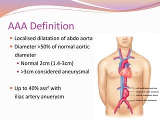
 What are AAA?
 Management Options
 Endovascular Repair
 Procedure
 Complications
 Post-Operative Management
Anatomy Revisited
 Abdominal aorta
 Hiatus of diaphragm  bifurcation into common iliac
arteries (L4)
 Paired and unpaired visceral branches
 Common iliac a.
 Internal iliac a.
 External iliac a.
 Common femoral a (after passing below inguinal
ligament)
AAA Definition
 Localised dilatation of abdo aorta
 Diameter >50% of normal aortic
diameter
 Normal 2cm (1.4-3cm)
 >3cm considered aneurysmal
 Up to 40% assd with
iliac artery anuerysm
Indications for Repair
 Symptomatic
 Tenderness, abdo or back pain
 Embolization
 Rupture
 AAA ≥ 5.5cm
 >0.5cm expansion withing 6-months
Management Options
 Open AAA repair
 EVAR
 Conservative
What is EVAR?
 Nicholas Volodos, Kiev, 1987
 Endovascular aneurysm repair
 Folded graft components inserted through femoral
artery into aorta and then deployed
 Graft expands contacting the aorta wall
 Excludes aneurysm sac from aortic blood flow and
pressure
 Suitable for 2/3 of pts with infrarenal AAA
Benefits of EVAR
 Accounts for nearly half of all AAA repairs
 Significant  peri-op mortality
 No open exposure or aortic clamping
  incidence of ruptured AAA
 Can be offered to pts not suitable for open repair
Disadvantages
 Not suitable for all
 Cost
 £3,000 - £10,000 more expensive
 May need conversion to open repair
 Lifetime surveillance
 ?radiation risk
 Does not completely eliminate future risk of rupture
Assessment
 Pre-Operative Assessment
 Technical Assessment
Pre-Operative Assessment
 As for open repair
 POAC
 IHD is leading cause of early & late mortality
 COPD
 Renal insufficency
Technical Assessment
 CT angiography with 3-D reconstruction
 (Arteriography in emergencies)
 Measurements:
 Aortic neck diameter
 Aortic neck length
 Aortic neck angulation
 Infrarenal aortic length
 Common iliac a. Diameter
 Renal or artery anomalies
Technical Aspects
 Aortic neck diameter
 Size + 15-20%
 Sufficient radial force
 Oversize - Kinking,
thrombus formn, endoleak
 Aortic neck
 10-15mm (sufficient landing zone)
 Normal appearance
 Aortic neck angulation
 <60°
 Iliac Arteries
 Minimal calcification / tortuosity
 No stenosis or mural thrombus
 Sufficient diameter & length
 If external Iliac art. = landing
zone, internal iliac art. should
be embolised (prevent
backflow)
Pre-Operative Preparation
 Thromboprophylaxis
 Prophylatic antibiotics
 Cephalosporin or vancomycin for <24hrs
 Prevent Contrast-Induced Nephropathy
Procedure
1. Anaesthetise
2. Gain vascular access
3. Place guidewires & sheaths
4. Confirm anatomy
5. Main body deployment
6. Gate cannulation
7. Iliac limb deployment
8. Graft ballooning
9. Completion imaging
1. Anaesthetise
 GA, regional, LA with sedation
2. Gain vascular access
 Bilateral femoral access
 Surgical cutdown or percutaneous
3. Place guidewires & sheaths
4. Confirm anatomy
 Aortography
5. Main body deployment
 Proximal radiopaque markers
 Below renal artery
6. Gate cannulation
 Guidewire through contralateral femoral vessel
 Into graft gate
7. Iliac limb deployment (bilateral)
8. Graft ballooning
 Angioplasty of attachment sites and endograft
junctions
9. Completion imaging
 Renal artery patency & exclude endoleak
Post-Operative Care
 Ward care
 Eat & drink
 Analgesia
 IV fluids (prevent contrast nephropathy)
 D1 – mobile
 LOWER LIMB PULSE MONITORING
Surveillance
 Contrast CTA
 Duplex US
 1 month
 12 month
 Annual thereafter
Complications
 Renal damage
 IV contrast, emboli
 DVT/PE
 5.3% develop DVT despite thromboprophylaxis
 MI
 Lower limb ischaemia/emboli
 Endoleak
Complications
 Overall complication rate ~ 10%
 Device-related
 Open conversion (<2%)
 30-day all cause mortality:
 1.6% vs 4.8% (all)
 4.7% vs 19.2% (ASA IV)
 3-4 year mortality equal
EndoleakType Aetiology
I Incompetent seal at proximal (Ia) or distal (Ib) attachment
sites
- Ongoing risk of rupture, correct promptly
II Flow into and out of aneurysm sac via patent branch vessels
(lumbar, IMA)
- 10-25%
III Separation of graft components (IIIa) or fabric tear (IIIb)
- Ongoing risk of rupture, correct promptly
IV Egress of blood through fabric pores
V Continued aneurysm sac expansion without demonstrable
leak
Thank You

EVAR - Nicola Tanner

  • 2.
     What areAAA?  Management Options  Endovascular Repair  Procedure  Complications  Post-Operative Management
  • 3.
    Anatomy Revisited  Abdominalaorta  Hiatus of diaphragm  bifurcation into common iliac arteries (L4)  Paired and unpaired visceral branches  Common iliac a.  Internal iliac a.  External iliac a.  Common femoral a (after passing below inguinal ligament)
  • 5.
    AAA Definition  Localiseddilatation of abdo aorta  Diameter >50% of normal aortic diameter  Normal 2cm (1.4-3cm)  >3cm considered aneurysmal  Up to 40% assd with iliac artery anuerysm
  • 6.
    Indications for Repair Symptomatic  Tenderness, abdo or back pain  Embolization  Rupture  AAA ≥ 5.5cm  >0.5cm expansion withing 6-months
  • 7.
    Management Options  OpenAAA repair  EVAR  Conservative
  • 8.
    What is EVAR? Nicholas Volodos, Kiev, 1987  Endovascular aneurysm repair  Folded graft components inserted through femoral artery into aorta and then deployed  Graft expands contacting the aorta wall  Excludes aneurysm sac from aortic blood flow and pressure  Suitable for 2/3 of pts with infrarenal AAA
  • 9.
    Benefits of EVAR Accounts for nearly half of all AAA repairs  Significant  peri-op mortality  No open exposure or aortic clamping   incidence of ruptured AAA  Can be offered to pts not suitable for open repair
  • 10.
    Disadvantages  Not suitablefor all  Cost  £3,000 - £10,000 more expensive  May need conversion to open repair  Lifetime surveillance  ?radiation risk  Does not completely eliminate future risk of rupture
  • 11.
  • 12.
    Pre-Operative Assessment  Asfor open repair  POAC  IHD is leading cause of early & late mortality  COPD  Renal insufficency
  • 13.
    Technical Assessment  CTangiography with 3-D reconstruction  (Arteriography in emergencies)  Measurements:  Aortic neck diameter  Aortic neck length  Aortic neck angulation  Infrarenal aortic length  Common iliac a. Diameter  Renal or artery anomalies
  • 14.
    Technical Aspects  Aorticneck diameter  Size + 15-20%  Sufficient radial force  Oversize - Kinking, thrombus formn, endoleak  Aortic neck  10-15mm (sufficient landing zone)  Normal appearance  Aortic neck angulation  <60°
  • 15.
     Iliac Arteries Minimal calcification / tortuosity  No stenosis or mural thrombus  Sufficient diameter & length  If external Iliac art. = landing zone, internal iliac art. should be embolised (prevent backflow)
  • 16.
    Pre-Operative Preparation  Thromboprophylaxis Prophylatic antibiotics  Cephalosporin or vancomycin for <24hrs  Prevent Contrast-Induced Nephropathy
  • 17.
    Procedure 1. Anaesthetise 2. Gainvascular access 3. Place guidewires & sheaths 4. Confirm anatomy 5. Main body deployment 6. Gate cannulation 7. Iliac limb deployment 8. Graft ballooning 9. Completion imaging
  • 18.
    1. Anaesthetise  GA,regional, LA with sedation 2. Gain vascular access  Bilateral femoral access  Surgical cutdown or percutaneous 3. Place guidewires & sheaths 4. Confirm anatomy  Aortography
  • 19.
    5. Main bodydeployment  Proximal radiopaque markers  Below renal artery 6. Gate cannulation  Guidewire through contralateral femoral vessel  Into graft gate 7. Iliac limb deployment (bilateral) 8. Graft ballooning  Angioplasty of attachment sites and endograft junctions 9. Completion imaging  Renal artery patency & exclude endoleak
  • 20.
    Post-Operative Care  Wardcare  Eat & drink  Analgesia  IV fluids (prevent contrast nephropathy)  D1 – mobile  LOWER LIMB PULSE MONITORING
  • 21.
    Surveillance  Contrast CTA Duplex US  1 month  12 month  Annual thereafter
  • 22.
    Complications  Renal damage IV contrast, emboli  DVT/PE  5.3% develop DVT despite thromboprophylaxis  MI  Lower limb ischaemia/emboli  Endoleak
  • 23.
    Complications  Overall complicationrate ~ 10%  Device-related  Open conversion (<2%)  30-day all cause mortality:  1.6% vs 4.8% (all)  4.7% vs 19.2% (ASA IV)  3-4 year mortality equal
  • 24.
    EndoleakType Aetiology I Incompetentseal at proximal (Ia) or distal (Ib) attachment sites - Ongoing risk of rupture, correct promptly II Flow into and out of aneurysm sac via patent branch vessels (lumbar, IMA) - 10-25% III Separation of graft components (IIIa) or fabric tear (IIIb) - Ongoing risk of rupture, correct promptly IV Egress of blood through fabric pores V Continued aneurysm sac expansion without demonstrable leak
  • 26.