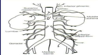
INTRODUCTIONTO EVAR & TEVAR
Dr Amrit Kumar
Senior Resident
Dept of CTVS
ENDOVASCULAR AORTIC REPAIR (EVAR)
• First Endovascular Abdominal aortic aneurysm repair done by Parodi,1990
• Performed by inserting graft components folded and compressed within a delivery sheath through the lumen of an
access vessel, usually the common femoral artery
• Upon deployment, the endograft expands, contacting the Aortic wall proximally and distally to exclude diseased
part
• Mainly Used in Treatment of
- Aortic Aneurysm (an abnormal dilatation of the aorta greater than 50% of the normal proximal segment)
- Aortic Dissection
- Penetrating Aortic Ulcer
Advantages over Open-surgical repair
• Avoidance of long incisions in the thorax or abdomen
• No cross-clamping of the aorta
• Lower incidence of visceral, renal, and spinal cord ischemia
• Shorter hospital stay
• Reduced recovery time
• Lower complications such as blood loss and wound infections.
• It is more suitable for patients who are considered too high-risk for open surgery due to factors such as age or
underlying health conditions.
INDICATION OF ANEURYSM REPAIR
Abdominal Aortic Aneurysm
- Symptomatic (tenderness or abdominal or back pain, evidence for embolization, rupture)
- Aneurysm size ≥5.5 cm
- AAA that has expanded by more than 0.5 cm within a six-month interval
Thoracic aortic aneurysm
- Size >6 cm
- Rapidly enlarging more than 0.5 cm within a six-month interval
- Symptoms such as chest pain, and diagnosis of aortic rupture or dissection
• Infrarenal– originates below the renal arteries
• Juxtarenal – originates at the level of the renal arteries
• Suprarenal – originates above the renal arteries
• Majority of Endovascular aneurysm repairs are performed on the Infrarenal aneurysm
• CT angiography with 3-D reconstruction preferred
• 2D CT- aortic diameter measurements will be overestimated if the aorta is Angulated
• DSA cannot evaluate the True lumen diameter, extent of thrombus, plaque, or degree of calcification
• MR angiography fails to depict vessel wall calcification, which has implications for vascular access
Pre-Operative Imaging
Aortic neck diameter
• Aortic diameter at the lowest renal artery
• The required endograft diameter 15 to 20% more than
the measured aortic neck diameter
• This provides sufficient radial force to prevent device
migration
• Over-sizing lead to kinking of the device, thrombus formation
or endoleak
• Under sizing- inadequate seal
- A conical neck is present when the diameter of the aorta 15 mm below the lowest renal artery is ≥10 percent larger
than the diameter of the aorta at the lowest renal artery
Solution-
• Supra-renal Fixation
• Balloon-expandable Stent
• Fenestrated or Branched Endograft
Conical/reverse tapered aortic neck
• The distance from the lowest renal artery to the origin of the
aneurysm
• Should be at least 13-15 mm to provide an adequate
proximal landing zone for endograft fixation
Aortic neck length
• The angle formed between points connecting the lowest
renal artery, the origin of the aneurysm, and the aortic
bifurcation
• Ideally, the aortic neck angle should be < 600
• Greater lead to difficulties in Implantation, Kinking,
Endoleak, and Distal device migration
• Angle >600 is generally considered to be a contraindication
Aortic neck angulation
ILIAC ARTERY & ACCESS VESSEL MORPHOLOGY
• Should have No significant Stenosis, Mural thrombus Calcification
and Tortuosity
• CIA is a preferred distal attachment site
• EIA diameter of 7mm is needed to allow safe passage of the endograft
delivery sheath
• CIA between 8 and 22 mm
• Length of normal diameter CIA into which the limbs of the endograft
will be fixed should be at least 15-20 mm to achieve an adequate seal
• In Case of Diffuse narrowing or significant calcification –an iliac conduit
can be created
• Proximal neck length >10mm
• Neck Diameter <28mm
• Aortic neck angulation <60
• Iliac artery diameter >7mm and <15mm
 Minimal to moderate tortuosity
 No mural thrombus at attachment sites
 Minimal calcification
• No associated mesenteric occlusive disease
Ideal case for EVAR
ENDOGRAFT
• Principle-Self-expanding metallic stent framework with a high outward radial force that allows attachment to the
artery wall and graft fabric to creates a new conduit for blood flow.
• There are significant variation in Endovascular graft design
• Three types of components are common to all:
A delivery system
Main body device
Extension (limb)
DELIVERY SYSTEM
• Typically delivered through the femoral artery, either percutaneously or by direct surgical cutdown
• If the femoral artery is too small to accommodate the delivery system, access can be obtaind by suturing
a synthetic graft to the iliac artery (ie, iliac conduit) through a retroperitoneal low abdominal incision
• The size of the delivery system varies depending upon the device diameter
ENDOGRAFTS MAIN DEVICE
• The main body device is usually bifurcated.
• Endovascular grafts rely primarily upon outward tension in the proximal graft to maintain the position of the graft
• Fixation system may also include barbs or a suprarenal uncovered extension
Endograft Extensions
• Bifurcated abdominal aortic grafts require adjunctive placement of iliac artery limbs to complete the graft
• Iliac Limbs on the main body device vary in length depending upon weather the graft is a 2 or 3 component graft
STENT-GRAFT DESIGN
• Commonaly used Graft system are
1. AneuRx (Medtronic, Inc., Minneapolis, MN)
2. Talent (Medtronic, Inc., Minneapolis, MN)
3. Zenith (Cook, Inc., Bloomington,IN)
4. Excluder (W.L Gore and Associates, Flagstaff, AZ)
5. Endurant (Medtronic, Inc., Minneapolis, MN)
6. Powerlink (Endologix, Irvine,CA)
A) AneuRx®device - A Bifurcated stent graft composed of a nitinol exoskeleton and polyester(Dacron) lining. It is
deployed just below the renal arteries and relies on radial force to fix the device into place. Distal and proximal
extension cuffs are available.
B) The Gore Excluder - Bifurcated device, with a Nitinol exoskeleton and a PTFE graft. It has proximal barbs to
anchor into the proximal infrarenal aorta.
C) The Powerlink®system- A unibody device made of PTFE and a Cobalt chromium alloy skeleton. It has a long
main body and sits on the anatomic bifurcation.
D) The Zenith®device- The bare proximal stents allow for suprarenal fixation. The device also has barbs to allow for
more secure attachment into the suprarenal aorta. It has a stainless steel exoskeleton.
A) Fenestrated Graft-
• Useful when Aneurysm involving the visceral vessels proximally or associated with hypogastric artery aneurysms
• Have openings in the fabrics of the endograft, which allow flow into the visceral arteries. Also Can be used when
the proximal aortic neck is short (ie, <10mm)
Special Design Graft
• B) Branched- Grafts have a separate small grafts sutured to the basic endovascular graft for deployment into a
vessel to preserve flow into it. Branched grafts have been designed to accommodate the hypogastric and renal
arteries.
C) Chimney grafts technique- Stent placed parallel to the aortic stent graft
• Used to preserve perfusion to branch vessels
• In the absence of available fenestrated or branched grafts, chimney grafts remain a feasible endovascular option for
high risk patients.
CHOICE OF GRAFT
• The choice is based upon multiple factors, including patient anatomy, operator preference and cost
• Bifurcated grafts are more often, but are not appropriate for patients with unilateral severe iliac stenosis or occlusion
• Unilateral iliac stenosis-unibody grafts known as aorta-uni-iliac (AUI) devices are used. Iliac artery occluded followed by
Distal Femoro-Femoro Bypass Grafting to maintain Distal limb perfusion.
PREPARATION
• Antithrombotic therapy- Moderate to high risk for DVT. The incidence of DVT following EVAR is 5.3% spite of
pharmacologic thromboprophylaxis
• Antibiotic prophylaxis- A first generation Cephalosporin or, in the case of penicillin allergy Vancomycin is
recommended
• Anesthesia- can be performed under GA or local anaesthesia with conscious sedation
• Vascular access- Bilateral femoral access is needed – via surgical cutdown or percutaneously.
PROCEDURE
1. Gaining vascular access
2. Placement of arterial guidewires and sheaths
3. Imaging to confirm aortoiliac anatomy
4. Main body deployment
5. Gate cannulation (bifurcated graft)
6. Iliac limb deployment
7. Graft ballooning
8. Completion imaginig
• Once vascular access is established and landmarks for positioning the device are obtain with angio, the main
device is positioned with particular attention paid to the location of the opening for the contralateral iliac limb (“
contralateral gate”)
• The aortic neck is imaged, slight degree of craniocaudal and left anterior oblique angulation may improve imaging
of the renal ostia
• With the proximal radiopaque markers of the graft positioned appropriately
• The body of the graft is deployed
• A guidewire is advanced through the contralateral access site into the contralateral gate
• Once the contralateral guidewire is positioned within the main body of the endograft, the deployment of the
endograft at the neck of the aneurysm is completed followed by deployment of the contralateral
• Once the Endograft component are in place, the attachment sites and endograft junctions are gently angioplastied with a
compliant or semi compliant balloon
• Completion angiography is performed to evaluate the patency of the renal arteries and evaluate for endoleak
• Guidewire access is maintained throughout the procedure but is particularly important when removing the main graft body
device sheath since disruption of the access vessels by an oversized sheath may not become apparent until after sheath has been
removed.
ENDOLEAK
• Type I -Persistent flow at proximal (a) or
distal (b) attachment sites
• Type II -Retrograde flow from side branches
• Inferior mesenteric
• Lumbar arteries
• Type III- Graft defect
• Type:-IV- Graft porosity
• Type V- Continued aneurysm sac expansion
without a demonstrable leak on any imaging
modality.
• Endoleak is a term that describes the presence of persistent flow of blood into the aneurysm sac after device
placement
• Untreated type I and III endoleaks are at high risk of rupture
• Type II are the commonest endoleaks, affecting up to 43% of cases, associated with low (0.52%) risk of rupture and
a significant rate of spontaneous closure. Treatment is required only for endoleaks that persist for more than a year
in an aneurysm of increasing size.
• Type IV endoleak typically resolves in 24 hours. It has not been associated with any long-term adverse events and
does not require any treatment.
COMPLICATION
DEPLOYMENT RELATED
• Failed deployment
• Arterial rupture
• Bleeding
• Dissection
• Retroperitoneal Heamorrhage
• Infection
• Embolization
IMPLANT RELATED
• Endoleaks
• Limb occlusion/stent graft kink
• Stent migration
• AAA rupture
SYSTEMIC COMPLICATION
• Cardiac
• Renal insufficiency, contrast-induced neuropathy
• Deep vein thrombosis
• Pulmonary embolism
• Coagulation
• Bowel ischemia
• Spinal cord ischemia
DEVICE MIGRATION
• Device movement of >10mm relative to anatomic landmark
• Multifactorial- Aortic neck length, Angulation, Thrombus in the aortic neck
• Short neck and Late neck dilation after EVAR is a major cause of concern because of the potential loss of proximal
fixation and seal
• Superstiff-Guidewire Technique: Maneuvers like bending the guidewire before introduction, in order to align it
with the axes of the aneurysm and the neck, could be helpful in prevention.
• The Anaconda device: undergoing clinical trials .This is the only graft system that enables repositioning of the
graft after deployment. It is highly flexible and has good torque control
POSTOPERATIVE SURVEILLANCE
• 30 days : CTA and X-ray Abdomen PA+Lateral
• 6 months: CTA can be omitted if no prior endoleak and good component overlap
• 1 year: CTA and X-ray Abdomen PA+lateral
• If No Endoleak and stable/shrinking Aneurysm
• Annual Doppler US with plain radiographs
• CTA if increasing diameter or new endoleak
THORACIC ENDOVASCULAR AORTIC REPAIR (TEVAR)
• TEVAR refers to the percutaneous placement of a stent graft in the descending thoracic or thoracoabdominal aorta in patient
• 1994, Dake first reported the initial Stanford experience with 13 patients undergoing endovascular therapy of
descending thoracic aortic aneurysms, had 100 percent technical success and no mortality.
• TEVAR was first approved by the FDA for Thoracic aneurysm repair following the Gore TAG pivotal trial in 2005
• Thoracic aortic aneurysm
• Penetrating aortic ulcer
• Blunt thoracic aortic injury
• Type B aortic dissection ( Type A Unsuitable for TEVAR due to involvement of Aortic valve and Proximity to Coronaries)
• Aortobronchial fistula
• Co-arctation
• Pseudoaneurysm
INDICATION
• The “gold standard” for preoperative evaluation is a computed tomography (CT) angiogram that includes the
thorax, abdomen, pelvis, and femoral arteries
• Procedure is typically done under GA
• A lumbar drain is placed in the L3-L4 disc space for drainage of CSF in cases where extensive coverage of the
thoracic aorta is anticipated where interruption of contributing blood supply is high
• Lumbar drainage of cerebrospinal fluid to decrease the pressure in the subarachnoid space and in increase the spinal
cord perfusion pressure
PROCEDURE
• The thoracic aorta is of larger Caliber than that of the infrarenal aorta so needs larger diameter stent grafts & Creation of a conduit
to the femoral or iliac artery may be necessary to achieve adequate access
• There is high force of blood flow in the thoracic aorta so requires a longer seal zone (20 mm both proximal and distal)
• In severe cases of tortuosity of Ileo-femoral vessel, Brachiofemoral access may be required to perform “Body Flossing” with
an stiff wire. A long, stiff wire is guided from the brachial artery and retrieved through the femoral artery. Gentle traction on
both the brachial and femoral sites will straighten out the tortuosity.
LANDING ZONE
• Unless revascularization is performed, proximal landing in zones 0 and 1 is
unacceptable because of the occlusion of the Left CCA in zone 1 & the
innominate artery in zone 0.
• Proximal landing in zone 2 is commonly used with either partial or total
occlusion of the left subclavian artery.
• Zone 3 landing is dependent on the exact anatomical neck at the arch. Proximal
landing in zone 3 can lead to angulation of the graft, which provides
inadequate sealing of the proximal graft.
• Zone 4 landing is usually straightforward because of the lack of angulation and
distance from the arch vessels
• To view the landing areas in the arch, it is necessary to view the arch
with a 60-to-90 degree left anterior oblique (LAO) projection
• A pigtail catheter via the femoral or brachial/radial artery is used to perform an angio, After the angiogram is
performed, the proximal neck is evaluated. The length and the diameter of the proximal and distal neck are
measured. Based on these measurements, the stent graft is chosen. The patient is heparinized to an activated
clotting time of 250 seconds.
.
• The stent graft is flushed with heparinized solution
and advanced into the proximal neck. A repeat
angiogram is commonly performed to reconfirm the
positioning of the device within the aorta and the
landing zone
• Prior to device deployment, different manufacturers of TEVAR devices recommend the induction of transient
cardiac arrest or a significant decrease in blood pressure to obtain precise deployment of the device, avoiding
migration secondary to forward arterial blood flow.
• During deployment, another option is to place a transfemoral venous pacing wire, and rapid pacing can be done to
prevent ejection.
• The ventilator is stopped shortly for device deployment in to eliminate motion artifact
• After the deployment, the stent graft is ballooned to decrease
the possibility of Endoleaks
• A completion angiogram is performed to confirm lack of
gross Endoleak
• The distal pulses are checked to assure they correspond with
the preoperative baseline. This maneuver is important as
embolic events to the lower extremities need to be realized
and acted on immediately
STENT GRAFTS
• Gore-TAG device-made of e-PTFE and an exoskeleton made of nitinol. The proximal and distal ends of the graft
have scalloped flares
• Medtronic Talent thoracic stent graft system- Four are available: proximal main, proximal extension, distal
main, and distal extension. The proximal configurations and the distal extension are offered with a bare-spring
design (Free-Flo design). The bare-spring design allows for placement of the device crossing the arch vessels
proximally and the celiac artery distally for suprasubclavian and infraceliac fixationIt consists of a woven polyester
graft with a nitinol endoskeleton
• The Cook TX2 stent graft- is a two-piece modular endograft system made of proximal and distal tubular
endografts. The proximal endograft is covered and has stainless steel barbs, allowing for active fixation to the aortic
wall. The distal component has at its distal end a bare metal. The TX2 is made of Dacron fabric covered by
stainless steel Z-stent
Gore-TAG device
The Cook TX2
stent-
Medtronic Talent
COMPLICATION
• Risk of Perioperative stroke 4% to 8%
• The risk of spinal cord ischemia 3 to 11%
• Viseral ischemia can occur with coverage of the celiac axis
• LOCAL
i. Groin hematoma
ii. Infection
iii. Lymphocele
• ACCESS ARTERY INJURY
i. Thrombosis
ii. Dissection
iii. Pseudoaneurysm
THANK YOU

Tevar & evar ppt by Dr. Amrit kumar CTVS

  • 1.
    INTRODUCTIONTO EVAR &TEVAR Dr Amrit Kumar Senior Resident Dept of CTVS
  • 2.
    ENDOVASCULAR AORTIC REPAIR(EVAR) • First Endovascular Abdominal aortic aneurysm repair done by Parodi,1990 • Performed by inserting graft components folded and compressed within a delivery sheath through the lumen of an access vessel, usually the common femoral artery • Upon deployment, the endograft expands, contacting the Aortic wall proximally and distally to exclude diseased part • Mainly Used in Treatment of - Aortic Aneurysm (an abnormal dilatation of the aorta greater than 50% of the normal proximal segment) - Aortic Dissection - Penetrating Aortic Ulcer
  • 3.
    Advantages over Open-surgicalrepair • Avoidance of long incisions in the thorax or abdomen • No cross-clamping of the aorta • Lower incidence of visceral, renal, and spinal cord ischemia • Shorter hospital stay • Reduced recovery time • Lower complications such as blood loss and wound infections. • It is more suitable for patients who are considered too high-risk for open surgery due to factors such as age or underlying health conditions.
  • 4.
    INDICATION OF ANEURYSMREPAIR Abdominal Aortic Aneurysm - Symptomatic (tenderness or abdominal or back pain, evidence for embolization, rupture) - Aneurysm size ≥5.5 cm - AAA that has expanded by more than 0.5 cm within a six-month interval Thoracic aortic aneurysm - Size >6 cm - Rapidly enlarging more than 0.5 cm within a six-month interval - Symptoms such as chest pain, and diagnosis of aortic rupture or dissection
  • 6.
    • Infrarenal– originatesbelow the renal arteries • Juxtarenal – originates at the level of the renal arteries • Suprarenal – originates above the renal arteries • Majority of Endovascular aneurysm repairs are performed on the Infrarenal aneurysm
  • 7.
    • CT angiographywith 3-D reconstruction preferred • 2D CT- aortic diameter measurements will be overestimated if the aorta is Angulated • DSA cannot evaluate the True lumen diameter, extent of thrombus, plaque, or degree of calcification • MR angiography fails to depict vessel wall calcification, which has implications for vascular access Pre-Operative Imaging
  • 8.
    Aortic neck diameter •Aortic diameter at the lowest renal artery • The required endograft diameter 15 to 20% more than the measured aortic neck diameter • This provides sufficient radial force to prevent device migration • Over-sizing lead to kinking of the device, thrombus formation or endoleak • Under sizing- inadequate seal
  • 9.
    - A conicalneck is present when the diameter of the aorta 15 mm below the lowest renal artery is ≥10 percent larger than the diameter of the aorta at the lowest renal artery Solution- • Supra-renal Fixation • Balloon-expandable Stent • Fenestrated or Branched Endograft Conical/reverse tapered aortic neck
  • 10.
    • The distancefrom the lowest renal artery to the origin of the aneurysm • Should be at least 13-15 mm to provide an adequate proximal landing zone for endograft fixation Aortic neck length
  • 11.
    • The angleformed between points connecting the lowest renal artery, the origin of the aneurysm, and the aortic bifurcation • Ideally, the aortic neck angle should be < 600 • Greater lead to difficulties in Implantation, Kinking, Endoleak, and Distal device migration • Angle >600 is generally considered to be a contraindication Aortic neck angulation
  • 12.
    ILIAC ARTERY &ACCESS VESSEL MORPHOLOGY • Should have No significant Stenosis, Mural thrombus Calcification and Tortuosity • CIA is a preferred distal attachment site • EIA diameter of 7mm is needed to allow safe passage of the endograft delivery sheath • CIA between 8 and 22 mm • Length of normal diameter CIA into which the limbs of the endograft will be fixed should be at least 15-20 mm to achieve an adequate seal • In Case of Diffuse narrowing or significant calcification –an iliac conduit can be created
  • 13.
    • Proximal necklength >10mm • Neck Diameter <28mm • Aortic neck angulation <60 • Iliac artery diameter >7mm and <15mm  Minimal to moderate tortuosity  No mural thrombus at attachment sites  Minimal calcification • No associated mesenteric occlusive disease Ideal case for EVAR
  • 14.
    ENDOGRAFT • Principle-Self-expanding metallicstent framework with a high outward radial force that allows attachment to the artery wall and graft fabric to creates a new conduit for blood flow. • There are significant variation in Endovascular graft design • Three types of components are common to all: A delivery system Main body device Extension (limb)
  • 15.
    DELIVERY SYSTEM • Typicallydelivered through the femoral artery, either percutaneously or by direct surgical cutdown • If the femoral artery is too small to accommodate the delivery system, access can be obtaind by suturing a synthetic graft to the iliac artery (ie, iliac conduit) through a retroperitoneal low abdominal incision • The size of the delivery system varies depending upon the device diameter
  • 16.
    ENDOGRAFTS MAIN DEVICE •The main body device is usually bifurcated. • Endovascular grafts rely primarily upon outward tension in the proximal graft to maintain the position of the graft • Fixation system may also include barbs or a suprarenal uncovered extension Endograft Extensions • Bifurcated abdominal aortic grafts require adjunctive placement of iliac artery limbs to complete the graft • Iliac Limbs on the main body device vary in length depending upon weather the graft is a 2 or 3 component graft
  • 17.
    STENT-GRAFT DESIGN • Commonalyused Graft system are 1. AneuRx (Medtronic, Inc., Minneapolis, MN) 2. Talent (Medtronic, Inc., Minneapolis, MN) 3. Zenith (Cook, Inc., Bloomington,IN) 4. Excluder (W.L Gore and Associates, Flagstaff, AZ) 5. Endurant (Medtronic, Inc., Minneapolis, MN) 6. Powerlink (Endologix, Irvine,CA)
  • 18.
    A) AneuRx®device -A Bifurcated stent graft composed of a nitinol exoskeleton and polyester(Dacron) lining. It is deployed just below the renal arteries and relies on radial force to fix the device into place. Distal and proximal extension cuffs are available. B) The Gore Excluder - Bifurcated device, with a Nitinol exoskeleton and a PTFE graft. It has proximal barbs to anchor into the proximal infrarenal aorta. C) The Powerlink®system- A unibody device made of PTFE and a Cobalt chromium alloy skeleton. It has a long main body and sits on the anatomic bifurcation. D) The Zenith®device- The bare proximal stents allow for suprarenal fixation. The device also has barbs to allow for more secure attachment into the suprarenal aorta. It has a stainless steel exoskeleton.
  • 19.
    A) Fenestrated Graft- •Useful when Aneurysm involving the visceral vessels proximally or associated with hypogastric artery aneurysms • Have openings in the fabrics of the endograft, which allow flow into the visceral arteries. Also Can be used when the proximal aortic neck is short (ie, <10mm) Special Design Graft
  • 20.
    • B) Branched-Grafts have a separate small grafts sutured to the basic endovascular graft for deployment into a vessel to preserve flow into it. Branched grafts have been designed to accommodate the hypogastric and renal arteries.
  • 21.
    C) Chimney graftstechnique- Stent placed parallel to the aortic stent graft • Used to preserve perfusion to branch vessels • In the absence of available fenestrated or branched grafts, chimney grafts remain a feasible endovascular option for high risk patients.
  • 22.
    CHOICE OF GRAFT •The choice is based upon multiple factors, including patient anatomy, operator preference and cost • Bifurcated grafts are more often, but are not appropriate for patients with unilateral severe iliac stenosis or occlusion • Unilateral iliac stenosis-unibody grafts known as aorta-uni-iliac (AUI) devices are used. Iliac artery occluded followed by Distal Femoro-Femoro Bypass Grafting to maintain Distal limb perfusion.
  • 23.
    PREPARATION • Antithrombotic therapy-Moderate to high risk for DVT. The incidence of DVT following EVAR is 5.3% spite of pharmacologic thromboprophylaxis • Antibiotic prophylaxis- A first generation Cephalosporin or, in the case of penicillin allergy Vancomycin is recommended • Anesthesia- can be performed under GA or local anaesthesia with conscious sedation • Vascular access- Bilateral femoral access is needed – via surgical cutdown or percutaneously.
  • 24.
    PROCEDURE 1. Gaining vascularaccess 2. Placement of arterial guidewires and sheaths 3. Imaging to confirm aortoiliac anatomy 4. Main body deployment 5. Gate cannulation (bifurcated graft) 6. Iliac limb deployment 7. Graft ballooning 8. Completion imaginig
  • 27.
    • Once vascularaccess is established and landmarks for positioning the device are obtain with angio, the main device is positioned with particular attention paid to the location of the opening for the contralateral iliac limb (“ contralateral gate”) • The aortic neck is imaged, slight degree of craniocaudal and left anterior oblique angulation may improve imaging of the renal ostia • With the proximal radiopaque markers of the graft positioned appropriately • The body of the graft is deployed • A guidewire is advanced through the contralateral access site into the contralateral gate
  • 28.
    • Once thecontralateral guidewire is positioned within the main body of the endograft, the deployment of the endograft at the neck of the aneurysm is completed followed by deployment of the contralateral • Once the Endograft component are in place, the attachment sites and endograft junctions are gently angioplastied with a compliant or semi compliant balloon • Completion angiography is performed to evaluate the patency of the renal arteries and evaluate for endoleak • Guidewire access is maintained throughout the procedure but is particularly important when removing the main graft body device sheath since disruption of the access vessels by an oversized sheath may not become apparent until after sheath has been removed.
  • 30.
    ENDOLEAK • Type I-Persistent flow at proximal (a) or distal (b) attachment sites • Type II -Retrograde flow from side branches • Inferior mesenteric • Lumbar arteries • Type III- Graft defect • Type:-IV- Graft porosity • Type V- Continued aneurysm sac expansion without a demonstrable leak on any imaging modality. • Endoleak is a term that describes the presence of persistent flow of blood into the aneurysm sac after device placement
  • 31.
    • Untreated typeI and III endoleaks are at high risk of rupture • Type II are the commonest endoleaks, affecting up to 43% of cases, associated with low (0.52%) risk of rupture and a significant rate of spontaneous closure. Treatment is required only for endoleaks that persist for more than a year in an aneurysm of increasing size. • Type IV endoleak typically resolves in 24 hours. It has not been associated with any long-term adverse events and does not require any treatment.
  • 32.
    COMPLICATION DEPLOYMENT RELATED • Faileddeployment • Arterial rupture • Bleeding • Dissection • Retroperitoneal Heamorrhage • Infection • Embolization IMPLANT RELATED • Endoleaks • Limb occlusion/stent graft kink • Stent migration • AAA rupture
  • 33.
    SYSTEMIC COMPLICATION • Cardiac •Renal insufficiency, contrast-induced neuropathy • Deep vein thrombosis • Pulmonary embolism • Coagulation • Bowel ischemia • Spinal cord ischemia
  • 34.
    DEVICE MIGRATION • Devicemovement of >10mm relative to anatomic landmark • Multifactorial- Aortic neck length, Angulation, Thrombus in the aortic neck • Short neck and Late neck dilation after EVAR is a major cause of concern because of the potential loss of proximal fixation and seal • Superstiff-Guidewire Technique: Maneuvers like bending the guidewire before introduction, in order to align it with the axes of the aneurysm and the neck, could be helpful in prevention. • The Anaconda device: undergoing clinical trials .This is the only graft system that enables repositioning of the graft after deployment. It is highly flexible and has good torque control
  • 35.
    POSTOPERATIVE SURVEILLANCE • 30days : CTA and X-ray Abdomen PA+Lateral • 6 months: CTA can be omitted if no prior endoleak and good component overlap • 1 year: CTA and X-ray Abdomen PA+lateral • If No Endoleak and stable/shrinking Aneurysm • Annual Doppler US with plain radiographs • CTA if increasing diameter or new endoleak
  • 36.
    THORACIC ENDOVASCULAR AORTICREPAIR (TEVAR) • TEVAR refers to the percutaneous placement of a stent graft in the descending thoracic or thoracoabdominal aorta in patient • 1994, Dake first reported the initial Stanford experience with 13 patients undergoing endovascular therapy of descending thoracic aortic aneurysms, had 100 percent technical success and no mortality. • TEVAR was first approved by the FDA for Thoracic aneurysm repair following the Gore TAG pivotal trial in 2005
  • 37.
    • Thoracic aorticaneurysm • Penetrating aortic ulcer • Blunt thoracic aortic injury • Type B aortic dissection ( Type A Unsuitable for TEVAR due to involvement of Aortic valve and Proximity to Coronaries) • Aortobronchial fistula • Co-arctation • Pseudoaneurysm INDICATION
  • 38.
    • The “goldstandard” for preoperative evaluation is a computed tomography (CT) angiogram that includes the thorax, abdomen, pelvis, and femoral arteries • Procedure is typically done under GA • A lumbar drain is placed in the L3-L4 disc space for drainage of CSF in cases where extensive coverage of the thoracic aorta is anticipated where interruption of contributing blood supply is high • Lumbar drainage of cerebrospinal fluid to decrease the pressure in the subarachnoid space and in increase the spinal cord perfusion pressure PROCEDURE
  • 39.
    • The thoracicaorta is of larger Caliber than that of the infrarenal aorta so needs larger diameter stent grafts & Creation of a conduit to the femoral or iliac artery may be necessary to achieve adequate access • There is high force of blood flow in the thoracic aorta so requires a longer seal zone (20 mm both proximal and distal) • In severe cases of tortuosity of Ileo-femoral vessel, Brachiofemoral access may be required to perform “Body Flossing” with an stiff wire. A long, stiff wire is guided from the brachial artery and retrieved through the femoral artery. Gentle traction on both the brachial and femoral sites will straighten out the tortuosity.
  • 40.
    LANDING ZONE • Unlessrevascularization is performed, proximal landing in zones 0 and 1 is unacceptable because of the occlusion of the Left CCA in zone 1 & the innominate artery in zone 0. • Proximal landing in zone 2 is commonly used with either partial or total occlusion of the left subclavian artery. • Zone 3 landing is dependent on the exact anatomical neck at the arch. Proximal landing in zone 3 can lead to angulation of the graft, which provides inadequate sealing of the proximal graft. • Zone 4 landing is usually straightforward because of the lack of angulation and distance from the arch vessels • To view the landing areas in the arch, it is necessary to view the arch with a 60-to-90 degree left anterior oblique (LAO) projection
  • 41.
    • A pigtailcatheter via the femoral or brachial/radial artery is used to perform an angio, After the angiogram is performed, the proximal neck is evaluated. The length and the diameter of the proximal and distal neck are measured. Based on these measurements, the stent graft is chosen. The patient is heparinized to an activated clotting time of 250 seconds. .
  • 42.
    • The stentgraft is flushed with heparinized solution and advanced into the proximal neck. A repeat angiogram is commonly performed to reconfirm the positioning of the device within the aorta and the landing zone
  • 43.
    • Prior todevice deployment, different manufacturers of TEVAR devices recommend the induction of transient cardiac arrest or a significant decrease in blood pressure to obtain precise deployment of the device, avoiding migration secondary to forward arterial blood flow. • During deployment, another option is to place a transfemoral venous pacing wire, and rapid pacing can be done to prevent ejection. • The ventilator is stopped shortly for device deployment in to eliminate motion artifact
  • 44.
    • After thedeployment, the stent graft is ballooned to decrease the possibility of Endoleaks • A completion angiogram is performed to confirm lack of gross Endoleak
  • 45.
    • The distalpulses are checked to assure they correspond with the preoperative baseline. This maneuver is important as embolic events to the lower extremities need to be realized and acted on immediately
  • 46.
    STENT GRAFTS • Gore-TAGdevice-made of e-PTFE and an exoskeleton made of nitinol. The proximal and distal ends of the graft have scalloped flares • Medtronic Talent thoracic stent graft system- Four are available: proximal main, proximal extension, distal main, and distal extension. The proximal configurations and the distal extension are offered with a bare-spring design (Free-Flo design). The bare-spring design allows for placement of the device crossing the arch vessels proximally and the celiac artery distally for suprasubclavian and infraceliac fixationIt consists of a woven polyester graft with a nitinol endoskeleton • The Cook TX2 stent graft- is a two-piece modular endograft system made of proximal and distal tubular endografts. The proximal endograft is covered and has stainless steel barbs, allowing for active fixation to the aortic wall. The distal component has at its distal end a bare metal. The TX2 is made of Dacron fabric covered by stainless steel Z-stent
  • 47.
    Gore-TAG device The CookTX2 stent- Medtronic Talent
  • 48.
    COMPLICATION • Risk ofPerioperative stroke 4% to 8% • The risk of spinal cord ischemia 3 to 11% • Viseral ischemia can occur with coverage of the celiac axis
  • 49.
    • LOCAL i. Groinhematoma ii. Infection iii. Lymphocele • ACCESS ARTERY INJURY i. Thrombosis ii. Dissection iii. Pseudoaneurysm
  • 50.