2. 274 E. Fishman and P.L. Faries
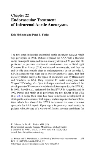
Fig. 22.1 (a) Introduction of delivery device (Schematic of Parodi JC stent—graft
1991). (b) Deployment of device (Schematic of Parodi JC stent—graft 1991)
3. 27522 Endovascular Treatment of Infrarenal Aortic Aneurysms
endovascular repair. As technology advances the percentage of
patients who undergo open repair is likely to become increasingly
smaller.
1 Anatomy
Understanding aorto-iliac anatomy is important for the successful
performance of EVAR.
Most AAA are infrarenal. However, up to 15% of AAA may have
a juxtarenal or suprarenal component. Currently available FDA-
approved devices can be used to treat most infrarenal AAA; however,
fenestrated grafts or hybrid procedures are required to treat suprare-
nal AAA. In the USA fenestrated grafts are currently available only
on a trial basis. Some of these devices are more readily available
outside the USA.
A second anatomic factor of importance relates to the landing
zone and the adequacy of the neck. A “neck” refers to the distance
from the origin of the most distal renal artery and the beginning of the
aneurysm. Currently most devices can treat AAA with a neck of up
to 3.2 cm in diameter and 15 mm in length. The talent device is
approved to treat shorter (10 mm) necks. The ideal neck is parallel,
with no angulation or eccentric thrombus in the wall. 45–60° angula-
tion is the maximum suggested by the device manufacturers
(Fig. 22.2). In cases where the diameter is not uniform throughout the
(conical) neck, up to a 3 mm difference in diameter is well tolerated.
The presence of an accessory or duplicated renal artery may reduce
the neck length. In these cases the possibility of covering an acces-
sory renal artery and the possibility of partial renal ischemia has to be
carefully considered. Other factors, which are important in terms of
the adequacy of the neck, are tortuosity of the aorta and the presence
and nature of thrombosis at the proximal seal zone. All devices today
are bifurcated and distally the stent is landed on the iliac arteries.
Distally adequate seal zones require adequate arterial length and
diameter to prevent a distal type I endoleak. 20–30% of infrarenal
AAA have common iliac artery aneurysms. If the common iliac arter-
ies are short or aneurysmal, the devices may require external iliac
artery deployment. In these cases the internal iliac artery or arteries
may need to be embolized to prevent a type II endoleak; an internal
iliac branched graft may be used in limited cases. Adequacy of
4. Fig. 22.2 (a) Treatment of AAA with talent device. Marked tortuosity of proxi-
mal aortic neck presented significant challenge to endovascular treatment. (b)
Treatment of AAA with Talent device. CTA 3D reconstruction. (c) Treatment of
AAA with Talent device. Use of proximal extension prosthesis permitted device
to conform to aortic neck and prevent endoleak at proximal fixation site
5. 27722 Endovascular Treatment of Infrarenal Aortic Aneurysms
collateral blood supply related to previous surgical history to the
colon and pelvic organs has to be taken into account. A large patent
IMA may be cause for a type II endoleak and in some centers this
vessel may be embolized preoperatively.
The third anatomic factor of importance relates to the access ves-
sels. Standard deployment of stent grafts requires placement of large
diameter sheaths in the common femoral arteries. Vessels of at least
6 mm are required for the current available devices. In addition, tor-
tuousity of femoral and iliac vessels may impede passage of the
device. Although the use of stiff wire may allow some straightening
of these vessels, care must be taken to prevent risk of arterial rupture.
Severe calcification (circumferential) of the vessels may also impede
advancement of the device. Techniques such the use of an iliac con-
duit, may allow for passage of the device. If one iliac vessel is simply
not amenable, an aorto-uni device and fem–fem bypass may be
considered.
Fig. 22.2 (continued)
6. 278 E. Fishman and P.L. Faries
2 Disease Definition
An aneurysm is defined as a 50% enlargement of a vessel. The nor-
mal infrarenal aorta measures 2 cm on average in men and 1.8 cm in
women. The normal common iliac artery measures approx 1.2 cm in
men. Most AAA are fusiform as opposed to saccular which are
slightly more frequent in thoracic aortic aneurysms (TAA).
In regards to etiology, more than 90% of aneurysm is degenera-
tive. Although the term “atherosclerotic” is still commonly applied to
such aneurysms, atherosclerosis per se is not a direct cause. Complex
processes are associated with the pathophysiology of these aneu-
rysms. Other causes of AAA are inflammatory or mycotic in nature.
Inflammatory aneurysms that are not infectious in etiology are poten-
tially amenable to EVAR. Mycotic aneurysms are not treated with
EVAR since the focus of infection is still present. The exception is an
emergency. EVAR can be used as a temporary bridge to definitive
repair for ruptured mycotic aneurysms. AAA is repaired to prevent
rupture which carries a high mortality rate. Size is the major factor
associated with aneurysm rupture. The relationship of AAA diameter
and rupture risks is described subsequently. In addition to size, the
aneurysm’s rate of increase over time, the patient’s cigarette usage,
and life expectancy play a role in the decision to operate as opposed
to the decision to observe. Clinical trials have been conducted com-
paring observation versus repair.
3 Disease Distribution
There are 27 million AAA patients worldwide. The prevalence of
AAA in the over 50 population ranges from 3% to 10% in multiple
screening and autopsy studies performed in the USA and internation-
ally. In a VA-screening study of 73,000 patients the prevalence of
AAA is 4.6% among patients from ages 50 to 79. The rupture of an
AAA is the 15th leading cause of death in the USA and the 10th lead-
ing cause of death in men older than 55. These numbers have steadily
increased since prevalence studies were performed starting 50 years
ago. The most significant risk factors for AAA are smoking, age,
gender, family history, and race. Men have a two to six times higher
frequency of AAA than women. Caucasians have a two to higher
7. 27922 Endovascular Treatment of Infrarenal Aortic Aneurysms
times frequency of AAA than non-Caucasians. Other less important
risk factors are hypertension and hypercholesterolemia.
The overall mortality of patients with ruptured AAA is 80–90%.
30–50% of patients with ruptured AAA die prior to reaching the
hospital. 30–40% of patients die after reaching the hospital without
any intervention. There are approximately 40,000 elective AAA
repair in the USA every year.
Operative mortality rates for ruptured AAA range from 40% to
50% though there is increasing evidence that mortality with endovas-
cular repair will be lower.
4 Diagnosis
Physical examination is very limited as a form of diagnosis in this
condition. The positive predictive value of physical examination is
only 15%. Most AAA are found either incidentally during the course
of radiologic examinations for other reasons or, more recently, from
screening programs. Multiple studies of ultrasound (U/S) screening
have shown benefit. Historically, most of these studies come from the
UK. Today in the USA, Medicare reimburses, a one time screening
for males 65 years and older. Screenings with a second ultrasound
have not proved to add benefit. Routine screenings for women are not
generally recommended in the USA though screenings of women
with specific risk factors, such as a positive family history or a history
of tobacco use are beneficial. The most common confirmatory test for
physical exam results is B-mode U/S. B-mode U/S is non-nvasive,
inexpensive and therefore commonly used for follow-up of small
AAA. When the AAA reaches a size where repair is considered, a
thin cut (3 mm) CT angiogram (CTA) is the test of choice. If a repair
is indicated, the CTA is useful in deciding upon whether the repair
should be open or endovascular. MRA is slightly more expensive
than CTA and requires more time for imaging, but it may be used in
lieu of CTA in specific circumstance such as when platinum coils
from a previous embolization limits the benefit of CTA, or if the
patient has an iodine dye allergy. Angiography is usually performed
when a specific preoperative intervention may be required, such as
pre-EVAR embolization of a hypogastric artery or the definition of
renal vascular anatomy in cases where the patient has more than one
renal artery. Digital subtraction angiography is rarely required at
8. 280 E. Fishman and P.L. Faries
present for diagnostic purposes and frequently underestimates the
aneurysm diameter.
Alternative approaches such as CT without contrast, CO2
as con-
trast or intravascular US (IVUS) may be utilized for preoperative
planning in patients with renal insufficiency. However the EVAR
procedure itself requires contrast, therefore the renal function itself
must be taken into account as comorbidity when evaluating a patient
for surgery.
5 Management
Repair of an asymptomatic AAA is prophylactic and elective. The
decision for repair must weigh the risk of AAA rupture on the one
hand with the operative risk and the patient’s life expectancy on the
other. A thorough discussion of each of these factors with the patient
is necessary for the patient to give informed consent.
Although not ideal, the primary determinant of AAA rupture used
in practice is aneurysm size. In the UK small aneurysm trial (UKSAT)
annual rupture risk was found to be 0.3% (3.9 cm or smaller), 1.5%
(4–4.9 cm), and 6.5% (5–5.9 cm). These finding apply mostly to men
who encompassed 85% of the study population. Other studies with
similar numbers document rupture rates of 10–20% (6–7 cm) and
20–40% (>7 cm). (Table 22.1) Advances in predicting the risk of
rupture include models of aneurysm wall stress and finite element
analysis.
Table 22.1 Range of
potential rupture rates for
a given size of abdominal
aortic aneurysm
AAA Diameter (cm) Rupture risk (%/year)
<4 0
4–5 0.5–5
5–6 3–15
6–7 10–20
7–8 20–40
>8 30–50
Brewster DC, Cronenwett JL, Hallett JW Jr,
et al. (2003) Guidelines for the treatment of
abdominal aortic aneurysms. Report of the sub-
committee of the Joint Council of the American
Association for Vascular Surgery and Society for
Vascular Surgery. J Vasc Surg.37:1106–1117
9. 28122 Endovascular Treatment of Infrarenal Aortic Aneurysms
Less significant factors influencing the risk of rupture include female
gender and family history, smoking (active as well as past history)
and the rate of the aneurysm’s expansion. Predicted rate of expan-
sion is 10% of size per year. An expansion rate beyond 10% may
assist in the recommendation for repair in smaller aneurysm.
Two major studies, The UK small aneurysm trial (UKSAT) and the
aneurysm detection and management study (ADAM), have demon-
strated a negligible risk of rupture in AAA less than 4 cm. These
aneurysms may be followed with U/S. There was a clear benefit of
AAA repair in aneurysms larger than 5.5 cm. Recommendation for
follow up versus operative intervention in aneurysms 4–5.5 cm may
vary according referral patterns, gender, and rate of expansion
In regards to endovascular repair, recently, two trials the CAESAR in
Europe and the PIVOTAL study in the USA are in the process of
studying the risk of rupture versus endovascular repair in aneurysms
smaller than 5.5 cm. Operative mortality of endovascular repair is
lower than for open repair and overall EVAR outcomes (in retro-
spective reviews) have been shown to be better in smaller AAA.
In terms of operative risk, open repair has had a steady 4% mortality
rate over the last few decades. EVAR has shown an operative mor-
tality rate of approximately 1% in multiple studies. For this rea-
son, the decision to intervene will be different if the patient is not
a candidate for endovascular repair. Given the low mortality rate
for EVAR, prediction algorithms, which take into account the
patient’s comorbidities, have not been found to contribute
significantly to the calculation of operative risk.
Calculating the life expectancy for candidates of AAA repair is not
simple. Factors such as the impact of the procedure itself, the patient’s
comorbidities (both associated and independent from AAA), and age
of the patient are all contributing factors. (Table 22.2)
Table 22.2 Life expectancy in years for patients surviving abdominal aortic
Aneurysm repair by age, gender, and race
Age (yr) Total
Male Female
White Black White Black
60 13 12 11 14 13
65 11 11 10 12 11
70 10 9 8 10 10
75 8 8 7 9 8
80 6 6 6 7 6
³85 5 4 4 5 5
Rutherford’s Vascular Surgery 7th Edition Page 1941
10. 282 E. Fishman and P.L. Faries
6 Types of Stent Grafts
Although there a many differences across stent-grafts, there are no
randomized studies comparing one device to another. The EuroSTAR
database provides information in the use and long-term follow-up of
a variety of devices. Use of one device versus the other may be more
related to the physician’s comfort with a specific device than to the
technical differences among devices. However, understanding the
differences among devices in terms of construction specifications,
benefits and failure modes is of importance. The EVAR market was
calculated at 370 million US$ in 2004 and is projected to be 1.7 bil-
lion US$ in 2012.
At present there are six FDA-approved stent graft devices in the
USA. Among the Medtronic devices (Santa Rosa, CA), the Talent
device has significantly replaced the use of the AneuRx device, which
is no longer manufactured. Other devices in use are the Gore Excluder
(W. L. Gore and Associates, Flagstaff, AZ), the Zenith (Cook, Inc.
Bloomington, IN), and the Powerlink (Endologix, Irvine, CA).
6.1 The Talent Device (Fig. 22.3)
The endovascular stent graft developed by World Medical and
Medtronic has been implanted in more than 15,000 patients world-
wide. The Talent graft is used in two configurations: tapered/aor-
touni-iliac and bifurcated/aortobi-iliac. It is self-expanding and
composed of a Dacron graft with a nitinol frame, which supports the
graft. The proximal aortic fixation device possesses a 1.5 cm of
uncovered nitinol frame proximal to the fabric portion of the device.
This uncovered portion permits transrenal fixation of the device,
thereby allowing the treatment of AAAs with relatively short or
angulated proximal necks. The Talent device may be used for proxi-
mal aortic neck sizes up to 32 mm and for iliac implantation site
diameters up to 22 mm. Deployment of the device is similar to that
of the AneuRx device, although runners are not required for delivery
of the Talent device. The main aortic component with the ipsilateral
iliac limb is delivered through a 22 or 24-French system. The second,
contralateral iliac limb module is then deployed via the contralateral
femoral artery using an 18 or 20-French delivery system.
11. 28322 Endovascular Treatment of Infrarenal Aortic Aneurysms
6.2 The Excluder
The Excluder is a modular endoprosthesis composed of PTFE bonded
to a nitinol exoskeleton. There are proximal covered flares, positive
fixation anchors, and a sealing cuff. Radiopaque markers are posi-
tioned at the very end of the graft material. The device comes in
aortic diameters of 23–31 mm and iliac diameters of 10–20 mm. The
ipsilateral limb sheath is 18–20 Fr and the contralateral limb sheath
is 12–18 Fr. The contraleral gate has a gold rim for opacification. The
device is deployed with one rapid pull of the deploying cord.
Connecting Bar
Connecting Bar
Connecting Bar
AORTIC
EXTENSION CUFF
CONTRALATERAL
LIMB
BIFURCATED
STENT GRAFT
ILIAC
EXTENSION CUFF
Mini-support Spring
Mini-support Spring
= Figur8 Radiopaque Marker
Fig. 22.3 Diagram of Medtronic Abdominal Endograft Components
12. 284 E. Fishman and P.L. Faries
6.3 The Zenith
The Zenith system from Cook is a modular bifurcated device but is
also available in an aortouni-iliac configuration. It consists of woven
polyester graft material supported throughout its length by self-
expanding stainless steel Z-stents. The introducer tip is tapered to
minimize trauma at the arterial insertion site and there are side holes
at the tip to allow angiography with the system in place. The Zenith
stent-graft system has a bare proximal stent that expands radially
upon deployment. There are barbs on this bare stent to secure the
device to the suprarenal aortic wall. The suprarenal bare stent is
deployed after being released by a trigger wire, which holds it in
place to avoid premature deployment. The main aortic body is
deployed using an 18, 20 or 22-Fr. Sheath (inner diameter). The iliac
limb is delivered using a 14 or 16-Fr. sheath (inner diameter). The
Zenith device can be used in aortic necks up to 32 mm and in iliac
arteries up to 20 mm. The system is designed to allow all components
to be used together and as a result a greater range of anatomic sizes
can be managed with the Zenith graft.
6.4 Endologix PowerLink System
The Endologix PowerLink system is a one-piece bifurcated graft
comprised of polytetrafluoroethylene (PTFE) supported by nitinol.
The one-piece design eliminates the risk of endoleaks seen at attach-
ment sites in modular devices. In addition, the frame is composed of
a self-expanding non-nitinol wire, which eliminates the need for
sutures to hold individual stents in place. The graft is thin-walled
PTFE, which may allow for downsizing of the delivery system. The
PTFE fabric is sewn to the stents only at the proximal and distal ends
of the device. This allows the fabric to move off the endoskeleton.
Aortic necks up to 26 mm may be treated.
6.5 Other Devices
There is a small group of stent grafts, which are currently used in
Europe and not available in the USA In addition to these, there is
constant development of newer devices with modifications that may
13. 28522 Endovascular Treatment of Infrarenal Aortic Aneurysms
allow use in patients who cannot undergo EVAR with the use of
current devices. These include prefabricated or in-vivo fenestrations,
branched grafts and larger aortic sizes.
6.5.1 The Anaconda System (Vascutek, Terumo,
Inchinnan, Scotland)
The Anaconda stent-graft system for aneurysm treatment is a fully
modular system made of woven material one-third thinner than con-
ventional graft material. The stents are made of nitinol. A unique
feature is the proximal ring stent, which is composed of multiple
turns of nitinol wire. The hoop strength that results from the radial
force of this ring stent allows the proximal end to anchor to the aortic
wall. Because of the saddle configuration of the proximal ring stent,
the device can be placed so that the graft is situated at and above the
renal ostia while the renal ostia themselves are uncovered. A system
of magnets is used to aid in cannulating the main body of the graft in
order to position the contralateral limb in place.
6.5.2 Trivascular Enovus and Ovation (Trivascular
Corporation, Santa Rosa, CA, USA)
The Trivascular graft employs a novel technology to provide support
to the vascular graft material. Rather than employing metallic stents
along the length of the graft material, the device contains longitudinal
channels that spiral along the course of the graft. After the device has
been positioned within the vascular system, the channels are filled
with a synthetic polymer that, when it subsequently hardens, provides
radial and longitudinal support to the graft material. In eliminating
stent support along the length of the graft, the profile of the device
has been lowered considerably. As a result, the device may be
deployed through a 14-French system. This reduction in the diameter
of the delivery system may ultimately allow for percutaneous appli-
cation of this endovascular technology.
6.5.3 Incraft (Cordis)
The Incraft device has recently started a phase II trial. The devices
uses an integrated sheath of braided construction which allows for a
14. 286 E. Fishman and P.L. Faries
small profile of 13 french. This ultra-low profile may allow for use in
a wider group of patients. The INNOVATION TRIAL is in progress
in Germany and Italy at this time.
6.5.4 Endurant (Medtronic)
The Endurant device is used in Europe at present time. The device is
undergoing evaluation in the USA for FDA approval. The device has
increased flexibility and smaller profile with 18–20 French for the
main device and 14–16 French for the contralateral iliac limb. The
delivery system is hydrophilic with an inner nitinol in delivery device
for support. Proximally, the device has suprarenal bare stent and cov-
ered M-shaped stent for increased radial force and improved proxi-
mal fixation. There is availability of straight, tapered, or flared limbs
for variable iliac anatomy.
6.5.5 Aorfix (Lombard UK)
The Aorfix device is designed for complex proximal aortic neck with
an angulation of up to 90°. The fishmouth design contributes to effec-
tive sealing. This device has been used in small group of patients in
Europe, Russia, and Brazil.
6.5.6 Aptus (Aptus Endosystems INC.)
The Aptus device uses helical endostapling technology for indepen-
dent endograft fixation. This device is being used on a trial basis in
Europe, where it is used in primary or secondary intervention for
complex necks.
6.5.7 Nellix (Nellix Endovascular)
The Nellix device uses a fully contained, polymer filled (PEG based)
endobag, which conforms to aneurismal sac and theoretically reduces
15. 28722 Endovascular Treatment of Infrarenal Aortic Aneurysms
likelihood of endoleak. This device is being used investigationally in
New Zealand and California.
7 Stent Graft Design
Stent grafts must create a seal for successful treatment of AAA to
take place. One of the factors, which may prevent this seal, initially
or in the long term, is migration of the graft distally. Other distal
components may also become separated from the normal aortic wall
by forces created by the pulses pressure of blood flow. There are dif-
ferent methods of fixation to prevent migration.
Positive fixation refers to the use of anchoring devices such as
barbs or hooks which embed into the aortic wall, which may provide
high fixation force and prevent migration. Of note, some circum-
stances such as proximal neck calcification may prevent embedding
of barbs. The, proximal covered flares and sealing cuffs may provide
further apposition. Stent stiffness along the length of the graft may
use a good seal in the iliac arteries to prevent migration of the proxi-
mal end of the device. Radial force may keep the device in place.
Suprarenal fixation in which a bare stent extends proximal to the
renal arteries may provide a theoretical advantage to prevent migration.
Radio-opaque markers are present in different forms in all devices.
It’s important to be aware of the different relationship of these mark-
ers to the actual end of the stent-graft (which is not radio-opaque). In
this manner deployment, proximally and opening of the contra-lateral
gate will be performed in the exact location and angle desired.
Since the initial development of stent grafts, there has been
significant improved in ease of the deployment mechanism. Although
a device may be more “simple” to deploy, the only real important fac-
tor in successful deployment is the understanding of device function.
Each device has a different type, use, and profile of sheath. Each
sheath has a different type of valve and may or may not allow for injec-
tion of contrast through the device at time of deployment. Improved
flexibility and trackability of devices has reduced injury to the access
arteries. In the future an even lower device profile may allow for per-
cutaneous access to the femoral vessels. It’s important to keep in mind
that as devices evolve in minor characteristics, we must renew our
long-term follow-up to confirm the improvement of EVAR results.
16. 288 E. Fishman and P.L. Faries
8 Intervention: Technique and Pitfalls
We perform all procedures in the operating room, where the utmost
sterility and nursing as well as instrument access is present in case of
the need for conversion to open repair. Anesthetic technique is mostly
based on the surgeon’s preference. The primary choice is between
spinal anesthesia with mild sedation or general anesthesia. Exceptions
are severe pulmonary disease where spinal anesthesia may be a better
option and situations when the need for iliac conduit makes general
anesthesia the better choice. We have used local with sedation, at
least initially, in cases of aneurysm rupture.
Historically access to the femoral vessels has been performed in
an open manner. With rare exceptions we perform a transverse inci-
sion above the femoral crease. This may prevent slightly the rate of
wound complications. More recently the availability of closure
devices, such as the Proglide device with the preclose technique, has
allowed us to perform a percutaneous access in certain group of
patients. CTA characteristics of femoral vessels (occlusive disease;
calcification), U/S and micropuncture-guided access and body mass
index (BMI) of patients are important factors to consider when utiliz-
ing percutaneous access.
If CTA evaluation of the external or common iliac arteries shows
severe tortuousity, circumferential calcification, or severe occlusive
disease, a few maneuvers may be considered. If the anatomy is con-
sidered prohibitive in terms of femoral access, an iliac artery conduit
using a retroperitoneal incision may be used. The anastomosis is per-
formed in an end-to-side fashion with a 10-mm graft. After placement
and deployment of the stent graft, the conduit stump may be over-
sewed or may be anastomosed to the common femoral artery in an
end-to-side fashion. This maneuver may allow for increased flow in
cases of severe occlusive iliac disease or more importantly for the pos-
sibility of need for re-intervention in the future. In our institution, in
specific groups of patients, we may perform an internal endoconduit.
This procedure requires deployment of a covered stent (to prevent
iliac artery rupture) with subsequent angioplasty to allow for a
sufficient diameter. Sewing an 8-mm PTFE graft to the distal end of
the covered stent may allow for anastomosis to the common femoral
artery at the end of the procedure. After access is acquired, stiff wires
will be required for delivery of the device. This type of wire will
straighten a tortuous iliac artery and allow for easier delivery of the
17. 28922 Endovascular Treatment of Infrarenal Aortic Aneurysms
device. Stiff wires must be inserted with the use of catheters to prevent
arterial injury or plaque disruption. Initially a stiff wire is placed
through the side of the main limb to the level of the descending tho-
racic aorta. A pigtail is placed right above the renal arteries (L1–L2).
Prior to starting to deploy, is important to plan the orientation of
the contralateral gate, for ease of access of the gate. For proximal
deployment, a magnified view is used. Parallax is avoided by center-
ing the gantry over the renal arteries. Cranio-caudad angulation of the
gantry is important to compensate for the possible anterior angulation
of the aortic neck. In the same fashion visualizing the take-off of the
lowest renal artery at right angle will allow for exact deployment
(within 2 mm of lowest renal artery). This will limit the risk of type
I endoleak in short necks and minimize the risk for device migration
in the long-term.
In terms of deployment, the Cook Zenith and Medtronic Talent
devices use a slow deployment. For these devices we begin with
deployment 1–2 cm proximal to intended deployment site. For the
Gore excluder, rapid deployment, the device is placed at the exact site
of intended placement. For the Gore or Medtronic device the main
device is deployed until the ipsilateral limb is open. In specific cases
the ipsilateral limb may remain constrained to allow ease of contral-
ateral gate access. For the Cook device the ipsilateral limb remains
constrained. At this point the contralateral gate maybe accessed or
deployment of the suprarenal fixation may be done first.
For access of the contra-lateral gate, putting the main device up
the more tortuous iliac (if safe from the profile standpoint), may
allow for a straight shot at time of cannulation. For the Medtronic and
Gore device the limbs may be crossed or parallel depending on the
angles of the iliac in question. For the Cook device crossing the limbs
is not recommended.
Access of the contra-lateral gate is successful in 99% of cases. In
case of inability to access the gate, conversion to an aorto-uni device
with a femoral–femoral bypass is performed. To access the contra-
lateral gate, an angle glidewire and catheters of different angles may
be used. If unsuccessful the wire may be snared from the main limb
(up and over), or using brachial access. Deployment of the iliac limbs
must be performed with maximum coverage (as close to hypogastric
as possible) to prevent migration and a distal type I endoleak in the
future. After complete deployment of the device, a compliant balloon
is used to complete apposition of the graft to the aortic wall. Care
18. 290 E. Fishman and P.L. Faries
must be taken to avoid inflating the balloon in the uncovered distal
iliac arteries in order to prevent arterial injury or rupture. Completion
arteriography is performed with an overall image and magnified
proximal and distal views. Endoleaks must be ruled-out, patency of
renals and hypogastric must be evaluated and exact landing of the
device proximally and distally must be confirmed.
For treatment of a proximal type I endoleak, after using the bal-
loon for a second time, a proximal cuff and, if necessary, a large
(uncovered) Palmaz stent may be used (Chap. 17—Endovascular
treatment of symptomatic abdominal aortic aneurysms). This some-
times requires extending the coverage proximally at or above the
renal arteries. To preserve the renal arteries the chimney graft tech-
nique has been suggested.
8.1 Chimney Stent
This is a stent placed parallel to the aortic stent graft to maintain flow
to the visceral side branches including the renal arteriesm which were
overstented to ensure an adequate seal. Depending upon the extent to
which the stent graft is deployed, this might require stenting of the
superior mesenteric artery (SMA), one or both renals.
Typically covered stents are used; however, uncovered stents can
also be used in certain circumstances. Both can be either balloon
expandable or self expanding, depending upon the need, whether the
need is for radial strength or flexibility respectively.
In preplanned procedures, a juxta-aneurysmal branch is cannu-
lated in an antegrade fashion by brachial artery access. A hydrophilic
glidewire is used to cannulate the branch. This is exchanged for a stiff
wire such as an Ampltaz Super Stiff (Boston Scientific, Natick, MA,
USA). Over this a long sheath with a stent is introduced into the side
branch. The aortic stent graft is then released, and the renal stet is
deployed in the side branch as a chimney stent.
For a distal type I endoleak, limb extensions may be used. If the
size of an ectatic common iliac artery does not allow for a proper seal,
intra-op embolization of the hypogastric artery with limb extension
into the external iliac artery may be performed. Newer branched
devices are being developed to preserve flow into the internal iliac.
They are not FDA approved yet. A femoral–femoral bypass, or
19. 29122 Endovascular Treatment of Infrarenal Aortic Aneurysms
external-to-internal iliac bypass may be performed to preserve flow
to the pelvis; although there is good evidence that preoperative, early
embolization of one or both hypogastric arteries is well tolerated.
9 Complications and Management
(a) Endoleaks:ThesearedescribedindetailinChap.23—Endoleaks—
Types and Management (Figs. 22.4 and 22.5).
(b) Renal Artery Occlusion: Renal artery occlusion is primarily
caused by a deployment issue.Awareness of parallax, renal artery
angle, distance of covered graft to opacified markers is of para-
mount importance. If partial renal artery coverage is diagnosed at
time of procedure, attempt at stenting through brachial artery
access should be considered.
(c) Graft Limb Occlusion: With the recent modifications of using
short evenly distributed stents within the limb grafts, and ade-
quate preoperative measurement, limb occlusion is uncommon.
However, close early postoperative clinical surveillance must
take this potential complication into account. In case of suspicion
of this, the patient should undergo a rapid CT angiogram.
(d) Stent-Graft Infection: Prevention requires keeping strict sterile
surgical technique. Reported stent graft infection is low at less
than 0.5%. However, if this does develop, this requires explanta-
tion of part or all of the graft.
(e) Pelvic Ischemia: Occlusion of one or both internal iliac arteries
may lead to pelvic ischemia. Buttock claudication is the most
common consequence; at approximately 30% of patients with
bilateral hypogastric artery occlusion. 50% of these patients’
symptoms will usually resolve within 6 months, other patients’
symptoms may improve. Other potential consequences of internal
iliac artery occlusion are erectile dysfunction, buttock necrosis,
ischemic colitis, colon necrosis, and spinal ischemia. Previous
colonic surgery where the SMAdoes not provide collaterals to the
colon and diseased profunda femoris artery with decreased pelvic
collaterals are some of the risk factors.At our institution we embo-
lize one side, wait 2 weeks looking for signs of ischemia of pelvic
ischemia and then wait another 2 weeks prior to EVAR. In specific
cases we perform routine sigmoidoscopy the day after surgery.
20. 292 E. Fishman and P.L. Faries
(f) Complications of VascularAccess: Rupture of the iliac artery at
time of the delivery of the device is potentially catastrophic and
maintaining wire access when starting the closure of the artery
may be life saving. Alternatively, in case of suspicion an angio-
gram with the sheath tip just inside the arteriotomy site can be
done. We routinely mark pulses or Doppler signals prior to start-
ing the procedure. A dissecting flap of a diseased access artery or
embolization may cause distal vessel occlusion.
Fig. 22.4 As in the figure
21. 29322 Endovascular Treatment of Infrarenal Aortic Aneurysms
10 Follow-up Protocol
At this time, there is wide institutional variability in terms of EVAR
follow-up. Initial rigorous follow-up with contrast-enhanced CT was
required to confirm ongoing procedural success. Many institutions
still used the initial protocol of a 1-month, 6-months, 1-year, and then
yearly CTA. A CTA with a non-contrast phase, an early phase (type I
and III endoleaks), and late phase (type II endoleaks) with 3D recon-
struction. Recent studies have shown that up to 25% of endoleaks
may not be adequately characterized by CTA. To distinguish the
Fig. 22.5 As in the figure
22. 294 E. Fishman and P.L. Faries
source of the endoleak, a duplex U/S (with or without contrast) or a
time resolved MRA (dynamic 3D) may be helpful. MRA will not be
useful in stents made with stainless steel.
After initial 1-month and 12-month follow-up with CTA some
institutions advocate follow-up with a duplex U/S. A CT without IV
contrast can also be used and is accurate in terms of aneurysm size
growth. U/S is being used in many institutions as part of the follow-
up protocol in attempts to minimize the use of CT or MR. However,
U/S is operator dependent and may not be as accurate in obese
patients. Further studies are required to clearly determine a standard
multi-institutional protocol.
11 Outcomes
EVAR has replaced Open AAA repair as the procedure of choice in
treatment of infrarenal abdominal aortic aneurysm. This is based on
the fact that in both level I randomized trials (EVAR I and DREAM)
and both major registries (EuroSTAR and Lifeline), as well as in
industry-supported studies the 30-day mortality was 1.5% in average,
and the 30-day mortality for open repair is 4.5–5%. Questions of
whether or not this benefit will hold up over the long term are difficult
to answer at the present time. Although the overall mortality curves
meet around the 2-year mark and aneurysm related death meet at the
4-year mark. In addition, in some of the studies, there may have been
an implicit bias against EVAR since healthier patients were more
likely to undergo open repair while patients who were less healthy
and were not candidates for open repair underwent EVAR.
One of the questions that have arisen has been whether EVAR
requires more re-interventions and more readmissions to the hospital
and whether this explains the potential loss of benefit when compared
to open surgery. However in a recent study, readmission and opera-
tions for complications of open surgery (such as bowel obstruction
9.7% and abdominal wall hernias 14.2%) were taken into account.
The study demonstrated that EVAR patients did well in terms of long-
term re-interventions.
23. 29522 Endovascular Treatment of Infrarenal Aortic Aneurysms
References
1. Parodi JC, Palmaz JC, Barone HD. Transfemoral intraluminal graft implantation
for abdominal aortic aneurysms. Ann Vasc Surg. 1991;5:491–9.
2. Lederle FA, Wilson SE, Johnson GR, et al. Immediate repair compared with
surveillance of small abdominal aortic aneurysms. N Engl J Med. 2002;346:
1437–44.
3. The UK Small Aneurysm Trial Participants. Mortality results for randomized
controlled trial of early elective surgery or ultrasonographic surveillance for
small abdominal aortic aneurysms. Lancet. 1998;352:1649–55.
4. EVAR trial participants. Endovascular aneurysm repair versus open repair in
patients with abdominal aortic aneurysm (EVAR trial 1): randomized con-
trolled trial. Lancet. 2005;365:2179–86.
5. Prinssen M, Verhoeven EL, Buth J, et al. Dutch Randomized Endovascular
Aneurysm Management (DREAM) Trial Group. A randomized trial compar-
ing conventional and endovascular repair of abdominal aortic aneurysms.
N Engl J Med. 2004;351(16):1607–18.
6. Schermerhorn ML, O’Malley AJ, Jhaveri A, et al. Endovascular vs. open
repair of abdominal aortic aneurysms in the Medicare population. N Engl J
Med. 2008;358:464–74.
7. Cao P. CAESAR trial collaborators. Comparison of surveillance vs. aortic
endografting for small aneurysm repair (CAESAR) trial:study design and
progress. Eur J Vasc Endovasc Surg. 2005;30:245–51.