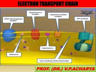
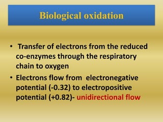
The document summarizes electron transport chain and oxidative phosphorylation. It discusses:
1) The four complexes of the electron transport chain located in the inner mitochondrial membrane that facilitate the transfer of electrons from NADH and FADH2 to oxygen. This creates a proton gradient used by ATP synthase to generate ATP.
2) The enzymes, electron carriers like cytochromes and iron-sulfur proteins, and redox reactions involved in electron transport.
3) How the proton gradient is used by ATP synthase to drive ATP synthesis via chemiosmosis.
4) Inhibitors and uncouplers that disrupt the proton gradient or electron transport.
Biological oxidation (part - III) Oxidative PhosphorylationAshok Katta
Biological oxidation (part - III) Oxidative Phosphorylation
- Mechanism of Oxidative Phosphorylation
-- Chemiosmotic theory
-P:O Ratio
Substrate Level Phosphorylation
Shuttle Systems for Oxidation of Extramitochondrial NADH
Biological oxidation (part - III) Oxidative PhosphorylationAshok Katta
Biological oxidation (part - III) Oxidative Phosphorylation
- Mechanism of Oxidative Phosphorylation
-- Chemiosmotic theory
-P:O Ratio
Substrate Level Phosphorylation
Shuttle Systems for Oxidation of Extramitochondrial NADH
The electron transport chain is comprised of a series of enzymatic reactions within the inner membrane of the mitochondria, which are cell organelles that release and store energy for all physiological needs.
As electrons are passed through the chain by a series of oxidation-reduction reactions, energy is released, creating a gradient of hydrogen ions, or protons, across the membrane. The proton gradient provides energy to make ATP, which is used in oxidative phosphorylation.
Pentose phosphate pathway is an alternative pathway to glycolysis and TCA cycle for oxidation of glucose. It is a shunt of glycolysis. It is also known as hexose monophosphate (HMP) shunt or phosphogluconate pathway. It occurs in cytoplasm of both prokaryotes and eukaryotes. While it involves oxidation of glucose, its primary role is anabolic rather than catabolic. It is an important pathway that generates precursors for nucleotide synthesis and is especially important in red blood cells (erythrocytes).
This presentation is targeted for MBBS, MD and BDS students that describes briefly about aetiopathogenesis, tumour markers, anti cancer agents, apoptosis
Glycine is an aliphatic amino acid which gives rise to many vital derivatives. This is a non-essential amino acid. This presentation is targeted for MBBS, MD, BDS and general Biochemistry students.
Report Back from SGO 2024: What’s the Latest in Cervical Cancer?bkling
Are you curious about what’s new in cervical cancer research or unsure what the findings mean? Join Dr. Emily Ko, a gynecologic oncologist at Penn Medicine, to learn about the latest updates from the Society of Gynecologic Oncology (SGO) 2024 Annual Meeting on Women’s Cancer. Dr. Ko will discuss what the research presented at the conference means for you and answer your questions about the new developments.
MANAGEMENT OF ATRIOVENTRICULAR CONDUCTION BLOCK.pdfJim Jacob Roy
Cardiac conduction defects can occur due to various causes.
Atrioventricular conduction blocks ( AV blocks ) are classified into 3 types.
This document describes the acute management of AV block.
Ozempic: Preoperative Management of Patients on GLP-1 Receptor Agonists Saeid Safari
Preoperative Management of Patients on GLP-1 Receptor Agonists like Ozempic and Semiglutide
ASA GUIDELINE
NYSORA Guideline
2 Case Reports of Gastric Ultrasound
Couples presenting to the infertility clinic- Do they really have infertility...Sujoy Dasgupta
Dr Sujoy Dasgupta presented the study on "Couples presenting to the infertility clinic- Do they really have infertility? – The unexplored stories of non-consummation" in the 13th Congress of the Asia Pacific Initiative on Reproduction (ASPIRE 2024) at Manila on 24 May, 2024.
Prix Galien International 2024 Forum ProgramLevi Shapiro
June 20, 2024, Prix Galien International and Jerusalem Ethics Forum in ROME. Detailed agenda including panels:
- ADVANCES IN CARDIOLOGY: A NEW PARADIGM IS COMING
- WOMEN’S HEALTH: FERTILITY PRESERVATION
- WHAT’S NEW IN THE TREATMENT OF INFECTIOUS,
ONCOLOGICAL AND INFLAMMATORY SKIN DISEASES?
- ARTIFICIAL INTELLIGENCE AND ETHICS
- GENE THERAPY
- BEYOND BORDERS: GLOBAL INITIATIVES FOR DEMOCRATIZING LIFE SCIENCE TECHNOLOGIES AND PROMOTING ACCESS TO HEALTHCARE
- ETHICAL CHALLENGES IN LIFE SCIENCES
- Prix Galien International Awards Ceremony
Title: Sense of Smell
Presenter: Dr. Faiza, Assistant Professor of Physiology
Qualifications:
MBBS (Best Graduate, AIMC Lahore)
FCPS Physiology
ICMT, CHPE, DHPE (STMU)
MPH (GC University, Faisalabad)
MBA (Virtual University of Pakistan)
Learning Objectives:
Describe the primary categories of smells and the concept of odor blindness.
Explain the structure and location of the olfactory membrane and mucosa, including the types and roles of cells involved in olfaction.
Describe the pathway and mechanisms of olfactory signal transmission from the olfactory receptors to the brain.
Illustrate the biochemical cascade triggered by odorant binding to olfactory receptors, including the role of G-proteins and second messengers in generating an action potential.
Identify different types of olfactory disorders such as anosmia, hyposmia, hyperosmia, and dysosmia, including their potential causes.
Key Topics:
Olfactory Genes:
3% of the human genome accounts for olfactory genes.
400 genes for odorant receptors.
Olfactory Membrane:
Located in the superior part of the nasal cavity.
Medially: Folds downward along the superior septum.
Laterally: Folds over the superior turbinate and upper surface of the middle turbinate.
Total surface area: 5-10 square centimeters.
Olfactory Mucosa:
Olfactory Cells: Bipolar nerve cells derived from the CNS (100 million), with 4-25 olfactory cilia per cell.
Sustentacular Cells: Produce mucus and maintain ionic and molecular environment.
Basal Cells: Replace worn-out olfactory cells with an average lifespan of 1-2 months.
Bowman’s Gland: Secretes mucus.
Stimulation of Olfactory Cells:
Odorant dissolves in mucus and attaches to receptors on olfactory cilia.
Involves a cascade effect through G-proteins and second messengers, leading to depolarization and action potential generation in the olfactory nerve.
Quality of a Good Odorant:
Small (3-20 Carbon atoms), volatile, water-soluble, and lipid-soluble.
Facilitated by odorant-binding proteins in mucus.
Membrane Potential and Action Potential:
Resting membrane potential: -55mV.
Action potential frequency in the olfactory nerve increases with odorant strength.
Adaptation Towards the Sense of Smell:
Rapid adaptation within the first second, with further slow adaptation.
Psychological adaptation greater than receptor adaptation, involving feedback inhibition from the central nervous system.
Primary Sensations of Smell:
Camphoraceous, Musky, Floral, Pepperminty, Ethereal, Pungent, Putrid.
Odor Detection Threshold:
Examples: Hydrogen sulfide (0.0005 ppm), Methyl-mercaptan (0.002 ppm).
Some toxic substances are odorless at lethal concentrations.
Characteristics of Smell:
Odor blindness for single substances due to lack of appropriate receptor protein.
Behavioral and emotional influences of smell.
Transmission of Olfactory Signals:
From olfactory cells to glomeruli in the olfactory bulb, involving lateral inhibition.
Primitive, less old, and new olfactory systems with different path
Ethanol (CH3CH2OH), or beverage alcohol, is a two-carbon alcohol
that is rapidly distributed in the body and brain. Ethanol alters many
neurochemical systems and has rewarding and addictive properties. It
is the oldest recreational drug and likely contributes to more morbidity,
mortality, and public health costs than all illicit drugs combined. The
5th edition of the Diagnostic and Statistical Manual of Mental Disorders
(DSM-5) integrates alcohol abuse and alcohol dependence into a single
disorder called alcohol use disorder (AUD), with mild, moderate,
and severe subclassifications (American Psychiatric Association, 2013).
In the DSM-5, all types of substance abuse and dependence have been
combined into a single substance use disorder (SUD) on a continuum
from mild to severe. A diagnosis of AUD requires that at least two of
the 11 DSM-5 behaviors be present within a 12-month period (mild
AUD: 2–3 criteria; moderate AUD: 4–5 criteria; severe AUD: 6–11 criteria).
The four main behavioral effects of AUD are impaired control over
drinking, negative social consequences, risky use, and altered physiological
effects (tolerance, withdrawal). This chapter presents an overview
of the prevalence and harmful consequences of AUD in the U.S.,
the systemic nature of the disease, neurocircuitry and stages of AUD,
comorbidities, fetal alcohol spectrum disorders, genetic risk factors, and
pharmacotherapies for AUD.
- Video recording of this lecture in English language: https://youtu.be/lK81BzxMqdo
- Video recording of this lecture in Arabic language: https://youtu.be/Ve4P0COk9OI
- Link to download the book free: https://nephrotube.blogspot.com/p/nephrotube-nephrology-books.html
- Link to NephroTube website: www.NephroTube.com
- Link to NephroTube social media accounts: https://nephrotube.blogspot.com/p/join-nephrotube-on-social-media.html
Lung Cancer: Artificial Intelligence, Synergetics, Complex System Analysis, S...Oleg Kshivets
RESULTS: Overall life span (LS) was 2252.1±1742.5 days and cumulative 5-year survival (5YS) reached 73.2%, 10 years – 64.8%, 20 years – 42.5%. 513 LCP lived more than 5 years (LS=3124.6±1525.6 days), 148 LCP – more than 10 years (LS=5054.4±1504.1 days).199 LCP died because of LC (LS=562.7±374.5 days). 5YS of LCP after bi/lobectomies was significantly superior in comparison with LCP after pneumonectomies (78.1% vs.63.7%, P=0.00001 by log-rank test). AT significantly improved 5YS (66.3% vs. 34.8%) (P=0.00000 by log-rank test) only for LCP with N1-2. Cox modeling displayed that 5YS of LCP significantly depended on: phase transition (PT) early-invasive LC in terms of synergetics, PT N0—N12, cell ratio factors (ratio between cancer cells- CC and blood cells subpopulations), G1-3, histology, glucose, AT, blood cell circuit, prothrombin index, heparin tolerance, recalcification time (P=0.000-0.038). Neural networks, genetic algorithm selection and bootstrap simulation revealed relationships between 5YS and PT early-invasive LC (rank=1), PT N0—N12 (rank=2), thrombocytes/CC (3), erythrocytes/CC (4), eosinophils/CC (5), healthy cells/CC (6), lymphocytes/CC (7), segmented neutrophils/CC (8), stick neutrophils/CC (9), monocytes/CC (10); leucocytes/CC (11). Correct prediction of 5YS was 100% by neural networks computing (area under ROC curve=1.0; error=0.0).
CONCLUSIONS: 5YS of LCP after radical procedures significantly depended on: 1) PT early-invasive cancer; 2) PT N0--N12; 3) cell ratio factors; 4) blood cell circuit; 5) biochemical factors; 6) hemostasis system; 7) AT; 8) LC characteristics; 9) LC cell dynamics; 10) surgery type: lobectomy/pneumonectomy; 11) anthropometric data. Optimal diagnosis and treatment strategies for LC are: 1) screening and early detection of LC; 2) availability of experienced thoracic surgeons because of complexity of radical procedures; 3) aggressive en block surgery and adequate lymph node dissection for completeness; 4) precise prediction; 5) adjuvant chemoimmunoradiotherapy for LCP with unfavorable prognosis.
New Directions in Targeted Therapeutic Approaches for Older Adults With Mantl...i3 Health
i3 Health is pleased to make the speaker slides from this activity available for use as a non-accredited self-study or teaching resource.
This slide deck presented by Dr. Kami Maddocks, Professor-Clinical in the Division of Hematology and
Associate Division Director for Ambulatory Operations
The Ohio State University Comprehensive Cancer Center, will provide insight into new directions in targeted therapeutic approaches for older adults with mantle cell lymphoma.
STATEMENT OF NEED
Mantle cell lymphoma (MCL) is a rare, aggressive B-cell non-Hodgkin lymphoma (NHL) accounting for 5% to 7% of all lymphomas. Its prognosis ranges from indolent disease that does not require treatment for years to very aggressive disease, which is associated with poor survival (Silkenstedt et al, 2021). Typically, MCL is diagnosed at advanced stage and in older patients who cannot tolerate intensive therapy (NCCN, 2022). Although recent advances have slightly increased remission rates, recurrence and relapse remain very common, leading to a median overall survival between 3 and 6 years (LLS, 2021). Though there are several effective options, progress is still needed towards establishing an accepted frontline approach for MCL (Castellino et al, 2022). Treatment selection and management of MCL are complicated by the heterogeneity of prognosis, advanced age and comorbidities of patients, and lack of an established standard approach for treatment, making it vital that clinicians be familiar with the latest research and advances in this area. In this activity chaired by Michael Wang, MD, Professor in the Department of Lymphoma & Myeloma at MD Anderson Cancer Center, expert faculty will discuss prognostic factors informing treatment, the promising results of recent trials in new therapeutic approaches, and the implications of treatment resistance in therapeutic selection for MCL.
Target Audience
Hematology/oncology fellows, attending faculty, and other health care professionals involved in the treatment of patients with mantle cell lymphoma (MCL).
Learning Objectives
1.) Identify clinical and biological prognostic factors that can guide treatment decision making for older adults with MCL
2.) Evaluate emerging data on targeted therapeutic approaches for treatment-naive and relapsed/refractory MCL and their applicability to older adults
3.) Assess mechanisms of resistance to targeted therapies for MCL and their implications for treatment selection
Pulmonary Thromboembolism - etilogy, types, medical- Surgical and nursing man...VarunMahajani
Disruption of blood supply to lung alveoli due to blockage of one or more pulmonary blood vessels is called as Pulmonary thromboembolism. In this presentation we will discuss its causes, types and its management in depth.
2. Biological Oxidation
Oxidation- removal of electrons
Reduction- gain of electrons
Electron donator- reducing agent/ reductant;
gets oxidized itself
Fe++ (reduced) Fe+++ (oxidized) + e-
Electron acceptor- oxidizing agent/ oxidant; gets
reduced itself
Two important e- carriers in metabolism: NAD+
& FAD
3. NAD+, Nicotinamide Adenine Dinucleotide,
is an electron acceptor in catabolic
pathways.
The Nicotinamide ring, derived from the
vitamin niacin, accepts 2 e- & 1 H+ (a
hydride) in going to the reduced state,
NADH.
NADP+/NADPH is similar except for Pi.
NADPH is e- donor in synthetic pathways.
4. The electron transfer reaction may be
summarized as :
NAD+ + 2e- + H+ NADH
It may also be written as:
NAD+ + 2e- + 2H+ NADH + H+
5. Redox couple
• When a substance exists both in the reduced
and oxidized state, the pair is called a redox
couple
• Redox potential- Electromotive force
measured by (EMF)
• Positive redox potential- higher affinity for e
than H+
• Negative redox potential- lower affinity for e
than H+
6. Redox potential
Analogous expression of standard free energy
Eo’
Redox couple
Electron flows from one redox couple to
another in the direction of more positive
system
↑negativity- ↑tendency to lose electrons
(more affinity towards H)
↑positivity- ↑ tendency to accept electrons
7. The more negative redox potential represents a greater tendency to
lose electrons
8. Substrate level phosphorylation
• Energy from a high energy compound is
directly transferred to nucleoside
diphosphate to form NTP
• 3 steps
• 1,3 –BPG (Glycolysis)
• Phosphoenolpyruvate (Glycolysis)
• Succinyl CoA (TCA cycle)
12. Biological oxidation
• Transfer of electrons from the reduced
co-enzymes through the respiratory
chain to oxygen
• Electrons flow from electronegative
potential (-0.32) to electropositive
potential (+0.82)- unidirectional flow
13. Oxidative Phosphorylation
• Energy released during biological
oxidation- trapped to form ATP
• Oxidation + phosphorylation= Oxidative
phosphorylation
14. How it all happened???
• Eugene Kennedy & A.
Lehninger – Discovered that
mitochondria is the site of
Ox. Phosphorylation
Helmut Beinert - FeS
proteins-
John E Walker-
Crystallographic str of F1
structure
16. Why Electron transport chain?
• Gradual flow of electrons through a sequence
of dehydrogenases- Electron transport chain
• NADH→ H2O; ∆ G0’= 53 Kcal/ mol
• The amount of energy is so huge that if
produced at one stretch then body may not be
able to use it
• Through ETC this energy is released in small
increments- trapped in Chemical bond energy-
ATP
17. Location of enzymes in mitochondria
Mitochondria outer membrane:
MAO
Acyl CoA synthatase
Phospholipase A2
Inter membrane space:
Adenylate kinase
Creatine kinase
Inner membrane outer surface:
Glycerol-3- P dehydrogenase
Inner membrane inner surface:
Succinate dehydrogenase
Enzymes of ETC
Soluble matrix:
TCA cycle enzymes
Beta oxidation of FA enzymes
18. • Groups of redox proteins
– Found on inner mitochondrial membrane
– Binding sites for NADH and FADH2
• On matrix side of membrane
• Electrons transferred to redox proteins
But NADH is impermeable to
mitochondrial membrane !!!
19. How NADH crosses the
mitochondrial membrane??
• Malate - aspartate shuttle- liver, kidney & heart
• Glycerol-3- Phosphate shuttle
21. Creatine phosphate shuttle
• Carries active phosphate from mitochondria
to extra-mitochondrial sites
• Skeletal mm & Heart
• MtCK - mitochondrial CK involved in the
shuttle
• Ubiquitous mtCK (umtCK) & Sarcomeric mtCK
(smtCK)
22. Enzymes, coenzymes and electron carriers
Oxidoreductases (Enzymes)
1. Oxidases- removal of H using O as a H acceptor; cyt
oxidase, tyrosinase, MAO
2. Dehydrogenases- Removal of H but O not as an
acceptor; Require co-enzymes NAD, NADP, FMN, FAD
3. Hydroperoxidases- Peroxidases & Catalases;
Hydrogen peroxide or organic peroxide are
eliminated
4. Oxygenases- Addition of 1 or both of the atoms of O2
a) Monooxygenases (mixed function oxidases)
b) dioxygenases (true oxygenases)
24. Oxygenases
Mono-oxygenases- Incorporate one atom of oxygen (1/2
O2)
• NADPH provides the reducing equivalents
• Ex- Cyt P450 monooxygenase system in microsome –
drug metabolism (morphine, aniline, aminopyrine)
• Biosynthesis of steroid hormones
Dioxygenases- Incorporates both atoms of oxygen
Ex- Homogentisate oxidase, L-Tryptophan pyrrolase
25. Electron carriers
• Cytochromes - Fe containing electron
transferring proteins
• 3 classes- a, b , c
• Cytochrome oxidase (Cyt aa3)- oxidase enz
• Rest all- Dehydrogenase
• ETC b→ c1 → c → aa3
• Cyt c- water soluble
• Cytochromes are also found in ER
27. Iron-sulfur protein (Fe4S4)
• Fe not in heme form
• 8 Fe-S proteins participate
• One electron transfer and 2 H+ transferred
• Fe+++
3, Fe++
1 (oxidized) + 1 e- Fe+++
2, Fe++
2
• (reduced)
• About 6 FeS proteins are discovered
• Mechanism not very clear
• Associated with FMN→ CoQ and Cyt b and C1
Helmut Beinert
28. Ubiquinone
O
O
CH3O
CH3CH3O
(CH2 CH C CH2)nH
CH3
OH
OH
CH3O
CH3CH3O
(CH2 CH C CH2)nH
CH3
2 e-
+ 2 H+
coenzyme Q
coenzyme QH2
FAD FeS
FeS
FeS
FMN
NAD+
ubiquinone
Cyt b
ubiquinone
29. • Coenzyme Q (CoQ, Q or Ubiquinone) is lipid-
soluble.
• Accepts 2e- via complex I & II
• Q→ QH2
• It stays dissolved in the hydrocarbon core of a
membrane.
• Only electron carrier not bound to a protein.
• it can accept/donate 1 or 2 e-
30. If an idea presents itself to us, we must
not reject it simply because it does not
agree with the logical deductions of a
reigning theory.
—Claude Bernard
32. NAD+
FMN
FeS
ubiquinoneFAD FeS
Cyt b
FeS Cyt c1 Cyt c Cyt a Cyt a3
1/2 O2
ubiquinone
I
II
III IV
NADH Dehydrogenase
Succinate
dehydrogenase
Cytochrome Oxidase
CoQ-cyt c Reductase
36. Complex I (NADH→ Ubiquinone)
• NADH dehydrogenase complex
• Has NADH binding site
– NADH reductase activity
• NADH - NAD+
– NADH ---> FMN--->FeS --> ubiquinone
– Ubiquinone ---> Ubiquinone H2
– 4 H+ pumped/NADH
25 different proteins
Energy released- 12 Kcal/mol
Energy utilised to pump out protons
1 ATP generated
37. Substrates for complex I
• Glyceraldehyde - 3 P
• Isocitrate
• Malate
• Glutamate
• Beta hydroxy acyl CoA
• Pyruvate
• Alpha keto glutarate lipoate→ FAD
38. Complex II
Succinate-Q-Reductase
• Succinate →FAD → FeS → CoQ
– FADH2 binding site
• FAD reductase activity
• FADH2 -- FAD
Substrates are- 1. Succinate
2. Acyl CoA
3. Glycerol-3- P
39. FADH2 bypasses complex I
• FADH2 doesn’t produce enough energy
• 1 ATP less produced than NADH
40. Complex III- Cytochrome reductase
• Cluster of FeS proteins, Cyt b
& c1
• CoQ → FeS → Cyt b c1 →
Cyt C
• FeS- Rieske’s FeS (bound to
His residues instead of 2Cys
residues)
• Free energy change- -10 Kcal
/ Mol
• 1 ATP synthesised
• 4H+ pumped out
41. Complex IV-Cytochrome oxidase
• Reduction of oxygen
• Cytochrome oxidase
• Cyt a+a3 red → oxidized state
• oxygen → water
– 2 H+ + 2 e- + ½ O2 -- 2 H2O
– transfers e- one at a time to oxygen
• Pumps 2H+ out
– Total of 10 H+ / NADH
– Total of 6 H+ / FADH2
1 ATP generated
44. Complex V- ATP Synthase complex
• ATP synthase
• Chemiosmotic theory
• Proton gradient-
electrochemical potential
• pH outside- 1.4 units lower
• Outside is positive relative
to the inside- +0.14 v
• Only 40% energy trapped
45. Organisation of ATP synthase complex
• 2 units
• Fo and F1
• Fo – o for Oligomycin
• Fo embedded in the inner mitochondrial
membrane
• F1- towards matrix
• 3α and 3β, 1 γ and 1 δ subunit
46. Binding change mechanism
• Water driven hammer minting
coins
• F1 has 3 confirmations- O, L & T
• O- doesn’t bind to substrates /
products
• L- Loosely binding
• T- Tightly binding
Paul D Boyer
N.P. 1997
47. Protons pumped to inter-membrane space
↓
Inner membrane impermeable to H+
↓
Protons induce rotation of γ subunits
↓
Induces conformational changes in β subunits
↓
ADP and Pi bind in L conformation
↓
L changes to T and ATP is formed
↓
T changes to O and ATP is released
48. An energy dependent process
2.5 ATP generated from NADH and 1.5 ATP from FADH2
49.
50. O2 consumption as a measure of electron transport
An oxygen electrode can measure O2 consumption in respiring mitochondria.
53. Babies do not shiver- Brown fat contains
Thermogenin (Non-shivering thermogenesis)
Thermogenin acts as a channel and allows H+
↓
Proton gradient disrupted
↓
Phosphorylation uncoupled; Energy dissipated as
heat
54. Other inhibitors of Oxidative
phosphorylation
• Atractyloside – Inhibition of ATP- ADP
exchange, inhibits translocase
• Doxorubicin- cardiotoxic; inhibits Ox-Phos
and also damages Mitochondria by free
radical generation
55. Valinomycin is an ionophore
• Works by changing K+
gradient
• Disrupts the gradient
across the membrane
56. MtDNA is maternally inherited
• OXPHOS diseases
• Lack of introns in MtDNA makes it 10 times more
susceptible to mutation
• Occurs in tissues having high rate of Ox-Phos
• CNS, skeletal mm, cardiac mm, Liver
58. • Myoclonic epilepsy and ragged
red fibre disease (MERRF)-
proteins for tRNA synthesis are
not synthesised properly
• MELAS (Mito. Encephalo-
myopathy lactic acidosis stroke
like episodes)
• DM- Mutations in the
mitochondrial lysyl-tRNA gene
59.
60. Mitochondria is involved in Apoptosis and
oxidative stress
• MPTP- Mitochondrial permeability transition
pore- escape of Cyt C and activating caspase 9
• Free radical damage by superoxide formation